Giant Frontal Mucocele in Child: about a Case
Introduction: The frontal mucocele is the most frequent of facial sinus mucoceles. It can expand and cause a facial distortion as well as endocrinal and orbital signs which require an urgent treatment. We hereby describe a case of a left frontal sinus mucocele which diagnostic and therapeutic approach we are discussing according to the data mentioned in this literature. Observation: It is the case of a 13 years old female patient who was, admitted for a frontal tumefaction on the left side. The physical examination revealed a large, renitent and painless supraorbital frontal tumefaction on the left side involving the whole superolateral angle and the upper edge socket with a limited upward movement including a ptosis. As a result, the facial and cranial encephalic computed tomography (CT) scan a diagnostic showed a cystic mass located in the frontal sinus with an osteolysis of the external table. The treatment consisted in an exeresis of the mass via external approach. The post-surgery history was simple and uneventful. Conclusion: Frontal sinus mucoceles diagnosis in our case is often late. This may cause some complications related to the compression of the noble organs along with a facial deformation. In our practice the therapy is surgical via external means.
Introduction
Facial sinus mucoceles are pseudo cystic benign and expansive tumors which substance is mucous, sticky and aseptic [1]. It is an uncommon pathology that mainly affects adults [2]. Mucoceles lesions develop slowly and are relatively locally aggressive. Frontal sinus mucoceles are more common and can expand towards the eye socket and the endocranium [3, 4]. Risks of complications with kinds of blindness or brain abscess through a superinfection require from then an urgent surgery. We report the case of a large frontal mucocele which diagnostic and therapeutic approach will be reviewed with data from the literature.
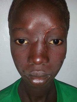
The patient is a 13 year old girl from Burkina Faso who was admitted for a frontal tumefaction on the left side developing for 1 year and 4 months. This tumefaction increased gradually and slowly in volume without any fever or health condition alteration. In her past history there is no notion of trauma, nasal or sinus surgery, or notion of sinus infection. The physical examination showed a renitent and painless fronto-orbital tumefaction on the left side, involving the whole superolateral angle and the entire upper edge eye socket with a skin looking healthy (Figure 1).

Figure 1: Left fronto-orbital tumefaction with ptosis. The ophthalmological examination revealed a reduction in the eye motion with a limited upward movement and a ptosis. Facial and Cranioencephalic TDM showed a cystic mass occupying the left frontal sinus. This cystic mass is homogeneous, well limited, blowing the sinus wall with osteolysis areas of the outer table (Figure 2) and it is also associated with bilateral maxillary sinusitis and ethmoid without parenchymal extension. The diagnostic hypothesis of a mucocele was asked. The cystic mass was removed by a first track of the brow Jacques under general anesthesia (Figure 3). After an incision of the upper left brow and detachment of subcutaneous flaps, we discover a lysis of the anterior wall of the frontal sinus and highlight a cystic mass in the left frontal sinus supraorbital. The spontaneous opening of the cystic mass was deaf left a thick mucoid whitish liquid. After suction of secretions, curettage of the mucocélique pocket we find a communication of both sinuses. The other walls of the frontal sinus in particular the posterior and inferior walls were unremarkable.
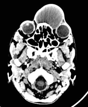
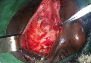
Figure 3: Per-operative image showing a ruptured cystic mass. Unclogging the nasal-frontal canal was conducted in order to ensure the ventilation and drainage of the frontal sinus. We performed the opening of the channel at the aid of a hemostat pliers placed in the nasal cavity with which it opens the meatus of nasofrontal channel as much as possible. A fatty wick was placed into the left frontal sinus, which is exited through the naso-front channel in homolateral nostril with the aid of pliers. An anterior nasal packing was placed. The postoperative care consisted in an antibiotic therapy (Ceftriaxone1g / day), corticosteroids 40mg / 24H and washing the sinus cavity with SSI 0,9% serum. The removal of the wick was performed at postoperative day 2. The postoperative course was uneventful. The patient was discharged on day 2. Histological examination of cystic pocket found a mucocele. Bacteriological research content was negative. On a decline of 6 months we have not noticed recurrence (Figure 4).
Discussion
Mucoceles are rare benign tumors of the facial sinus that sit most often in the frontal sinuses [3, 5]. They are the result of an obstruction of the ostium which causes a blockage of secretions and a gradual extension of the sinus cavity volume [6] and this may lead to the giant mucocele. It along with a thinning of the bone walls. The high frequency of the affection of the frontal sinus may be due to the confinement of naso frontal channel. The sinus expansion can then lead to a compression of the surrounding structures. Several triggering factors are involved in the onset and development of mucocele. The most common are traumatic and surgical such as facial sinus and sinonasal endoscopic surgery. Inflammatory factors (chronic rhinosinusitis) are also reported and radiotherapy in tumors of the facial structure as well. In our case, the pathogenesis of frontal mucocele was unclear. But it would probably be related to a chronic sinusitis. The highest incidence is between the ages of 30 and 40 years old [4]. However, our patient is relatively younger than the age reported in the literature. The mucocele is symptomatic pauci in general. As for the giant mucocele, it shows signs of borrowing depending on the topography of the sinus. If the clinic enables to evoke a mucocele, imaging plays an important role in the diagnosis and therapeutic decision making. Thus, TDM is a first line examination, allowing the diagnosis [7]. The mucocele is presented as an expansive and convex density developed at the expense of a sinus cavity having an "egg shape". The walls of the mucocele are reshaped, classically refined and repress adjacent structures without infiltrating them. They can be partial or eroded [7]. TDM enables to take stock of extension to neighboring organs, noting the preoperative anatomic variations and to look for a likely cause. In our case, TDM of the facial sinus has been of major interest in the diagnostic and therapeutic approach, because it allowed us to specify the frontal topography of the mucocele, affection of adjacent organs and find the likely cause. Resonance Imaging (MRI), for better tissue contrast resolution, to better appreciate the relationship with sinus adjacent noble organs [3]. The diagnosis of a mucocele is radio-clinical particularly in the giant form. Giant mucocele form (ENT, eye doctor, neurosurgeon) therapy is always surgical with a multidisciplinary approach. Two therapeutic attitudes coexist. Endonasal endoscopic functional surgery (FESS) and the first external channel. Some authors [7, 8] recommend a first immediate endonasal approach which will be supplemented if necessary by external means. Indeed endoscopy is currently the method of choice in the surgical management of chronic rhino-sinus diseases. In the mucocele, the principle is based on the marsupialization while respecting the mucosa health state and expanding natural drainage channels [7]. This is what we did with our patient by external approach. Other authors [4, 7, 9] also defend the external approach in the most aggressive, compressive or invasive forms. Indeed, it is on the one hand the most common location and on the other hand the one where morbidity and recurrence are the most frequent [7, 10]. In our context, the lack of technical equipment requires us an external approach first. But the major drawback is the aesthetic ransom. So, for benign lesions should we carry on with the external approach as endoscopic surgery is now well codified.
Conclusion
Giant frontal mucocele is a rare pseudo- cystic benign tumor. However it can compress adjacent organs when it is large. The effect of mass on the noble organs requires an urgent and adequate care. This care should be multidisciplinary. The diagnosis must be early to avoid a giant mucocele. Hence, the importance of educating parents and equipping the technical platform of our health facilities for an efficient management of cases.
Conflict of interest
The authors declare that there is no conflict of interest regarding the publication of this paper.
References
-
Aderdour L, Fakiri MMEL, Nouri H, Hassani R, Raji A (2010) Les mucocèles sinusiennes : aspects diagnostiques et thérapeutiques à propos de 16 cas. Journal marocain des sciences médicales. 2010; 17(3): 2010.
-
Alamia F, Benchekroun N, Berdaoui El N, Oumelal J, Berraho A (2013) Mucocèle sphénoïdale bilatérale révélée par une paralysie du nerf abducens : à propos d’un cas. Journal Française d’ophtalmologie 36(5) : 87-91.
-
Ba ML, Tall A, Hossini A, Ly Ba A, Ndoye N, et al. (2005) Les mucocèles du sinus frontal en milieu neurochirugical. A propos de 16cas Dakarois. AJNS 24(2): 40-47.
-
Abdülcemal ÜI, Selçuk A, Erhan A and Süleyman B (2015) A giant frontoethmoid mucocele with intracranial extension. SCOTTISH Medical journal 60(1): 1-3.
-
Conboy PJ, Jones NS (2003) The place of endoscopic sinus surgery in the treatment of paranasal sinus mucoceles. Clin.Otolaryngol 28(3): 207-210.
-
Klossek J M, Dufour X, Ferrie JC, Fontanel JP (2003) Pneumo sinus dilatans et mucocèles des cavités naso- sinusienne. EMC (Elsevier Masson SAS, Paris) Oto- Rhino-Laryngologie 20-730-A-10, 8p.
-
Facon F, Nicollas R, Paris J, Dessi P (2008) La chirurgie des mucocèles sinusiennes : notre expérience à propos de 52 cas suivis à moyen terme. Rev Laryngol Otol Rhinol 129(3): 167-173.
-
Nao EEM, Ndiaye M, Diom S, Toure S, Deguenonvo R, et al. (2012) Mucocèle fronto-ethmoïdal: A propos d’un cas. Journal Africain de chirurgie 2(2): 101-104.
-
Khong JJ, Malhotra R, Wormald PJ, D Selva (2004) Endoscopic sinus surgery for paranasal sinus mucocèle with orbital involvement. Eye 18: 877-881.
-
May M, Schaitkin B (1995) Frontal sinus surgery: Endonasal drainage instead of an external osteoplastic approach. Operative Tech Otolaryngol Head Neck Surg 6(3): 184-192.
- 4th Branchial Cleft Sinus Anomaly Presenting as Recurrent Thyroid Abscess in A Child: A Case Report
- Parotid Duct Injury Repaired Using an Angiocatheter Stent: A Case Report
- Organization and Functionality of the Referral and Counter-Referral System for ENT Disorders in District Hospitals of N’Djamena, Chad: A Cross-Sectional Analytical Study
- Facial Metastases from a Gastrointestinal Stromal Tumor: A Case Report
- Panorama of Ent Cancers and Literature Review: Epidemiological Profile and Therapeutic Management
- Could Antimicrobial Resistance Prove to Be Both a Threat and an Opportunity for us?