Schwannoma of Tongue an Unusual and Rare Entity
Introduction: Schwannoma, also called neurilemmoma, was first described by Verocay in 1908. It is a slow growing benign tumor arising from the schwann cells of nerve sheath. Approximately 25-45% of all schwannomas occur in the head and neck region. In the head and neck region only 1% occur intraorally. Case report: We report the rare case of scahwannoma of tongue in 17 year-old male patient who presented with progressive swelling of tongue since last 1 year. Local examination revealed 2x3 cm circumscribed mucosal covered swelling on the right lateral border of mobile tongue with no other significant findings. It was excised completely via peroral approach. Histopathological examination confirmed it to be schawnnoma of tongue. Conclusion: Schwannoma should be considerd as possible differential diagnosis of any progressive intraoral painless swelling irrespective of age and sex.
Introduction
Schwannoma, also called neurilemmoma, was first described by Verocay in 1908 [1, 2]. It is a slow growing benign tumor arising from the schwann cells of nerve sheath. It is a solitary and encapsulated tumor which can arise from any nerve with two notable exceptions, the optic and olfactory nerves both of which lack Schwann cell encasement hence are not involved [3]. Approximately 25–45% of all schwannomas occur in the head and neck region [4]. The most commonly affected nerve by schwannoma is the vestibulocochlear (VIII) nerve (acoustic neurinomas) [2, 5, 6]. Schwannomas are rarely found perorally (only 1%) with tongue being the most common peroral structure involved [3, 7, 8]. It has no predilection for sex or race and it is usually seen between the third and sixth decades of life [9]. Schwannoma tongue usually appear as a painless and slowly enlarging mass, but when it grows to a certain size it may lead to dysphagia, voice changes, and breathing difficulties [10]. Given the close proximity of hypoglossal, glossopharyngeal and lingual nerves in tongue it is difficult to know the exact origin of Schwannoma [9]. Though the malignant transformation of head and neck schwannomas is rare several cases have been reported [11, 12] in the literature, including one occurrence in the tongue [13]. The goal of treatment is complete excision of the tumor, which results in low rates of recurrence [14]. The most common approach used is transoral approach, with a very low rate of morbidity and recurrence even in the large lesions [15]. Recently CO2 laser has also been used for excision of a base of tongue schwannoma [16].
Case report
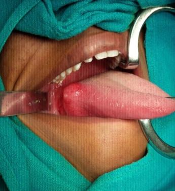
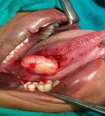
We report a rare and unusual case of schwannoma of tongue in a young male.A non smoker and non alcholic male patient of 17 years age presented with history of swelling of tongue from last 1 year.It was slowly progressive and painless with no other significant history. There was no significant past medical or surgical history.Local examination revealed 2x3 cm circumscribed mucosal covered swelling on the right lateral border of mobile tongue, which was firm in consistency and non tender as shown in (Figure 1) below. The base of tongue, floor of mouth, retromolar trigone were free and apparantly normal.No restriction of tongue movements was seen nor was any cervical lymphadenopathy felt.All the blood investigations were normal.Fine needle aspiration of the swelling was done which revealed benign mesenchymal nature of the lesion, with possibility of neurogenic tumor. The tumor was excised perorally en mass, shown in (Figure 2). The sample was sent for histopathological examination which revealed the presence of elongated tumor cells arranged in eosinophilic cords and compact oviod bodies( verocay bodies) with palisading ,confirming the diagnosis of Schwannoma as shown in (Figure 3).
![Figure 3: Histopathology of schwannoma, showing eosinophilic cords and compact oviod bodies (verocay bodies) with palisading. Discussion Schwannoma is a slow growing benign tumor arising from the schwann cells of nerve sheath. Approximately 25-45% of all schwannomas occur in the head and neck region [4]. An intraoral site israre, constituting only about 1% [7]. The peak incidence of Schwannoma is between the third and sixth decades of life, with no predilection for sex or race [10]. In our case it was young male of 17 years of age only. Although the tongue is the most common site of intraoral schwannoma there are, only 44 cases of schwannoma of tongue reported in the English literature in the last 20 years [17]. Clinically, schwannomas may be indistinguishable from other encapsulated benign tumors, like lipoma, haemengioma, fibroma, lingual thyroid, benign salivary gland tumors, rhabdomyoma, leiomyoma and lymphangioma, so biopsy and histological examination are essential for correct diagnosis.](/fulltextimages/350/fig_3.jpeg)
Figure 3: Histopathology of schwannoma, showing eosinophilic cords and compact oviod bodies (verocay bodies) with palisading. Discussion Schwannoma is a slow growing benign tumor arising from the schwann cells of nerve sheath. Approximately 25-45% of all schwannomas occur in the head and neck region [4]. An intraoral site israre, constituting only about 1% [7]. The peak incidence of Schwannoma is between the third and sixth decades of life, with no predilection for sex or race [10]. In our case it was young male of 17 years of age only. Although the tongue is the most common site of intraoral schwannoma there are, only 44 cases of schwannoma of tongue reported in the English literature in the last 20 years [17]. Clinically, schwannomas may be indistinguishable from other encapsulated benign tumors, like lipoma, haemengioma, fibroma, lingual thyroid, benign salivary gland tumors, rhabdomyoma, leiomyoma and lymphangioma, so biopsy and histological examination are essential for correct diagnosis.
Conclusion
Schwannoma should be considerd as possible differential diagnosis of any progressive intraoral painless swelling irrespective of sex and age.Complete excision forms the main treatment modality with low rate of recurrence.
References
-
Chiapasco M, Ronchi P, Scola G (1993) Neurilemmoma (Schwannoma) of the oral cavity: a report of 2 clinical cases. Minerva Stomatol 42(4): 173-178.
-
Enzinger FM, Weiss SW (1995) Soft tissue tumors 3 St Louis MO: Mosby-Year Book Inc Pp. 821-888.
-
Wayne LK, Pollak N, Daniel Liess B, Miick R, Zitsch R (2010) Schwannoma of the hard palate. Am J Otolaryngol 31(2): 139-140.
-
Katz AD, Passy V, Kaplan N (1971) Neurogenous neoplasms of major nerves of head and neck. Arch Surg 103(1): 51-56.
-
Bansal R, Trivedi P, Patel S (2005) Schwannoma of the tongue. Oral Oncol Extra 41(2): 15-17.
-
Robert OG (1997) The oral cavity. In: Steven GS (Eds.) Principle and practice of surgical pathology and cytology. New York: Churchill Livingstone pp. 1399- 1460.
-
Pfeifle R, Baur DA, Paulino A, Helman J (2001) Schwannoma of the tongue: report of 2 cases. Journal of Oral and Maxillofacial Surgery 59(7): 802-804.
-
Hatziotis JC, Asprides H (1967) Neurilemoma (schwannoma) of the oral cavity. Oral Surg Oral Med Oral Pathol 24(4): 510-526.
-
Sawhney R, Carron MA, Mathog RH (2008) Tongue base schwannoma: report, review, and unique surgical approach. Am J Otolaryngol 29(2): 119-122.
-
Das Gupta TK, Brasfield RD, Strong EW, Hajdu SI (1969) Benign solitary schwannomas (neurilemmomas). Cancer 24(2): 355-366.
-
Kragh LV, Soule EH, Masson JK (1960) Benign and malignant neurilemmomas of the head and neck. Surg Gynecol Obstet 111: 211-218.
-
Colreavy MP, Lacy PD, Hughes J et al (2000) Head and neck Schwannomas-a 10 year review. J Laryngol Otol 114(2): 119-124.
-
Piatelli A, Angelone A, Pizzicannella G, Piatelli M (1984) Malignant schwannoma of the tongue: report of a case and review of the literature. Acta Stomatol Belg 81(3): 213-225.
-
Cohen M, Wang MB (2009) Schwannoma of the tongue: two case reports and Review of the literature. Eur Arch Otorhinolaryngol 266(11): 1823- 1829.
-
Mehrzad H, Persaud R, Papadimitriou N, Kaniyur S, Mochloulis G (2006) Schwannoma of the tongue base treated with transoral carbon dioxide laser. Lasers Med Sci 21(4): 235-237.
-
Lira RB, Gonçalves Filho J, Carvalho GB, Pinto CA, Kowalski LP (2013) Lingual schwannoma: case report and review of the literature. Acta Otorhinolaryngol Ital 33(2): 137-140.
- 4th Branchial Cleft Sinus Anomaly Presenting as Recurrent Thyroid Abscess in A Child: A Case Report
- Parotid Duct Injury Repaired Using an Angiocatheter Stent: A Case Report
- Organization and Functionality of the Referral and Counter-Referral System for ENT Disorders in District Hospitals of N’Djamena, Chad: A Cross-Sectional Analytical Study
- Facial Metastases from a Gastrointestinal Stromal Tumor: A Case Report
- Panorama of Ent Cancers and Literature Review: Epidemiological Profile and Therapeutic Management
- Could Antimicrobial Resistance Prove to Be Both a Threat and an Opportunity for us?