Significance of Posterior Septectomy in the Management of Choanal Atresia in Young Adults
Unilateral choanal atresia is more common than bilateral cases. Unilateral cases may present later in life with unilateral nasal congestion and mucoid rhinorrhea. Pure bony atresia represents 30% of cases while mixed bony and membranous atresia is more common (70%) [1].
Waleed Ragab Jabri* and Mohamed Qotb
Egypt, Tel: 00201005128946; E-mail: waleedrajabri@hotmail.com
Introduction
Unilateral choanal atresia is more common than bilateral cases. Unilateral cases may present later in life with unilateral nasal congestion and mucoid rhinorrhea. Pure bony atresia represents 30% of cases while mixed bony and membranous atresia is more common (70%) [1]. The ideal procedure for management of choanal atresia should restore the normal nasal passage, be safe, prevent damage to any growing structures, have short operative time, short hospitalization and convalescence with minimal morbidity and mortality. Endoscopic trans- nasal management is a challenging procedure and is considered the standard route in choanal atresia surgery [2]. The aim of this study is to present our experience with a transnasal endoscopic posterior septectomy without postoperative stenting for repair of choanal atresia in relatively young adult patients.
Material and Methods
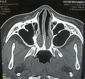
Fifteen patients were enrolled in this study with 9 females and 6 males. Age ranges from (8 to 17years) with mean of 13.5 (Table 1). Two cases presented with bilateral atresia (Figure 1) and gave a history of respiratory distress at birth. No revision cases were included.
Perspective
All surgeries were performed in Faculty of Medicine, Fayoum university hospital, Egypt, a tertiary referral hospital, between 2009 and 2012.
Clinical and Radiological Assessment
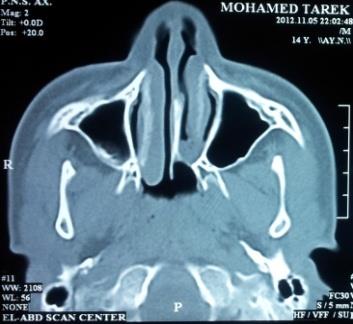
Cases were examined endoscopically by 4mm, 0◦ telescope (Karl Storz-Germany) and the atresia was documented. CT scan was performed for all cases to detect and carefully evaluate the anatomic site with a focus on the bone width of the vomer, the degree of membranous, bony, or mixed atresia, the choanal airspace distance and to delineate bony abnormalities within the nasopharynx or the nasal cavity (Figure 2).
| Patient no | Sex | Age | Uni or Bilateral | Follow up | Recurrence | Complications | ||||||||||||||
|---|---|---|---|---|---|---|---|---|---|---|---|---|---|---|---|---|---|---|---|---|
| 1 | F | 17 | Bilateral | 26 month | No | No | ||||||||||||||
| 2 | F | 15 | Unilateral | 18 month | No | No | ||||||||||||||
| 3 | M | 14 | Unilateral | 22 month | No | No | ||||||||||||||
| 4 | F | 9 | Unilateral | 18 month | No | Synechia | ||||||||||||||
| 5 | M | 12 | Unilateral | 12 month | No | No | ||||||||||||||
| 6 | M | 8 | Unilateral | 26 month | No | Synechia | ||||||||||||||
| 7 | M | 10 | Unilateral | 17 month | No | No | ||||||||||||||
| 8 | F | 12 | Bilateral | 20 month | No | No | ||||||||||||||
| 9 | F | 16 | Unilateral | 16 month | No | No | ||||||||||||||
| 10 | F | 10 | Unilateral | 19 month | No | No | ||||||||||||||
| 11 | F | 11 | Unilateral | 18 month | No | No | ||||||||||||||
| 12 | F | 15 | Unilateral | 12 month | No | No | ||||||||||||||
| 13 | M | 8 | Unilateral | 10 month | No | Synechia | ||||||||||||||
| 14 | M | 8 | Unilateral | 9 month | No | No | ||||||||||||||
| 15 | F | 10 | Unilateral | 8 month | No | Synechia |
Table 1: The cases enrolled in this study.
Surgical Technique
All surgical procedures were performed under general anesthesia with endotracheal intubation. The technique started by topical decongestion. 4mm, 0
- and 30
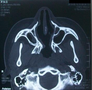
- rigid telescopes (Karl Storz-Germany) were used. A mucoperiosteal flap was elevated from the atretic plate and a mucoperichondrial flap was elevated from the posterior part of the septum (this was performed also bilaterally in bilateral case). A powered instrumentation system was used (Karl Storz intranasal drill) with 3mm diamond burrs for removing the atretic plate and vomer. It has the advantage of saving tissues due to the protection of its sheath. Drilling started infero-medially until the atretic plate was perforated, then the perforation was drilled laterally to the medial ptyregoid plate and upward until it is completely removed. The posterior part of the septum (the vomer) was drilled from the base superiorly creating one single choana. The elevated mucoperiosteal and mucoperichondrial flaps were excised up to the extent of the drilled bony septum (Figure 3). No stents were used in all cases but only nasal packing with alginates was inserted to stop postoperative bleeding and was left in place for 24 hours. Patients were discharged on next day and were instructed to use antibiotics and nasal douching with saline solution 3 times per day for 10 days. Follow up ranges from 8 to 26 months in some cases, using 4mm, 0
- and 30
- rigid telescopes (Karl Storz-Germany) as an office procedure. Removal of crusts and secretions were performed under local anesthesia in the office upon each visit.
Results
Follow up was satisfactory regarding nasal obstruction for all cases. No severe stenosis or complete closure of the common choana was noticed in all cases. The bilateral cases (3 cases) showed moderate stenosis in the new choana but none of them require either dilatation or reoperation. Four cases developed nasal synechiae. Two cases were lost during follow up. No other complications; as bleeding or skull base injury occurred either during or after the surgical procedure.
Discussion
Many approaches have been used for the repair of choanal atresia including transpalatal and transnasal routes [3]. However, [4] reported that the transpalatal approach might affect the hard palate and alveolar arch growth in 52% of patients. No doubt that the endoscopic transnasal route, is the most popular route nowadays [2, 5, 6]. It presents more excellent visualization of the nasal cavity and atretic area, good illumination and magnification, angled vision, minimal blood loss, can be applied for all ages and it has low restenosis rate [5, 7]. It is now considered the standard route in choanal atresia surgery [2]. Being congenital, this disease is rarely seen in young adults, nevertheless, many authors have published reports dealing with unilateral choanal atresia in older ages reaching 17-18 years in some reports [3, 4, 6, 7] and up to 54 year in others [6, 8]. On the other hand, bilateral cases are very difficult to present in young adult age, but few reports have been published on this issue [6, 9, 10, 11, 12]. In this study, we present eleven young adults and four children with 13 unilateral and 3 bilateral cases. We assumed that dealing with that age is different from dealing with infant and young children cases while using endoscopic approach with a drill system and aiming at the same time not to use stents. Removal of the posterior part of the nasal septum was recommended by most authors [5, 6, 12, 13], especially in revision cases [8]. It is performed by many tools either nasal instruments as backbiter forceps or drill. Only one report by Anderhuber & Stammberger [14] has mentioned that resection of the vomer was not necessary, probably because they planned to use a stent for a long period. On the other hand, the use of stents stills a controversial issue. Some authors use it for variable periods of time ranging from 1 or 2 days up to 6-8 weeks in selected cases as in children younger than 12 months while others do not [3, 5, 11, 12, 13, 15]. Its use may be associated with restenosis and foreign body sensation [4, 11]. The stents also may cause discomfort, localized infection, ulceration, circumferential scar tissue and injury to the surrounding normal tissue [9]. Stents might migrate backwards or blocked by crusts causing nasal obstruction. For these reasons, Pasquini, et al. [7] suggested to shorten the period of stenting to avoid these complications [7]. In this study, stents were not used, due to the creation of a large common posterior choana created by posterior septectomy. This study demonstrated the importance of excising the mucoperiosteal and mucoperichondrial flaps to minimize the incidence of postoperative restenosis or complete closure of the new common posterior choana.
Conclusion
The transnasal endoscopic complete posterior septectomy without stenting is an effective and safe technique in managing cases with unilateral or bilateral choanal atresia in young adult patients.
References
-
Brown OE, Pownell P, Manning SC (1996) Choanal atresia: a new anatomic classification and clinical management applications. Laryngoscope 106: 97- 101.
-
Haginomori SI, Nonaka R, Takenaka H (2005) Surgical technique in endoscopic posterior septoplasty for an adult patient with choanal stenosis. Auris Nasus Larynx 32(4): 365-368.
-
Samadi DS, Shah UK, Handler SD (2003) Choanal Atresia: a twenty-year review of medical co- morbidities and surgical outcomes. Laryngoscope 113(2): 254-258.
-
Freng A (1978) Surgical treatment of congental choanal atresia. Ann Otol Rhinol Laryngol 87: 346- 350.
-
Deutsch E, Kaufman M, Eilon A (1997) Transnasal endoscopic management of choanal atresia. Int J Pediatr Otorhinolaryngol 40(1): 19-26.
-
Van Den Abbeele T, Francois M, Narcy P (2002) Transnasal endoscopic treatment of choanal atresia without prolonged stenting. Arch Otolaryngol Head Neck Surg 128(8): 936-940.
-
Pasquini E, Sciarretta V, Saggese D, Cantaroni C, Marcri G, et al. (2003) Endoscopic treatment of congenital choanal atresia. Int J Pediatr Otorhinolaryngol 67(3): 271-276.
-
McLeod IK, Brooks DB, Mair EA (2003) Revision choanal atresia repair. Int J Pediatr Otorhinolaryngol 67(5): 517-524.
-
Josephson GD, Vickery CL, Giles WC, Gross CW (1998) Transnasal endoscopic repair of congenital choanal atresia: long-term results. Arch Otolaryngol Head Neck Surg 124(5): 537-540.
-
Kamel R (1994) Transnasal endoscopic approach in congenital choanal atresia. Laryngoscope 104 (5): 642-646.
-
Khafagy YW (2002) Endoscopic repair of bilateral congenial choanal atresia. Laryngoscope 112(2): 316- 319.
-
Durmaz A, Tosun F, Yldrm N, Sahan M, Kivrakdal C, et al. (2008) Transnasal endoscopic repair of choanal atresia: results of 13 cases and meta-analysis. J Cranio fac Surg 19(5): 1270-1274.
-
Holzmann D, Ruckstuhl M (2002) Unilateral choanal atresia: surgical technique and long-term results. J Laryngol Otol 116(8): 601-604.
-
Anderhuber W, Stammberger H (1997) Endoscopic surgery of uni-bilateral choanal atresia. Auris Nasus Larynx 24(1): 13-19.
-
Friedman NR, Mitchell RB, Bailey CM, Albert DM, Leighton SEJ (2000) Management and outcome of choanal atresia correction. Int J Pediatr Otorhinolaryngol 52(1): 45-51.
- 4th Branchial Cleft Sinus Anomaly Presenting as Recurrent Thyroid Abscess in A Child: A Case Report
- Parotid Duct Injury Repaired Using an Angiocatheter Stent: A Case Report
- Organization and Functionality of the Referral and Counter-Referral System for ENT Disorders in District Hospitals of N’Djamena, Chad: A Cross-Sectional Analytical Study
- Facial Metastases from a Gastrointestinal Stromal Tumor: A Case Report
- Panorama of Ent Cancers and Literature Review: Epidemiological Profile and Therapeutic Management
- Could Antimicrobial Resistance Prove to Be Both a Threat and an Opportunity for us?