Invasive Klebsiella Abscess Syndrome with Subsequent Acute Respiratory Distress Syndrome: a Case Report and Review
Background: Invasive Klebsiella Abscess Syndrome is a newly emerging syndrome, whereby an initial Klebsiella pneumonia liver abscess causes bacterial, extra hepatic complications. It has been well described in Asian countries because of the increased prevalence of highly virulent strains, and the increased diabetic population. However, only a handful of cases have been published in Europe. Also, a case has never been published where the syndrome has resulted in an Acute Respiratory Distress Syndrome, compounding the patient management. Case Report: A 48-year-old, diabetic Asian woman presented to a London Accident and Emergency Department with abdominal pain and fever. A CT abdomen scan showed a large liver abscess which aspirated Klebsiella pneumonia, K1 strain. The same pathogen was isolated from blood cultures and thus a diagnosis of Invasive Klebsiella Abscess Syndrome was made. The infection was managed with aspiration drainage and intravenous antibiotics. The patient developed Acute Respiratory Distress Syndrome, caused by the septicemia, which required respiratory support. After lengthy treatment the patient made a full recovery. Conclusions: This is the first case reporting these two concurrent syndromes in a country where Invasive Klebsiella Abscess Syndrome has only been described in a small number of patients in recent years. This is in contrast to Asia, where it has been reported numerous times, due to the increased pathogenic prevalence. Given increasing global migration, Invasive Klebsiella Abscess Syndrome is likely to become more prevalent in non-Asian countries.
Introduction
Klebsiella pneumonia is a pathogen most commonly associated with pneumonia and urinary tract infections [1, 2]. However, over the past three decades a new syndrome, associated with this well known pathogen, has emerged: Invasive Klebsiella Abscess Syndrome [3]. It is characterized by a primary liver abscess caused by Klebsiella that then develops an extra-hepatic complication e.g. disseminated bacteraemia, endophthalmitis, meningitis, and necrotising fasciitis [4, 5, 6]. Invasive Klebsiella Abscess Syndrome has been predominantly reported in Asian countries, the reason for which seems to be multi factorial: Klebsiella pneumonia has been shown to be the main cause of liver abscess in a number of Asian countries [7, 8] with the organism being isolated in 38% of liver abscess cases [9]. There are also an increased proportion of the highly virulent and invasive K1/K2 strains in these countries [10]. Thirdly, Diabetes Mellitus has been identified as a risk factor, with poor glycogenic control increasing the risk of metastatic complications [11]. We describe a novel clinical presentation of Invasive Klebsiella Abscess Syndrome that presented with fulminant Type 1 Respiratory Failure from Acute Respiratory Distress Syndrome (ARDS). ARDS is a serious, life-threatening condition where there is diffuse inflammation of the lung parenchyma, with interstitial degradation, and intra-alveolar oedema. The most common a etiology is severe infection, with numerous pathogens being responsible [12]. However, there has never been a case reported of Invasive Klebsiella Abscess Syndrome causing ARDS. Given the increasing prevalence of global migration, a diagnosis of Invasive Klebsiella Abscess Syndrome should be suspected in anyone with an isolated K1 or K2 stain of Klebsiella pneumonia from a liver abscess. This will facilitate prompt treatment and help prevent life threatening complications.
Case Report
A 48 year old female patient, of Asian descent, presented to our hospital’s Accident and Emergency department complaining of abdominal pain and fever for the previous two days. This was also accompanied by decreased appetite and vomiting.
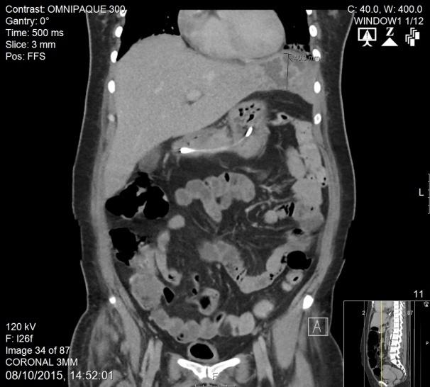
Subsequently, she was admitted to the Observation Unit with a diagnosis of sepsis from a suspected urinary tract infection (UTI); she had been treated for several UTI’s in the past year. On the day of admission, antibiotic treatment was started according to trust policy: Co- Amoxiclav 1.2g tds and Amikacin 15mg/kg stat dose. She also received 1.5l fluid resuscitation forhyperglycaemia as she was a known Type II Diabetic, normally treated with Insulin and Metformin. During the next 12 hours she did not respond to treatment, remaining tachycardia with poor blood pressure control, and tachypnoeic from trying to compensate a metabolic acidosis. Her oxygen requirements also increased, with a Chest X-Ray showing bilateral infiltrates. Her abdominal pain had increased which prompted an urgent CT abdomen scan with contrast. This showed significant hepatomegaly, 24cm, with an ill-defined hypo dense lesion in the left lobe, consistent with a liver abscess of size 6.5cm by 5.5cm, shown in (Figure 1). Significant bilateral consolidation was also noted. She was admitted to the Intensive Therapy Unit (ITU) for further monitoring and treatment. CT guided aspiration of the liver abscess was performed on Day 3 of admission which yielded a 15ml aspirate that cultured Klebsiella pneumonia. The same pathogen was also isolated from the blood cultures taken on admission, suggesting a bacteraemia following a primary liver abscess. Thus, based on the pathogen sensitivities, she received a 10 day course of Ceftriaxone 2g/12h IV, followed by Meropenem 1g/8h IV. The exact strain was subsequently typed and identified as K1 Klebsiella pneumonia which is a highly virulent strain, accounting for the majority of liver abscesses in Asian countries [10]. However, during her antibiotic treatment she still required increasing oxygen support for Type I Respiratory Failure. By Day 4 of admission a diagnosis of Acute Respiratory Distress Syndrome was made using the Berlin definition: acute onset, bilateral infiltrates, not explained by heart failure or fluid overload [13]. It was classed as severe ARDS, defined as PaO2/FiO2≥100mmHg with PEEP ≥5cm H2O. She also had a poor tolerance for Outflow™ and was tiring. Therefore, the patient agreed to be intubated. She was weaned off the ventilator and extubated after 9 days, during which time the septicemia resolved, with blood cultures showing no further growth.
The liver abscess was further drained, reducing it in size, and the patient was stepped down to the ward before being discharged home with 6 weeks of antibiotic cover: Ertapenem 1g IV, once a day.
Discussion
Klebsiella pneumonia is a Gram-negative bacterium that most commonly causes pneumonia, soft tissue, or urinary tract infections. It has been reported to have a large pathogenic reservoir in the community, often found within the gastrointestinal tract, skin, nose, and throat as a commensally [14]. As previously described, there has been an emergence of a new disease process associated with this pathogen: Invasive Klebsiella Abscess Syndrome, with a proposed mechanism for the formation of the primary Klebsiella pneumonia abscess being bacterial translocation across the intestinal endothelium, as shown in animal models [15]. This mechanism is further supported by the large number of Klebsiella pneumonia abscesses reported in Asian countries: over 900 cases reported in Asia, compared to 23 reported outside of Asia, from 1992 to 2002 [16]. This correlates to a higher incidence of gastrointestinal reservoirs in these countries [10, 17].
These gastrointestinal reservoirs also have a greater prevalence of the strains K1 and K2, which, owing to their phenotype, are more virulent and cause the majority of invasive phenomena [3]. The presence of the K1 stain, isolated from the patient, is thus consistent. The ability of a Klebsiella pneumonia abscess to cause extra-hepatic complications increases the difficulty in managing this syndrome. Vigilance is needed to identify early complications and treat accordingly. As previously mentioned, the extra-hepatic complications in Asia are well documented, where a number of retrospective studies and population-based studies have been published [16, 18, 19, 20]. The Americas have followed suit, in more recent years, detailing how the majority affected are of Asian descent [21, 22]. In contrast, the few European publications are case reports, detailing a variety of extra-hepatic complications: septic shock, bacteraemia, pneumonia were reported in a Spanish case [23]. In Croatia and Norway septicemia was reported as an extra-hepatic complication of a primary abscess [24, 25], while there are reports of the extra- hepatic complication, endophthalmitis, from both Belgium and the United Kingdom [26, 27]. However, there has never been a reported case of Invasive Klebsiella Abscess Syndrome causing ARDS. It should be noted that sepsis caused by Klebsiella pneumonia has been documented but these cases do not have a primary abscess and as such do not meet the criteria for the Syndrome [12]. It would appear that this syndrome has its origin in Asia, owing to the distribution of pathogen in its population, but now it is beginning to be noticed further afield, both in the Americas, and Europe. A question to be answered is whether this is because of already infected Asian migrants presenting in other countries, or because the gastrointestinal reservoirs are becoming more prevalent in the non-Asian population.
Conclusion
In this case report the patient was initially found to have a liver abscess caused by the virulent K1 strain of Klebsiella pneumonia. This subsequently caused septicemia, therefore meeting the criteria for Invasive Klebsiella Abscess Syndrome, as described in The Lancet by Siu et al. [3]. The case was further complicated by ARDS. This is the first published case of Invasive Klebsiella Abscess Syndrome causing ARDS, in a critically ill patient. It is also interesting as Invasive Klebsiella Abscess Syndrome has had limited cases reported in Europe compared to the vast number reported in Asia. However, given the incidence of global migration and the steep rise of the main identifiable risk factor, Diabetes Mellitus, this could be an emerging problem that may be increasingly encountered in the European health care setting.
References
-
Wang JL, Chen KY, Fang CT, Hsueh PR, Yang PC, et al. (2005) Changing bacteriology of adult community- acquired lung abscess in Taiwan: Klebsiella pneumoniae versus anaerobes. Clin Infect Dis 40(7): 915-922.
-
Shon AS, Bajwa RP, Russo TA (2013) Hypervirulent (hypermucoviscous) Klebsiella pneumoniae: a new and dangerous breed. Virulence 4(2): 107-118
-
Siu LK, Yeh KM, Lin JC, Fung CP, Chang FY (2012) Klebsiella pneumoniae liver abscess: a new invasive syndrome. The Lancet Infectious Diseases 12(11): 881-887
-
Liu YC, Cheng DL, Lin CL (1986) Klebsiella pneumoniae liver abscess associated with septic endophthalmitis. Arch Intern Med 146(10): 1913- 1916
-
Saccente M (1999) Klebsiella pneumoniae liver abscess, endophthalmitis, and meningitis in a man with newly recognized diabetes mellitus. Clin Infect Dis 29(6): 1570-1571.
-
Hu BS, Lau YJ, Shi ZY, Lin YH (1999) Necrotizing fasciitis associated with Klebsiella pneumoniae liver abscess. Clin Infect Dis 29(5): 1360-1361.
-
Chang FY, Chou MY (1995) Comparison of pyogenic liver abscesses caused by Klebsiella pneumoniae and non-K. pneumoniae pathogens. J Formos Med Assoc 94(5): 232-237.
-
Yeoh KG, Yap I, Wong ST, Wee A, Guan R, et al. (1997) Tropical liver abscess. Postgrad Med J 73(856): 89- 92.
-
Abbas MT, Khan FY, Muhsin SA, Al-Dehwe B, Abukamar M, et al. (2014) Epidemiology, Clinical Features and Outcome of Liver Abscess: A single Reference Center Experience in Qatar. Oman Medical Journal 29(4): 260-263.
-
Turton JF, Englender H, Gabriel SN, Turton SE, Kaufmann ME, et al. (2007)Genetically similar isolates of Klebsiella pneumoniae serotype K1 causing liver abscesses in three continents. J Med Microbiol 56(5): 593-597.
-
Lin JC, Siu LK, Fung C-P, Tsou H-H, Wang J-J, et al. (2006) Impaired Phagocytosis of Capsular Serotypes K1 or K2 Klebsiella pneumoniae in Type 2 Diabetes Mellitus Patients with Poor Glycemic Control. J Clin Endocrinol Metab 91(8): 3084-3087.
-
Yang SC, Liao KM, Chen CW, Lin WC (2013) Positive blood culture is not associated with increased mortality in patients with sepsis-induced acute respiratory distress syndrome. Respirology18(8): 1210-1216.
-
Ranieri VM, Rubenfeld GD, Thompson BT, Ferguson ND, Caldwell E, et al. (2012) Acute respiratory distress syndrome: the Berlin Definition. Jama 307(23): 2526-2533.
-
Podschun R, Ullmann U (1998) Klebsiella spp. as nosocomial pathogens: epidemiology, taxonomy, typing methods, and pathogenicity factors. Clin Microbiol Rev 11(4): 589-603.
-
Tu YC, Lu MC, Chiang MK, Huang SP, Peng HL, et al. (2009) Genetic requirements for Klebsiella pneumoniae-induced liver abscess in an oral infection model. Infect Immun 77(7): 2657-2671.
-
Wen-Chien K, David LP, Anthanasia JS, Dennis SH, Anne von G, et al. (2002) Community-Acquired Klebsiella pneumoniae Bacteremia: Global Differences in Clinical Patterns. Emerg Infect Dis 8(2): 160.
-
Lin YT, Siu LK, Lin JC, Chen TL, Tseng CP, et al. (2012) Seroepidemiology of Klebsiella pneumoniae colonizing the intestinal tract of healthy Chinese and overseas Chinese adults in Asian countries. BMC Microbiology 19: 12-13.
-
Chan KS, Yu WL, Tsai CL, Cheng KC, Hou CC, et al. (2007) Pyogenic liver abscess caused by Klebsiella pneumoniae: analysis of the clinical characteristics and outcomes of 84 patients. Chin Med J (Engl) 120(2): 136-139.
-
Lee SS, Chen YS, Tsai HC, Wann SR, Lin HH, et al. (2008) Predictors of septic metastatic infection and mortality among patients with Klebsiella pneumoniae liver abscess. Clin Infect Dis 47(5): 642-650.
-
Keller JJ, Tsai MC, Lin CC, Lin YC, Lin HC (2013) Risk of infections subsequent to pyogenic liver abscess: a nationwide population-based study. Clinical Microbioligy and Infection 19(8): 717-722.
-
Pastagia M, Arumugam V (2008) Klebsiella pneumoniae liver abscesses in a public hospital in Queens, New York. Travel Medicine and Infectious Disease 6(4): 228-233.
-
Rahimian J, Wilson T, Oram V, Holzman RS (2004) Pyogenic Liver Abscess: Recent Trends in Etiology and Mortality. Clin Infect Dis 39(11): 1654-1659.
-
Carrillo Esper R, Soto Hernandez JL, Pena Perez CA, Carrillo Cordova LD, Carrillo Cordova CA (2013) Liver abscess syndrome with lung involvement secondary to hypermucoviscosity Klebsiella pneumoniae. Gac Med Mex 149(1): 102-107.
-
Holmas K, Fostervold A, Stahlhut SG, Struve C, Holter JC (2014) Emerging K1 serotype Klebsiella pneumoniae primary liver abscess: three cases presenting to a single university hospital in Norway. Clinical Case Reports 2(4): 122-127.
-
Zoricic I, Vukusic D, Sever M, Lojo N, Baric M (2012) Pyogenic liver abscess caused by Klebsiella pneumoniae. Acta Med Croatica 66(4): 321-325.
-
Karama EM, Willermain F, Janssens X, Claus M, Van den Wijngaert S, et al. (2008) Endogenous endophthalmitis complicating Klebsiella pneumoniae liver abscess in Europe: case report. Int Ophthalmol 28(2): 111-113.
-
Abdul-Hamid A, Bailey S-J (2013) Klebsiella pneumoniae liver abscess and endophthalmitis. BMJ Case Reports bcr 2013008690.
- Editorial on Multimodal Analgesia
- Surgical Incision Site Local Anaesthetic Infiltration and Superior Hypogastric Plexus Block in Total Abdominal Hysterectomy Under General Anaesthesia- A Placebo-Controlled, Randomized Clinical Trial
- Supraglottic Airway Insertion in Semi Fowler Position Due to Severe Thoracic Hyperkyphosis: A Case Report
- Anaesthetic Management of Cardiac Myxoma Patient with Systemic Involvement: A Case Report
- Current Problems in Pulmonary Respiratory Distress Syndrome (Literature Review)
- Evolution of Perioperative Hemodynamic Monitoring from the Hand on Pulse to Hypotension Prediction Index