Effect of Neural Stem Cells Transplantation on the Paradoxical Sleep in the Rat
Introduction: Nowadays a lot of research is being done on neural stem cell (NSCs) transplantation to repair brain damage. Neural stem cells are found in the nervous system. These cells can differentiate interneurons, astrocytes, and oligodendrocytes. Noradrenergic cells in the locus coeruleus (LC) play an important role in paradoxical sleep (PS). The purpose of this study was the assessment of NSCs transplantation on paradoxical sleep in the unilateral lesion of LC. Methods: Forty adult male Wistar rats (225-250 gr), were categorized into four groups (Control, Lesion, experimental 1 (intravenous transplantation of NSCs), and experimental 2 (intravenous transplantation of NACs). NSCs were obtained from the subventricular zone (SVZ) of newborn rat brains. NSCs were differentiated into NACs in neurobasal medium, B-27, GDNF (30 ng/ ml) and BDNF (50 ng/ ml) for 5 days. We used 3,000,000 cells for cell transplantation. The animals received unilateral lesion of LC by injection of 6_hydroxydopamine. For paradoxical sleep recording, 2EMG and 3 EEG electrodes were placed in the neck and skull muscles, respectively. Results: In this study Tyrosine Hydroxylase (TH) was detected in NACs. A significant increase in paradoxical sleep was seen in the lesion group in comparison with the control group. After NSCs and NACs transplantation, a significant decrease in PS was seen in experimental groups in comparison with the lesion group. Conclusion: The results show that NSCs and NACs transplantation improve PS after unilateral lesion of LC.
Introduction
Nowadays it is known that our brain is very active during sleep and has a great impact on our mental and physical health and daily activities. Neurotransmitters stimulate different groups of nerve cells to induce sleep or wakefulness. Sleep is a specific behavior of the brain. Sleep is a reversible disconnection from the environment, without response and sensitivity to the environment. Sleep consists of five stages. Stages 1, 2, 3, and 4 are called non-rapid eye movement (NREM) sleep. Stages 3 and 4 are also called slow wave sleep (SWS), and stage 5 is called rapid eye movement (REM) sleep or paradoxical sleep. Typically, about 50% of the total sleep is stage 2, about 20%, Paradoxical sleep, and the remaining 30% include other stages of sleep [1].
Neural stem cells (NSCs) are found in the nervous system of growing mammals And also in the subventricular zone (SVZ) and the brain hippocampus [2, 3]. The SVZ is in the Sidewalls of the lateral ventricles, which is a neurogenesis site in the mature brain [4]. Previous studies have shown that NSCs can be broken down into nerve cells, NACs, and glial cells [4, 5]. GDNF and BDNF are both very important for the differentiation of NSCs into NACs. The GDNF is not effective individually, therefore requires a cofactor like brain-derived neurotrophic factor (BDNF) [6].
The noradrenergic system in LC modulates structures involved in sleep regulation. In the LC area, neuronal activity is increased during wakefulness, and completely cessations during REM sleep [7, 8]. Subcortical wake-promoting systems play an essential role in wakefulness [9]. Many studies show that serotonin, norepinephrine (NE), hypocretin, and histamine levels are low during sleep stages and high during wakefulness [10, 11, 12], but dopaminergic and acetylcholine levels are low during NREM sleep and increase during REM sleep and wakefulness [13, 14]. The studies show a causal relationship between awakenings and neuromodulatory activity in noradrenergic neurons in the LC [15, 16].
Materials and Methods
Animals
Forty adult male Wistar rats (225-250 gr), were categorized into four groups (Control, Lesion, experimental
1 and, experimental 2). In experimental 1, and 2 groups, intravenous transplantation of NSCs and NACs was done, respectively. NSCs were obtained from the SVZ of newborn rat brains and kept each animal individually in a cage at 22 - 25 ° C with a 12-hour cycle: 12 hours and has free access to water and food [17]. This study has been conducted in the match with the ethical guidelines of the Ethics Commission of Iran University of Medical Sciences.
Culture and Differentiation of NSCs
Sub-ventricular areas were taken from the brains of the newborn rats. The SVZs was digested for 5 min at 37 °C in 0.002% deoxyribonuclease I (Sigma) plus 0.02% trypsin (Invitrogen), then were isolated into individual cells with a Pasteur pipette, It was then cultured in DMEM / F12 (Dulbecco’s Modified Eagle Medium F12) (Gibco) with 2% B-27 serum-free supplement (Gibco), 1% streptomycin (Invitrogen), 1% penicillin (Invitrogen) and 2 mML- glutamine (Gibco) for 24 h. the cells were grown for 2 weeks in 20 ng/ml hEGF (Calbiochem) and 20 ng/ml hFGF (Pasteur Institute). During culture, cells grew as spherical structures (neurospheres). The cells were passaged for 14 days [4]. For differentiation NSCs into Noradrenergic-like cells, NSCs were cultured in Neurobasal Culture medium (Gibco) with B-27serum-free supplement (Gibco) and 0.5 mM L-glutamine (Gibco). Trophic factors GDNF (30 ng/ml, Sigma) and BDNF (50 ng/ml; Sigma) Added for 5 days [6, 18].
Identification of NSCs & NACs
For identification NSCs and NACs, the cells were fixed with 4% paraformaldehyde (Sigma) for 20 minutes and washed in PBS (three times). After incubation with Hcl and washing with buffer borate, we used blocking solution (10% goat serum (Invitrogen) and 0.3% Triton X-100 (Sigma) in PBS for 30 min. Then primary antibodies were used overnight at 4°C and polyclonal antibody anti-goat rabbit Conjugated with FITC was used as a secondary antibody. The antibodies were polyclonal anti-Sox2 antibody(anti-Sox2) (Abcam) and polyclonal anti-Nestin antibody (Abcam) [5]. The differentiated cells were incubated overnight at 4 ° C with a primary anti-TH polyclonal antibody (Tyrosine hydroxylase) (Abcam) [6, 18, 19]. Finally, the samples were washed with PBS and examined under a fluorescent microscope.
Surgery
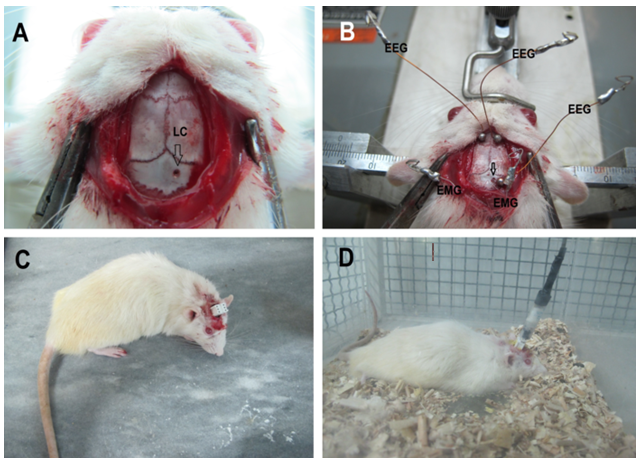
On the first day of study, the animals were anesthetized with 10% ketamine (100 mg/kg) and 2% xylazine (10 mg/ kg), and all were placed in a stereotaxic apparatus. In lesion and experimental groups, stainless steel guide cannula (23 G) was implanted unilaterally 1 mm above the injection site in LC (Figure 1A). The LC region was+ 1.3mm from the median suture,−9.8mm caudal to bregma and−7.2mm under the skull (Figure 1B) [20]. Two EMG electrodes were implanted in the dorsal neck muscle and 3 EEG electrodes were implanted on the skull (from bregma), one in the left occipital cortex (−11 and 4 mm L), one over the right parietal (−5 mm A and 6 mm L), and an electrode over the left frontal (5 mm A and 2 mm L) [8]. 7 days after electrode implantation the animals in the lesion and experimental groups received intra locus coeruleus (LC) injection of 2μg/0.5μl 6-OHDA in 0.1% ascorbic acid slowly at 5 min [7, 8]. For cell transplantation, 14days after electrode implantation we injected 3,000,000 cells through the tail vein.
Sleep Recordings
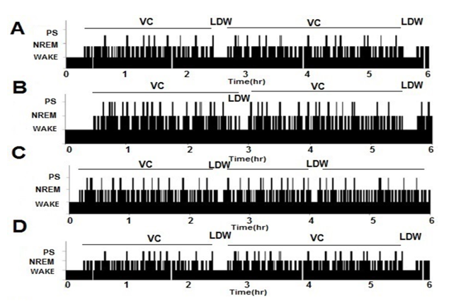
All records were made inside the faraday cage (Figures 1C&1D). The signals were reinforced by a polygraph booster (SienceBeam, Tehran, Iran) and it is filtered at low speed at 100 Hz for EEG and 1 kHz for EMG recording. EEG / EMG recordings were analyzed at 30-second intervals. If the wake- up episode is less than 300 seconds, the wake-up episode is assigned to the brief wake-up (BW), but if the episode is longer than 300 seconds, it is assigned to the Long Duration Wake (LDW). Then we used the LDW parts to separate one sleeping part from the other conscious cycling [21, 22].
For Paradoxical sleep recordings, 6-hour polygraph recordings (EMG, EEG) were performed from 9.00 am to 3.00 pm. Before each recording, compatibility with the recorded cage was performed for at least 24 hours. Recordings were made before the lesion (7th day) and cell transplantation (14th day), then 28, 49 days after the electrode implantation.
Sleep Analysis
Paradoxical sleep was defined by low amplitude, high- frequency activity in EEG, just similar to wake waves and inactivity of EMG [23, 24]. All visual scores were inspected to ensure correct alertness and eliminate any periods with artificial noise.
Statistical Analysis
Data analysis was carried out using SPSS 24. One-way analysis of variance (ANOVA) was used to analyze group differences in the data. Immunocytochemistry data are presented with at least three independent repeats. (P≤0.05).
Results
Characterization of NSCs and NACs
In all cultures, cells multiply in clusters or neurospheres (Figure 2A). The result shows that the proliferation of NSCs increase by hEGF and hFGF and the elimination of these factors cause of NSCs differentiation into neurons (Figures 2B&C). We did immunocytochemistry with anti-Sox-2 and Nestin antibodies as specific markers for NSCs. As shown in Fig. 3neurospheres were Nestin and Sox-2 positive (Figures 2D&E). Like other studies, this study shows that the combination of GDNF and BDNF is effective in increasing the differentiation of NSCs into NACs (Figures 2F&G) [6]. The results of immunocytochemistry after 5 days culture indicate that NSCs are differentiated into the TH positive cells (NACs) (Figure 2H).
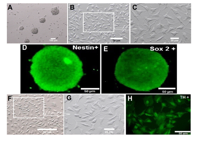
Figure 2: Isolation and culture of Neural Stem Cells. Phase-contrast images of neurospheres (A) and neurons after cell differentiation(B,C). Immunocytochemical analysis of neurosphere with the antibodies against Nestin (D) and Sox- 2(E) markers for neural stem cells. Phase-contrast images of noradrenergic- like cells (F, G) and Tyrosine hydroxylase (TH) positive cells with antibodies against TH (H).
The LC after NSCs &NACs Transplantation
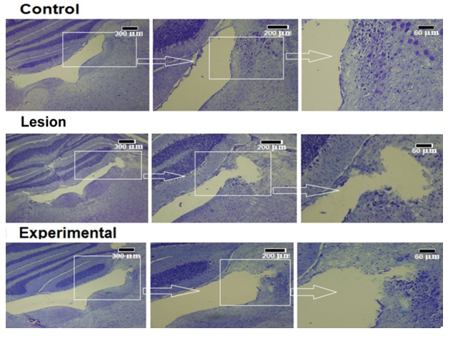
For estimation of the extent of damage in the LC area, the brains were cut 7μm thickness. Then we selected 15 sections of the LC area. After staining with cresyl violet, stereological studies were done. Finally, we estimated cavity volumes by the Cavalieri method (Figure 3). Cavity size caused by
6-OHDA in the lesion group was significantly less than the LC volume in the control group. The result shows that the Cavity volume in experimental groups is less than of LC and damage volume in control and lesion groups, respectively; that is due to the migration of transplanted cells to the damaged area. So, we can tell, cell transplantation decreases damage volume and repairs LC tissue (Figure 4).
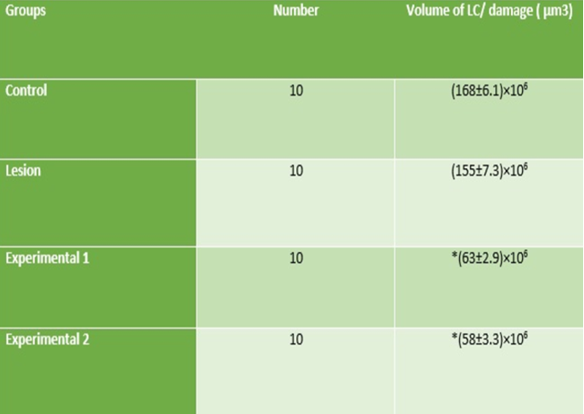
*: Significant differences with lesion group (P≤0.05). Figure 4: The mean volumes of the LC and damage areas in the study groups.
Effects of Unilateral Lesion of LC& Cells Transplantation on the Number of PS Episodes
Fragmentation of the sleep-wake cycle has been observed after LC lesions. The result indicates a unilateral lesion of LC enhances Paradoxical sleep, EEG activity consisted of high frequency and low voltage waves in Paradoxical sleep. The number of PS episodes increased after lesioning and decreased after cell transplantation. The number of PS episodes in the lesion group significantly increased 49 days after electrode implantation in comparison with the control group and also the amount of PS in experimental groups in comparison with the lesion group was significantly decreased. There were no significant differences in PS between experimental groups (Figures 5A-5D). The results of our study show that NSCs and NACs transplantation improve Paradoxical sleep after unilateral lesion of LC.
Evaluation of PS Percentage in the Study Groups
The results show that the percentage of PS after unilateral lesion of LC increased compared to the control group and this number decreased after cell transplantation compared to the lesion group. There was no significant difference in PS percentage between the groups on day 7. This difference on day 14 showed a significant difference between the control and other groups. After cell transplantation, the percentage of PS on day 28 in transplant groups showed a significant decrease compared to the lesion group. It shows that the percentage of PS improved after cell transplantation. On day 49 compared to day 28, the percentage of PS in transplanted groups showed a more significant difference than the lesion group. But still, the transplanted groups show a significant difference compared to the control group. There were no significant differences in the percentage of PS between experimental groups (Figure 6).
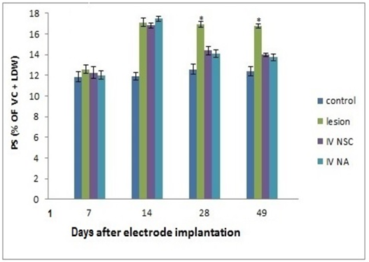
*: Significant differences with other groups (P≤0.05). Figure 6: Percentages of paradoxical sleep (PS) were obtained in the records of all rats. 1st day: Electrode implantation; 7th day: Lesion; 14th day: cells transplantation.
Discussion
There is a complex relationship between brain injury and sleep disorder. After brain injuries, in addition to related sensory and motor disorders, sleep-related problems occur for people [25]. Today, sleep disorders are treated with common methods such as medication, phototherapy, surgery, and etc. In this regard, the use of cell transplantation is a new method in the treatment of sleep disorders, which in addition to repairing damaged tissue, can also improve sleep disorders.
The NSCs obtained from the subventricular zone and cultured according to the method described in this study. Our study identifies NSCs from SVZs can be propagated as neurospheres in serum-free suspension cultures along with growth factors hEGF and hFGF. Neurospheres were positive for Sox-2 and Nestin markers. Our study shows that the percentage of TH-positive cells after 5 days of culture was significantly increased compared to the control group. It has been observed that GDNF has similar effects in distinction from mesencephalon dopamine neurons [26]. GDNF and BDNF are required for the production of dopaminergic primary sensory neurons in vivo [27].
There are several studies on LC lesion by electrolyte radio frequency [28], chemical, and 6-OHDA [2, 20, 29]. But none of these studies have been used as a model for studying Paradoxical sleep. According to a previous study, our study shows that unilateral LC lesion changes the amount of Paradoxical sleep. The mean percentage of PS in the lesion group increased significantly compared to the control group. In agreement with previous data [30], our results showed that an increase of PS is observed after unilateral lesion LC.
On the other hand, after cell transplantation mean percentages of PS were significantly decreased in comparison to the lesion group. Previous studies of unilateral LC lesions in the cat have confirmed that inactivation of the LC tail increases PS production [31]. In these studies, the results show the inhibitory role of the LC tail for PS appearance [32].
Conclusion
Collectively, our study shows that the size of LC lesions was decreased after cell transplantation. And also, we showed that in experimental groups amount of Paradoxical sleep was significantly reduced in comparison with lesion groups. These findings could be useful for research into neurological diseases due to damage to other areas of the brain that contain noradrenergic cells.
Ethical Considerations
- Compliance with ethical guidelines The ethical approval of this study was obtained from the Iran University of Medical Science (no.: IR.IUMS.REC 1395.95-04- 222-30176).
- Funding This study is done with financial support from the Iran University of Medical Sciences (95-04-222-30176).
- Conflict of interest The authors declare that there is no conflict of interest in this study.
- Acknowledgments This study is done with financial support from the Iran University of Medical Sciences. In this study, sleep recording was performed at the Neuroscience Research Center, Iran University of Medical Sciences.
References
-
Barkoukis TJ, Matheson JK, Ferber R, Doghramji K (2011) Therapy in Sleep Medicine E-Book: Elsevier Health Sciences.
-
Wang Y, Pan J, Wang D, Liu JJCscr (2018) The use of stem cells in neural regeneration: a review of current opinion. Current Stem Cell Research & Therapy 13(7): 608-617.
-
Yu SJ, Airavaara M, Wu KJ, Harvey BK, Liu H, et al. (2017) 9-cis retinoic acid induces neuro repair in stroke brain. Scientific Reports 7(1): 1-12.
-
Mizrak D, Bayin NS, Yuan J, Liu Z, Suciu RM, et al. (2020) Single-cell profiling and SCOPE-Seq reveal lineage dynamics of adult ventricular-subventricular zone neurogenesis and NOTUM as a key regulator. Cell Reports 31(12): 107805.
-
Zhang L, Wang G, Chen X, Xue X, Guo Q, et al. (2017) Formyl peptide receptors promote neural differentiation in mouse neural stem cells by ROS generation and regulation of PI3K-AKT signaling. Scientific Reports 7(1): 1-16.
-
Mahabadi VP, Movahedin M, Semnanian S, Mirnajafi Zadeh J, Faizi M (2015) In vitro differentiation of neural stem cells into noradrenergic-like cells. International Journal of molecular and cellular medicine 4(1): 22.
-
Iwasaki K, Komiya H, Kakizaki M, Miyoshi C, Abe M, et al. (2018) Ablation of central serotonergic neurons decreased REM sleep and attenuated arousal response. Frontiers in Neuroscience 12: 535.
-
Pirhajati Mahabadi V, Mazaheri Z, Semnanian S, Mirnajafi_zadeh J, Faizi M (2015) Effects of bilateral lesion of the locus coeruleus on the sleep-wake cycle in the rat. Physiology and Pharmacology 19(1): 22-30.
-
Scammell TE, Arrigoni E, Lipton JO (2017) Neural circuitry of wakefulness and sleep. Neuron 93(4): 747- 765.
-
Takahashi K, Lin JS, Sakai K (2006) Neuronal activity of histaminergic tuberomammillary neurons during wake–sleep states in the mouse. Journal of Neuroscience 26(40): 10292-10298.
-
Sakurai T (2007) The neural circuit of orexin (hypocretin): maintaining sleep and wakefulness. Nature Reviews Neuroscience 8(3): 171-181.
-
Hayat H, Regev N, Matosevich N, Sales A, Paredes Rodriguez E, et al. (2019) Locus-coeruleus norepinephrine activity gates sensory-evoked awakenings from sleep. Sci Adv 6(15): eaaz4232.
-
Lee MG, Hassani OK, Alonso A, Jones BE (2005) Cholinergic basal forebrain neurons burst with theta during waking and paradoxical sleep. Journal of Neuroscience 25(17): 4365-4369.
-
Eban Rothschild A, Rothschild G, Giardino WJ, Jones JR, de Lecea L (2016) VTA dopaminergic neurons regulate ethologically relevant sleep-wake behaviors. Nature Neuroscience 19(10): 1356-1366.
-
Carter ME, Yizhar O, Chikahisa S, Nguyen H, Adamantidis A, et al. (2010) Tuning arousal with optogenetic modulation of locus coeruleus neurons. Nature Neuroscience 13(12): 1526.
-
Li Y, Hickey L, Perrins R, Werlen E, Patel AA, et al. (2016) Retrograde optogenetic characterization of the pontospinal module of the locus coeruleus with a canine adenoviral vector. Brain Research 1641(PtB): 274-290.
-
Eslahi N, Shakeri Zadeh A, Ashtari K, Pirhajati Mahabadi V, Moghadam TT, et al. (2019) In vitro cytotoxicity of folate-silica-gold nanorods on mouse acute lymphoblastic leukemia and spermatogonial cells. Cell Journal (Yakhteh) 21(1): 14-26.
-
Masood MI, Schäfer KH, Naseem M, Weyland M, Meiser PJPo (2020) Troxerutin flavonoid has neuroprotective properties and increases neurite outgrowth and migration of neural stem cells from the subventricular zone. Plos one 15(8): e0237025.
-
Chen WT, Hsu FT, Liu YC, Chen CH, Hsu LC, et al. (2019) Fluoxetine induces apoptosis through extrinsic/ intrinsic pathways and inhibits ERK/NF-κB-modulated anti-apoptotic and invasive potential in hepatocellular carcinoma cells in vitro. International Journal of Molecular Sciences 20(3): 757.
-
Iovino M, Messana T, De Pergola G, Iovino E, Guastamacchia E, et al. (2019) Vigilance states: central neural pathways, neurotransmitters and neurohormones. Endocrine, Metabolic & Immune Disorders - Drug Targets 19(1): 26-37.
-
Yaghouby F, Donohue KD, O’Hara BF, Sunderam S (2016) Noninvasive dissection of mouse sleep using a piezoelectric motion sensor. Journal of Neuroscience Methods 259: 90-100.
-
Phillips AJ, Klerman EB, Butler JP (2017) Modeling the adenosine system as a modulator of cognitive performance and sleep patterns during sleep restriction and recovery. PLoS Computational Biology 13(10): e1005759.
-
Manzella FM, Joksimovic SM, Orfila JE, Fine BR, Dietz RM, et al. (2020) Neonatal Ketamine Alters High-Frequency Oscillations and Synaptic Plasticity in the Subiculum But Does not Affect Sleep Macrostructure in Adolescent Rats. Frontiers in Systems Neuroscience 14.
-
Oishi Y, Takata Y, Taguchi Y, Kohtoh S, Urade Y, et al. (2016) Polygraphic recording procedure for measuring sleep in mice. Journal of Visualized Experiments 107: e53678.
-
Theodorou AA, Rice SA (2007) Is the silent epidemic keeping patients awake?. American Academy of Sleep Medicine 3(4): 347-348.
-
Conway JA, Ince S, Black S, Kramer ERJC, Research T (2020) GDNF/RET signaling in dopamine neurons in vivo. Cell and Tissue Research 382(1): 135-146.
-
Srivastav S, Neupane S, Bhurtel S, Katila N, Maharjan S, et al. (2019) Probiotics mixture increases butyrate, and subsequently rescues the nigral dopaminergic neurons from MPTP and rotenone-induced neurotoxicity. The Journal of Nutritional Biochemistry 69: 73-86.
-
Iranzo AJC (2018) The REM sleep circuit and how its impairment leads to REM sleep behavior disorder 373(1): 245-266.
-
Wang Y, Zhang QJ, Liu J, Ali U, Gui ZH, et al. (2010) Noradrenergic lesion of the locus coeruleus increases the firing activity of the medial prefrontal cortex pyramidal neurons and the role of α2-adrenoceptors in normal and medial forebrain bundle lesioned rats. Brain Research 1324: 64-74.
-
Aston Jones G, Gonzalez M, Doran S (2007) Role of the locus coeruleus-norepinephrine system in arousal and circadian regulation of the sleep-wake cycle. Brain Norepinephrine pp: 157-195.
-
Brown RE, Basheer R, McKenna JT, Strecker RE, McCarley RW (2012) Control of sleep and wakefulness. Physiological reviews 92(3): 1087-1187.
-
Khanday MA, Somarajan BI, Mehta R, Mallick BNJe (2016) Noradrenaline from locus coeruleus neurons acts on pedunculopontine neurons to prevent REM sleep and induces its loss-associated effects in rats. eNeuro 3(6).
- Are the Vaccines the Only Solution to Prevent the COVID-19 Pandemic? Part Two
- Clinical Characteristics of Women in this New Global Immunodeficiency
- Cell Dynamics in HIV Pathogenesis: Insights and Implications
- Determination of the CDR (CDR1, CDR2) « Complementary- Determining Region Invertebrate Primitive Antibody from Sea Star »
- Prioritizing Care for High-Risk COVID-19 Patients in the EU: 10 Civic Recommendations to the Institutions
- Comprehensive Insights into ModRNA Vaccines: Persistent PP-Spike Recombinant Protein, Hyperimmune/Inflammatory Reactions, Thrombotic Vasculopathy, Chronic Organ Complications and Excess Deaths