Characterisation Covid-19 IgG Antibody Kinetics Post Infection and Vaccination using the Boditech iCHROMA™ Fluorescence Immunoassay (FIA) Method
Background: Coronavirus disease 2019 (COVID-19) is an emerging threat affecting millions of people worldwide. This study aims to assess the serologic profiles and time kinetics of antibodies against severe acute respiratory syndrome coronavirus 2 (SARS-CoV-2) in patients with COVID-19 using the Boditech iCHROMA fluorescence immunoassay (FIA) method. Methods: Samples were collected from 43 subjects and analyzed for IgG antibodies against SARS-CoV-2 using the Boditech iCHROMA FIA. The iCHROMA processes the signal using a cut off index of 0.9 – 1.1, results <0.9 are interpreted as negative, results between 0.9 and 1.1 are interpreted as indeterminate and results >1.1 are interpreted as positive. Longitudinal samples were collected on days between day 1- day 100 post onset of symptoms in 1 subject, single samples collected on days 40-90 post infection in 30 subjects, and single samples collected at 3 time points (pre vaccination, 8-18 days post vaccination and 20-28 days post vaccination) collected from 12 subjects non-infected and previously infected. Results: The Covid-19 IgG concentration was detected (positive and cut off index above 1.1) on day 11 and remained consistently elevated for another 100 days of measurement in the sera of the single infected subject. The IgG cut off indices between day 20 and 100 ranged between 26.8 and 46.7, with a mean of 36.5. In the sera of the 30 patients infected with Covid-19 collected 40 – 90 days post infection, IgG antibodies were detected in all the samples. The IgG cut off indices ranged between 14.0 and 32.60, with a mean of 20.65. Of the twelve subjects who were vaccinated, eight had not been previously infected, while 4 had been previously infected. All 4 (100%) previously infected subjects seroconverted by the first time point (8-18 days post vaccination), whilst only 1/8 (12.5%) of the non-infected subjects seroconverted by the first time point (8-18 days post vaccination). By the second time point (20 - 28 days post vaccination), 2/12 subjects (17%) had failed to seroconvert, whilst 10/12 (83%) of the subjects had seroconverted. In addition, a previously infected subject whose IgG antibodies had dropped to a very low-level, after receiving the first dose of vaccination his IgG antibodies had gone back to his previous antibody levels at the first time point (8-18 days post vaccination). Conclusion: The Boditech iCHROMA FIA method for antibody testing is useful in detecting SARS-CoV-2 in a variety of situations at different time points but has its limitations with regards to determining the difference between previously infected and vaccinated individuals.
Introduction
Coronavirus disease 2019 (COVID-19), the lung disease caused by severe acute respiratory syndrome coronavirus 2 (SARS-CoV-2), is an emerging threat to individual and public health. Accurate diagnosis of COVID-19 is important for appropriate treatment and to limit the spread of the virus. The current gold standard diagnosing tool for SARS-CoV-2 infection is real-time reverse transcription-polymerase chain reaction (rRT-PCR) using respiratory tract specimens [1]. Lateral flow methods for testing have now become more widely used as they are convenient and time efficient. However, these can often be quite uncomfortable and painful for patients as they require the insertion of a swab deep into the nasal and throat cavities [2]. Serologic testing is currently emerging as an additional diagnostic tool for COVID-19. Understanding the kinetics of the immune response and antibody dynamics against SARS-CoV-2 is vital for serologic testing.
Based on current available data, the IgG antibodies to SARS-CoV-2 have been shown to develop between 5 –15 days post disease onset [3, 4, 5, 6, 7, 8, 9, 10]. The median seroconversion time for IgG antibodies were day-14 post symptom onset. The presence of antibodies was detected in <40% among patients within 1 week from 79.8% (IgG) from day-15 after onset [8]. Researchers found that 90% of SARS-CoV-2–positive individuals had detectable antibodies from 40 days up to 7 months post-infection, with higher levels in patients with more severe disease. Over half patients who had antibodies measured 30-40 days after onset of symptoms were positive for both IgM and IgG antibodies [11]. A previously conducted study determined the positive percent agreement (PPA) and negative percent agreement (NPA) between the Boditech iCHROMA IgG FIA antibody assay and the Abbott Architect SARS-Cov-2 IgG assay, to detect IgG antibodies to the nucleocapsid protein of SARS-Cov-2. The study concluded that there was 100% agreement of the presence of IgG antibodies in the samples to Covid-19 between the Boditech iCHROMA Covid 19 IgG antibody assay method and the Abbot Architect SARS-CoV-2 IgG method (Clinical Sensitivity). In addition, there was 90% agreement in the absence of Covid-19 IgG antibodies in the samples between the Boditech iCHROMA Covid 19 IgG antibody assay and the Abbot Architect SARS-CoV-2 IgG (Clinical Specificity) [12]. In another study, the performance of the Boditech iCHROMA Covid 19 IgG antibody assay was assessed using the NIBSC (National Institute for Biological Standards and Control) reference material. In this study, the Boditech iCHROMA SARS-CoV-2 IgG assay identified 100% of the positive control sample, as positive, with an index range from 19.50 – 29.00, with a mean of 23.75 and standard deviation of SD of 2.53. It also identified 100% of the negative control samples, as negative, with a consistent index range of 00.00. The range of the index estimated in s NIBSC positive control sample was 19.50-29.00, interestingly the index range observed in patient samples was between 22.8 and 74.6, indicating that the NIBSC positive control was within the range of the lower levels seen in clinical samples [13].Another study demonstrated the kinetics for the first 27 days post onset of infection in a mildly infected subject [14]. In this report, the kinetics of the Boditech iCHROMA FIA Covid-19 IgG assay was studied in: (i) a mildly infected subject who was followed up for 100 days post infection, (ii) 30 patients who were infected with Covid-19 at 3 time points (10-18 days, 20- 28 days and 40-90 days) post infection, (iii) 12 vaccinated subjects (not previously infected and previously infected).
Materials
Covid-19 Infected subject (Day 1-100 post infection): Blood samples had been taken from a subject who suffered mild symptoms confirmed by PCR and antigen tests on day 1,2,3,4,5,6,7,8,10,11,12,13,14,15,16,17,18,19,20,21,22,23, 24,25,26 and 27 post onset of symptoms. The subject was followed up with blood samples collected on day 28, 29, 30, 31, 38, 42, 55, 69, 76, 78, 83, 91, 99 and 100 post onset of infection. Consent taken from subject.
Convalescent sera of Covid-19 Infected patients (Day 40-90 post infection): Thirty Covid-19 unique donor panel obtained from Cambridge Biosciences obtaining fresh human blood service in partnership with London-based Research Donors. Research Donors is a HTA licenced and ISO 9001 2015 certified company with Research Ethics (REC) approval as a Research Tissue bank. The Covid-19 positivity of the donors was confirmed by positive PCR or MD diagnosis. SARS-CoV-2 IgG antibodies had been estimated using the GOLD ELISA and Diasorin Liaison methods, consent taken from subjects Table 1.
| Subject # | Donor No | Age | Gender | Race |
|---|---|---|---|---|
| 1 | 7048 | 38 | F | AA |
| 2 | 6850 | 45 | F | C |
| 3 | 6935 | 44 | F | C |
| 4 | 7045 | 48 | F | C |
| 5 | 7006 | 55 | F | C |
| 6 | 6819 | 65 | F | C |
| 7 | 6913 | 40 | F | AA |
| 8 | 6947 | 39 | M | AA |
| 9 | 6965 | 34 | F | C |
| 10 | 6852 | 46 | F | C |
| 11 | 6879 | 30 | M | C |
| 12 | 6940 | 47 | F | C |
| 13 | 6928 | 57 | F | AA |
| 14 | 6829 | 36 | M | C |
| 15 | 6836 | 30 | F | AA |
| 16 | 6844 | 60 | F | AA |
| 17 | 6878 | 38 | M | C |
| 18 | 7027 | 30 | M | C |
| 19 | 6983 | 50 | F | C |
| 20 | 6901 | 35 | F | AA |
| 21 | 6830 | 47 | F | C |
| 22 | 7023 | 45 | M | NA |
| 23 | 7061 | 42 | F | AA |
| 24 | 6900 | 66 | M | C |
| 25 | 5884 | 53 | F | C |
| 26 | 6989 | 50 | M | AA |
| 27 | 7071 | 25 | F | C |
| 28 | 7066 | 63 | M | C |
| 29 | 6891 | 30 | F | AA |
| 30 | 7063 | 57 | F | C |
Table 1: Demographics of donors M-Male, F-Female, C-Caucasian, AA-African American, NA-not available.
Vaccinated Subjects
Twelve subjects who have been followed up with antibody measurements since April last year were followed up after their first vaccination with their blood samples taken for IgG antibodies analysis done at two time points: 10-18 days and 20-28 days post vaccination. Consent taken from subjects.
Method
Boditech iCHROMA Method Principle: The test uses a sandwich immune detection method; fluorescence labelled conjugates in a dried detection buffer binds to antibody in sample, forming antibody-antigen complexes, and migrates onto nitrocellulose matrix to be captured by the other immobilized anti-human IgG on test strip. The more antigen- antibody complexes lead to stronger fluorescence signal by the detector antigen which is processed by iCHROMA. The iCHROMA processes the signal using a cut off index of 0.9- 1.1, results <0.9 are interpreted as negative, results between 0.9 and 1.1 are interpreted as indeterminate and results >1.1 are interpreted as positive. The blood samples from the Covid-19 infected subject, the 30 Covid-19 unique donor panels and the vaccinated subjects were analysed using the Boditech iCHROMA Fluorescence Immunoassay (FIA) IgG assay described below:
- Transfer 150μL of detector diluent using a pipette into the detector tube containing a granule. When the granule is completely dissolved it becomes the detection buffer.
- Aspirate 10μL of whole blood/serum/plasma/control with a pipette, and add into the detector tube, close and shake the tube at least 10 times.
- Pipette out 75μL of the content of the tube and load it into the sample well on the test cartridge and leave for 10 minutes.
- Insert the test cartridge into the cartridge holder in iCHROMA II device and press start.
- Read the result on the display screen of the iCHROMA II device.
Results
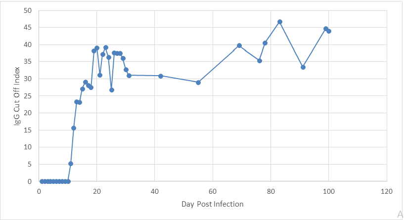
The cut off indices of the blood samples on Days 1,2,3,4,5,6,7, 8 and 10 were 0 and were interpreted as negative for the IgG antibodies. On day 11 the samples cut off index rose to 5.2 and were interpreted as positive, rising to 39.1 on day 20 post onset of symptoms and plateaued. The IgG cut off index between day 20 and 100 ranged between 26.8 and 46.7, with a mean of 36.5 (Figure 1).
In the convalescent sera of patients with Covid-19, all 30 patients had IgG present in their serum, the IgG cut off indices ranged between 14.0 and 32.60, with a mean of 20.65 Table 2.
| Age | Gender | Days after onset of symptoms | iCHROMA IgG | |
|---|---|---|---|---|
| 1 | 38 | F | 73 | 19.8 |
| 2 | 45 | F | 66 | 19.7 |
| 3 | 44 | F | 80 | 20.1 |
| 4 | 48 | F | 49 | 20.3 |
| 5 | 55 | F | 74 | 20 |
| 6 | 65 | F | 87 | 21.2 |
| 7 | 40 | F | 76 | 21.6 |
| 8 | 39 | M | 77 | 20.7 |
| 9 | 34 | F | 58 | 20.6 |
| 10 | 46 | F | 72 | 20.4 |
| 11 | 30 | M | 65 | 21.9 |
| 12 | 47 | F | 75 | 20.2 |
| 13 | 57 | F | 80 | 21.5 |
| 14 | 36 | M | 73 | 23 |
| 15 | 30 | F | 67 | 22.8 |
| 16 | 60 | F | 66 | 17.4 |
| 17 | 38 | M | 67 | 23 |
| 18 | 30 | M | 48 | 18.6 |
| 19 | 50 | F | 68 | 15.4 |
| 20 | 35 | F | 80 | 17.8 |
| 21 | 47 | F | 80 | 22.6 |
| 22 | 45 | M | 61 | 23.5 |
| 23 | 42 | F | 48 | 15.9 |
| 24 | 66 | M | 63 | 14 |
| 25 | 53 | F | 93 | 21.5 |
| 26 | 50 | M | 73 | 25.4 |
| 27 | 25 | F | 78 | 32.6 |
| 28 | 63 | M | 40 | 19.6 |
| 29 | 30 | F | 76 | 19.5 |
| 30 | 57 | F | 75 | 19 |
Table 2: Convalescent serum of Covid-19 Infected patients (Day 40-90 post infection):
Vaccinated Subjects
Of the twelve subjects who were vaccinated, eight had no IgG antibodies present Table 3, these workers had probably not been exposed to Covid-19 infection previously. Of these 8 subjects with no IgG antibodies prior to vaccination, only 1 (12.5%) had IgG antibodies present with a cut off index of 2.5 present when tested at the first point (8-10 days post vaccination), by the second time point (20-28 days post vaccination), 6 had developed IgG antibodies. However, two out of the 8 (25%) of the subjects failed to develop any IgG antibodies by the second time point (20-28 days post vaccination), one of these subjects had a haematological malignancy.
| Pre-vaccination | 8-18 days post vaccination | 20-28 days post vaccination | |
|---|---|---|---|
| 1 | 0 | 0 | 18.5 |
| 2 | 0 | 0 | 0 |
| 3 | 0 | 0 | 4.2 |
| 4 | 0 | 2.5 | 10.8 |
| 5 | 0 | 0.1 | 4.3 |
| 6 | 0 | 0 | 0 |
| 7 | 0 | 0 | 12.6 |
| 8 | 0 | 0 | 38.1 |
Table 3: IgG cut off indices in 8 subjects with antibodies present prior to vaccination, 8-18 days post vaccination and 20-28 day
Of the twelve subjects who were vaccinated, four had IgG antibodies present (cut off indices 1.8-24.4) prior to their first dose vaccination Table 4, these workers had developed Covid-19 earlier in the year and were having their antibodies measured routinely. Interestingly, subject 2’s IgG antibodies had dropped to a cut off index of 1.8 from previous values with cut off index values of >20. All the subjects had their IgG antibodies sustained at the first time point (8-10 days post vaccination) and the second time point (20-28 days post vaccination). Subject 2’s IgG cut off index rose from 1.8 to 22.4 by the first time point (8-10 days post vaccination) and remained through to the second time point (20-28 days post vaccination).
| Pre-vaccination | 8-18 days post vaccination | 20-28 days post vaccination | |
|---|---|---|---|
| 1 | 23.7 | 13.3 | 18 |
| 2 | 1.8 | 22.4 | 20.5 |
| 3 | 24.4 | 26.6 | 28 |
| 4 | 18 | 23.6 | 28 |
Table 4: IgG cut off indices in 4 subjects with antibodies present prior to vaccination, 8-18 days post vaccination and 20-28 day
Discussion
The Boditech iCHROMA Covid-19 IgG assay successfully allows for the assessment of the kinetics of the Covid-19 IgG antibody, in a variety of situations. In this study, 3 situations were assessed: i) IgG levels of an infected patient were measured on day 1 through 100 post infection, ii) 30 infected patients IgG levels were measured at some point between 40-90 days post infection, iii) IgG levels of 12 vaccinated subjects’ pre-vaccination, 8-18 days post vaccination and 20-28 days post vaccination. In the infected patient, the IgG started to rise on day 11 (cut off index 5.2) and remained consistently elevated for another 100 days (cut off index between 26.8 and 46.7). This is consistent with the median seroconversion time for IgG on day-14 post symptom onset described in the literature [15]. IgG levels have also been described to remained stay stable, and similar dynamic changes have been reported in another study [16]. In the serum of the 30 patients infected with Covid-19 collected 40 – 90 days post infection, IgG antibodies (cut off index ranged between 14.0 and 32.60) were detected in all (100%) of the samples. This data is consistent with other data where researchers have found that 90% of SARS-CoV-2–positive individuals had detectable antibodies from 40 days up to 7 months post-infection, with higher levels in patients with more severe disease [11]. In the vaccinated population, of the twelve subjects who were vaccinated, eight had no IgG antibodies present, these workers had probably not been exposed to Covid-19 infection previously, only 1 (12.5%) had IgG antibodies present with a cut off index of 2.5 present when tested at the first point (8-10 days post vaccination). By the second time point (20-28 days post vaccination), 6 out of the 8 (75%) had developed IgG antibodies. All four vaccinated subjects who had IgG antibodies and therefore previously infected had IgG antibodies present at the first point (8-10 days post vaccination). This study showed a quicker rate of sera conversion and higher cut off indices in the subjects with prior infection. This data is consistent with that seen in the literature where post-vaccine antibody responses showed that positive anti-spike IgG results increased over the 2-4 weeks after the first vaccination.
In addition, subjects without evidence of prior infection showed a slower seroconversion rate compared to subjects with prior infection [17]. In this study, two out of the 12 (17%) of the subjects failed to develop any IgG antibodies by the second time point (20-28 days post vaccination), one of these subjects had a haematological malignancy. This finding on non-response was not surprising because research has shown that there are group of patients such as those with haematological malignancies or immune suppressing conditions that do not produce antibodies and are therefore considered vaccine non-responders [18, 19, 20, 21].
In this study, we have also described the IgG kinetics in a situation where a previously infected subject whose IgG antibodies had dropped to a very low-level (cut off index 1.8), who then went on to receive the first vaccine dose. This study showed that at the first time point (8-18 days post vaccination) his IgG antibodies went back to his previous antibody (cut off index 22.4). This response was like those subjects (Subject 1,3 and 4) Table 4 who had been previously infected, but their antibodies had not dropped. In addition, we can also see that this subject’s seroconversion was earlier than that seen in the subjects with no previous infection, most of them seroconverted at the second time point (20- 28 days post vaccination). This suggest that although the IgG antibody is waning the body is already primed to produce antibodies in response to the infection or vaccination. The Boditech iCHROMA Covid-19 IgG assay is a successful tool for analysing antibodies in a variety of situations. However, the limitation of this assay using a cut off index would be the ability to distinguish between a sustained immune response from previous infection and vaccination, as seen by the estimations taken at the first time point (8-10 days post vaccination) and second time point (20-28 days post vaccination) in subjects 1,3 and 4 Table 4. Data from this study has shown to be in line with current seroconversion data from other studies.
Declaration of Competing Interest
JB as an independent Consultant for several diagnostic companies including Boditech Med Inc, Korea the manufacturer of the iCHROMA Covid-19 IgG and IgM antibody system, supporting their clinical development and commercial activities.
References
-
Kevadiya BD, Machhi J, Herskovitz J, Oleynikov MD, Blomberg WR, et al. (2021) Diagnostics for SARS-CoV-2 infections. Nat Mater 20(5): 593-605.
-
Mardian Y, Kosasih H, Karyana M, Neal A, Lau CY (2021) Review of Current COVID-19 Diagnostics and Opportunities for Further Development. Front Med (Lausanne) 8: 615099.
-
Huang AT, Garcia-Carreras B, Hitchings MDT, Yang B, Katzelnick L, et al. (2020) A systematic review of antibody mediated immunity to coronaviruses: antibody kinetics, correlates of protection, and association of antibody responses with severity of disease. Nat Commun 11(1): 4704.
-
Kellam P, Barclay W (2020) The dynamics of humoral immune responses following SARS-CoV-2 infection and the potential for reinfection. J Gen Virol 101(8): 791-797.
-
Lou B, Li TD, Zheng SF, Su YY, Li ZY, et al. (2020) Serology characteristics of SARS-CoV-2 infection since exposure and post symptom onset. Eur Respir J 56(2): 2000763.
-
Borremans B, Gamble A, Prager K, Helman S, McClain A, et al. (2020) Quantifying antibody kinetics and RNA shedding during early-phase SARS-CoV-2 infection by time since symptom onset. Elife 9: 60122.
-
Ma H, Zeng W, He H, Zhao D, Jiang D, et al. (2020) Serum IgA, IgM and IgG responses in COVID-19. Cellular and Molecular Immunology 17(7): 773-775.
-
Zhao J, Yuan Q, Wang H, Liu W, Liao X, et al. (2020) Antibody responses to SARS-CoV-2 in patients of novel coronavirus disease 2019. Clin Infect Dis 71(16): 2027- 2034.
-
Wang B, Wang L, Kong X, Geng J, Xiao D, et al. (2020) Long-term coexistence of SARS-CoV-2 with antibody response in COVID-19 patients. J Med Virol 92(9): 1684- 1689.
-
Qu J, Wu C, Li X, Zhang G, Jiang Z, et al. (2020) Profile of IgG and IgM antibodies against severe acute respiratory syndrome coronavirus 2 (SARS-CoV-2). Clin Infect Dis 71(16): 2255-2258.
-
Figueiredo-Campos P, Blankenhaus B, Mota C, Gomes A, Serrano M, et al. (2020) Seroprevalence of anti-SARS- CoV-2 antibodies in COVID-19 patients and healthy volunteers up to 6 months post disease onset. Eur J Immunol 50(12): 2025-2040.
-
Bolodeoku J, Bass M, Anyaeche C, Kim TK, Retnasingham V (2020) Performance of the Boditech iCHROMA Covid 19 IgG antibody assay with the external quality control from UK NIBSC (National Institute of Biological Standards and Control). J, Clin Med Rev and Rep 2(8).
-
Bass M, Bolodeoku J, Stevenson E, Anyaeche C, Kim TK, et al. (2020) Agreement of the Point of Care Test (POCT) Boditech iCHROMA Covid-19 IgG antibody assay with the Abbott Architect SARS-CoV-2 IgG antibody assay. Ann Immunol Immunther 2(2): 000121.
-
Bolodeoku J, Bass M, Anyaeche C, Retnasingham V (2021) A Mild Case of COVID-19 Infection: An Observational Longitudinal Study 27 Days Post Symptom of Antigen, Antibodies (IgM & IgG), IL-6 and D-Dimer. A Review. Am J Biomed Sci & Res 11(6).
-
Orner EP, Rodgers MA, Hock K, Tang MS, Taylor R, et al. (2021) Comparison of SARS-CoV-2 IgM and IgG seroconversion profiles among hospitalized patients in two US cities. Diagn Microbiol Infect Dis 99(4): 115300.
-
Jin Y, Wang M, Zuo Z, Fan C, Yeet F, et al. (2020) Diagnostic value and dynamic variance of serum antibody in coronavirus disease 2019. Int J Infect Dis 94: 49-52.
-
Wei J, Stoesser N, Matthews PC, Ayoubkhani D, Studley R, et al. (2021) Antibody responses to SARS-CoV-2 vaccines in 45,965 adults from the general population of the United Kingdom. Nat Microbiol 6(9): 1140-1149.
-
Thakkar A, Gonzalez-Lugo JD, Goradia N, Gali R, Shapiro LC, et al. (2021) Seroconversion rates following COVID-19 vaccination among patients with cancer. Cancer Cell 39(8): 1081-1090.
-
Mehta V, Goel S, Kabarriti R, Cole D, Goldfinger M, et al. (2020) Case fatality rate of cancer patients with COVID-19 in a New York Hospital System. Cancer Discov 10(7): 935-941.
-
Ollila TA, Lu S, Masel R, Zayac A, Paiva K, et al. (2021) Antibody response to COVID-19 vaccination in adults with hematologic malignant disease. JAMA Oncol 7(11): 1714-1716.
-
Agha M, Blake M, Chilleo C, Wells A, Haidar G (2021) Suboptimal response to COVID-19 mRNA vaccines in hematologic malignancies patients. medRxiv.
- Are the Vaccines the Only Solution to Prevent the COVID-19 Pandemic? Part Two
- Clinical Characteristics of Women in this New Global Immunodeficiency
- Cell Dynamics in HIV Pathogenesis: Insights and Implications
- Determination of the CDR (CDR1, CDR2) « Complementary- Determining Region Invertebrate Primitive Antibody from Sea Star »
- Prioritizing Care for High-Risk COVID-19 Patients in the EU: 10 Civic Recommendations to the Institutions
- Comprehensive Insights into ModRNA Vaccines: Persistent PP-Spike Recombinant Protein, Hyperimmune/Inflammatory Reactions, Thrombotic Vasculopathy, Chronic Organ Complications and Excess Deaths