Evidence of Sea Star Monocytes Evolving in Phagocytes: Asterias Rubens T.E.M Observations
Sea star Axial organ (A.O) cells were observed in T.EM: Evidence of primitive monocytes was done: They contained azurophile particles and a reniform nucleus, sometimes a phagosome appeared and amoeboid images were seen. These cells, in a second time, evolve mainly in phagocytes which may be assimilated to Vertebrate Macrophages, with functional phagosomes and azurophile particles. The sea star monocytes are smaller in diameter (4 to 5µ) than Vertebrate ones.
Introduction
Observation of sea star Asterias rubens T and B lymphocytes have already been performed in TEM [1, 2]. It was asserted by biochemistry and biophysical assays. Second sea star platelets were so observed [3]. In a third time, we confirm the existence of sea star Monocytes which, mainly, evolves in Phagocytes corresponding to vertebrate macrophages.
Materials and Methods
Sea star Asterias rubens were obtained from the Marine Institute of Arcachon (France) Axial organs (A.O) were excised and:
- Either the whole cellular population was conserved and so observed in TEM
- Or the whole A.O was separated into B and T cell subpopulations according the well-known method of Julius and al [4]. Cells were fixed with glutaraldehyde in cacodylate buffer as precedently described No post-coloration was operated [1].
Observations were done with a Hitachi Microscope.
Results
As seen in Figure 1:
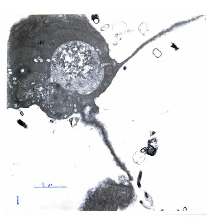
We observe a reniform Nucleus (N). Besides of it: azurophiles particles smaller than 0, 2 µ, these particles may correspond to lysosomes villous digitations which evoke ciliatures (2) and a small phagosome are present.
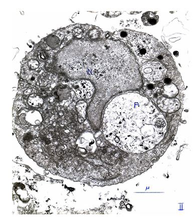
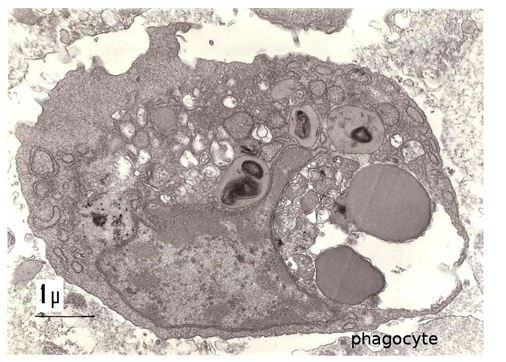
In Figure 2 We see a circular cell with a nucleus containing fine chromatine. The cell diameter is about 4-5 µ. Azurophile particle are always present. In the last figure III, the cell is longer than the two precedent ones and culminates at 8µ. .There is no azurophile particles in the cytoplasme. The phagosome is greatly opened on the outside: it is a true phagocyte Figure 3.
Conclusion
The sea star Asterias rubens presents T and B lymphocytes, Monocytes, Macrophages and Platelets, in T.E.M observations. The evolution of Monocytes in Phagocytes, resembling to evolution of macrophages, in Vertebrates was shown in this work through 3 Figures.
References
-
Anteunis A, Leclerc M, Vial M, Brillouet C, Luquet G, et al. (1985) Immunocompetent cells in the starfish Asterias rubens. An ultrastructural study. Cell Biol Int Rep 9(7): 663-670.
-
Leclerc M (2012) Humoral Immune Responses to Various Antigens in the Asterids: A. gibbosa and A. rubens. American Journal of Immunology 8(4): 196-199.
-
Leclerc M (2019) Role of Sea Star Platelets, of Sea Star Lymphocytes in Invertebrates. Acta Scientific Pharmaceutical Science 3(11): 32-33.
-
Tokuda Y, Toyohara H, Kina T, Sakaguchi M (1999) Characterization of Distinct Subpopulations of Japanese Flounder Lymphocytes with Monoclonal Antibody against Serum Immunoglobulin. Fisheries Science 65(3): 347-352.
- Are the Vaccines the Only Solution to Prevent the COVID-19 Pandemic? Part Two
- Clinical Characteristics of Women in this New Global Immunodeficiency
- Cell Dynamics in HIV Pathogenesis: Insights and Implications
- Determination of the CDR (CDR1, CDR2) « Complementary- Determining Region Invertebrate Primitive Antibody from Sea Star »
- Prioritizing Care for High-Risk COVID-19 Patients in the EU: 10 Civic Recommendations to the Institutions
- Comprehensive Insights into ModRNA Vaccines: Persistent PP-Spike Recombinant Protein, Hyperimmune/Inflammatory Reactions, Thrombotic Vasculopathy, Chronic Organ Complications and Excess Deaths