Mucinous Cystadenoma of Liver with Mesenchymal Stroma
Hepatobiliary cystadenoma is a rare benign cystic tumor of the liver. These are predominantly located in right hepatic lobe. A 45 year old female presented with abdominal pain, epigastric discomfort, fever and vomiting. On radiological evaluation USG showed large multiloculated cystic mass in right lobe of liver. Contrast Enhanced CT examination exhibited exophytically growing multiloculated cytic lesion with differential content and peripheral capsular and central enhancement with imaging diagnosis of hydatid cyst was given. The surgical excision of hepatic cyst was done. On histopathology reported as mucinous cystadenoma of liver with mesenchymal stroma. We are presenting this case for its rarity, clinical, radiological and histopathological findings.
Introduction
Hepatobiliary Cystadenoma (HBC) is a rare cystic tumor of the liver which is derived from the biliary epithelium [1]. However the histogenesis of these neoplasms has been debated. The recent studies suggest that ultrastructural and immunochemical findings support that HBCs arise from ectopic rests of embryonic gallbladder tissue within the liver. They are benign tumors but high rate of recurrence. Also these are potential for neoplastic transformation into cystadenocarcinoma in about 10% of cases [2]. Hepatobiliary cystadenomas constitute less than 5% of intrahepatic cysts [3]. Most of the patients presented with nonspecific abdominal pain. On radiological imaging cystic hepatic lesions are reported. The histopathological confirmation is important for diagnosis and further management of patients.
A 45 year old female presented to surgery department with complaints of abdominal pain, epigastric discomfort, fever and vomiting. On physical examination, right hypochondriac tenderness was noted. There was no history of any previous operative procedures. No history of any hormonal use. The other systemic examination was normal. On multimodality radiological evaluation, ultrasonography showed large multiloculated cystic mass in right lobe of liver. Contrast Enhanced CT examination also exhibited exophytically growing multiloculated cystic lesion measuring 14.9 x 10.1 x 10.5 cm (CC x AP x T) with differential and high density content and peripheral capsular and central septal ehnacement. Mild ascites and small bowel dilatation also seen - preoperative likely diagnosis of hydatid cyst with probable rupture and resultant peritonitis was given.
Liver preserving procedure of marsupalisation was performed. We received specimen of cyst measuring 11.5 x 10.5 x 6.0 cm. External surface was smooth. There was an associated focal area of congestion. On cut section single large cyst with multilocular lesions filled with mucinous material was noted. Larger cyst was 3cms in diameter. The wall of cyst was thickened at places (Figures 1A,B). The inner lining was smooth.
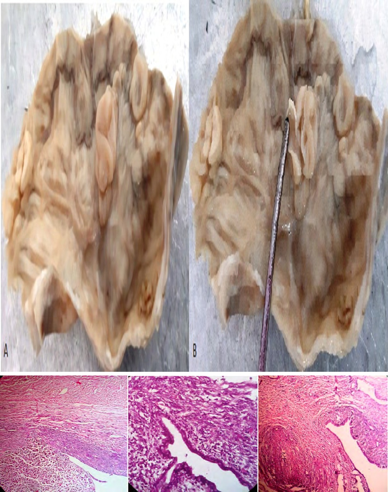
Figures 1(A,B): Single large cyst with multilocular lesions filled with mucinous material. Larger cyst was 3 cm in diameter. The wall of cyst was thickened at places.
On microscopic examination, showed a cyst lined by single layered cuboidal to columnar epithelium having basal nuclei and apical mucin. At places, epithelial hyperplasia was noted. The lumen was filled with mucinous material. The wall of cyst was formed by dense mesenchymal stroma with focal atypia (Figure 2). Also areas of fibrosis, congestion and focal haemorrhage were noted. There was no evidence of malignant transformation. On histopathology reported as mucinous cystadenoma of liver with mesenchymal stroma.
Discussion
Hepatobiliary cystadenomas are rare cystic tumor of the liver. These are of 2 types – mucinous and serous. Mucinous cystadenomas are predominant type and constitute about 95% of cases. The location is usually intrahepatic region (80%), then at common bile duct (10%) and other are at hepatic duct and gallbladder [4, 5]. The other histopathological type is serous cystadenoma. These are lined by cuboidal epithelium with no mucin and stroma. Clinically hepatobiliary cystadenomas occurs in age from 2-87 year. The peak incidence at 40-50 years with female preponderance having male: female ratio of 4:1 [6].
These patients remain asymptomatic or detected incidentally. The common clinical presentation is abdominal pain, vague tenderness in upper right abdomen, jaundice, vomiting, etc [7]. Hepatobiliary cystadenomas are slow growing tumor with good prognosis.
Radio Imaging Evaluation: On ultrasonography (USG) a biliary cystadenoma appears as a unilocular or multilocular cyst with postacoustic enhancement. The content of the cysts may range from completely anechoic to having low-level echoes due to hemorrhage, mucin, or proteinaceous fluid. Mural nodules and papillary projections may project into the cyst lumen. Septal or peripheral wall calcification may also be seen [8].
Computed Tomography (CT): The appearance of the cyst fluid on CT is variable depending on its composition. It can range from that of water (0 HU) to high density if the cyst has been complicated by acute hemorrhage. Calcifications of septa or cyst wall may be seen. Additionally, the septa and peripheral wall may enhance following administration of contrast.
Magnetic Resonance Imaging (MRI): The MR signal intensity of biliary cystadenoma is variable on both T1- and T2- weighted images, depending on the content of the cyst fluid. Also evaluate for any associated polycystic liver disease in few cases. On histopathological evaluation the mucinous hepatobiliary cystadenomas are of three types- Hepatobiliary cystadenomas with mesenchymal stroma.Others are Hepatobiliary cystadenoma without mesenchymal stroma and Hepatobiliary cystadenoma with polypoidal epithelial component projecting into cyst lumen .In our case it was of hepatobiliary cystadenoma with mesenchymal stroma. There was no evidence of any malignant transformation. As reported in literature, these are precancerous lesions and transformations to cystadenocarcinoma are noted. Careful examination of multilocular cyst for any nodule, thickening, papillae should be done to rule out malignancy. The differential diagnosis are simple cyst, congenital cyst, cystic hamartomas, echinococcal cyst, cysadenocarcinomas, hepatic metastasis [9, 10].
Khanna G, et al. reported a case of intrahepatic biliary cystadenoma which on radiology mimicked a hydatid cyst and also caused a histological diagnostic dilemma [11]. It is crucial to differentiate biliary cystadenoma from other cystic lesions of liver. The estimation of serum and cyst fluid for CEA and CA19-9 levels in the cyst fluid can helps in such situations. The gold standard management of hepatobiliary cystadenoma are complete surgical resection. The fenestration of cystic component and partial tumor removal are also considered, but in these cases local recurrence is very high (60%) [12].
Conclusion
As hepatic cystic lesions are rare, its early recognition and diagnosis are essential part in patient care .Amongst it the mucinous cystadenoma of liver with mesenchymal stroma are extremely rare benign neoplasm. The histopathological evalution is crucial role in diagnosis and management of these cases for better prognosis.
References
-
Park SJ, Lee HY, Joo SH, Kim YW, Lee SM, et al. (2007) Biliary cystadenoma and cystadenocarcinoma. J Korean Surg Soc 73(1): 77-82.
-
Suh KS, Ahn SM, Kim SW, Lee KU, Park YH, et al. (1996) Biliary cystadenoma and cystadenocarcinoma. J Korean Surg Soc 50: 410-416.
-
Ishak KG, Willis GW, Cummins SD, Bullock AA (1977) Biliary cystadenoma and cystadenocarcinoma: report of 14 cases and review of the literature. Cancer 39(1): 322-338.
-
Ferrell L (2004) Benign and malignant tumors of the liver. In: Odze RD, Goldblum JR, Crawford JM (Eds.) Surgical Pathology of the Gastrointestinal Tract, Liver, Biliary Tract, and Pancreas. Saunders, Philadelphia, pp: 1015-1016.
-
Salerno S, Florena AM, Romano I, Miceli L, Casto AL (2006) Multifocal biliary cystadenocarcinoma of the liver: CT and pathologic findings. Tumori 92(4): 358- 360.
-
Joel JM, Jeyasingh SD, Kalyanaraman S (2016) Biliary Cystadenoma: A Case Report. J Clin Diagn Res 10(2): 19- 20.
-
Kinoshita H, Tanimura H, Onishi H, Kasano Y, Uchiyama K, et al. (2001) Clinical features and imaging diagnosis of biliary cystadenocarcinoma of the liver. Hepato- gastroenterology 48(37): 250-252.
-
Horton KM, Bluemke DA, Hruban RH, Soyer P, Fishman EK (1999) CT and MR imaging of benign hepatic and biliary tumors. Radiographics 19(2): 431-451.
-
Choi HK, Lee JK, Lee KH, Lee KT, Rhee JC, et al. (2010) Differential Diagnosis for Intrahepatic Biliary Cystadenoma and Hepatic Simple Cyst: Significance of Cystic Fluid Analysis and Radiologic Findings. J Clin Gastroenterol 44(4): 289-293.
-
Jagtap SV, Tele JS, Rajput SR, Jagtap SS (2019) Mucinous Adenocarcinoma of Gall Bladder-A Rare Histopathological Variant Presented as Perforation. Journal of Clinical & Diagnostic Research 13(5): 07-09.
-
Khanna G, Sharma P, Madhusudhan KS, Barwad A, Ranjan P, et al. (2017) Intrahepatic biliary cystadenoma with ciliary metaplasia: Report of a rare morphological variant. Indian J Pathol Microbio 60(2): 253-255.
-
Koffron A, Rao S, Ferrario M, Abecassis M (2004) Intrahepatic biliary cystadenoma: role of cyst fluid analysis and surgical management in the laparoscopic era. Surgery 136(4): 926-936.
- Genomic Landscape of Aggressive Penile Squamous Cell Carcinoma including TERT-p and NOTCH1 Mutations – An Institutional Experience
- Establishment of Baseline Haematological Values for Canine Population in North-Central Nigeria: A Cross-Sectional Study in the Federal Capital Territory
- Biochemical Assessment of Uroliths Extracted in Patients with Urolithiasis in a Tertiary Health Institution
- Update on Gastrointestinal Pecomas: Molecular Pathogenesis and Risk Stratification
- A Comparative Study of Serum C-reactive Protein Level Between Pre-eclampsia and Normal Pregnancy in Tertiary Level Hospital
- From Deformity to Alignment: Clinical Outcomes of the Schnepp Osteotomy in Hallux Valgus in 47 Feet