Protoconidal Cingulum/Protostylid in Permanent Mandibular First Molar-Report of a Rare Dental Morphological Trait
The present article shows the occurrence of a rare dental morphological trait, “Protoconidal Cingulum” or “protostylid” on the buccal surface of permanent mandibular first molar in a 14- year-old Indian male patient. This morphological dental variation was reported more frequently in primary dentition involving second molars. Prevalence, clinical implications, classification systems and various nomenclatures pertaining to protostylid is discussed in this paper.
Introduction
Permanent mandibular molars in humans usually consist of five cusps in which three are buccal and two lingual cusps. Occurrence of an extra cusp is an uncommon variation and represents the dental evolution and is of anthropological significance. “Protostylid” is the additional cusp like structure seen on the mesial half of the buccal surface of mandibular molars [1]. This is considered as an accessory cusp and as a remnant of a cingulum representing crest feature, a pit or a surface irregularity. Various definitions are given by different researchers regarding protostylid [1]. According to Arizona State University Dental Anthropology System (ASUDAS), protostylid can be defined as “a paramolar cusp found on the buccal surface of the protoconid or mesiobuccal cusp which is normally associated with the buccal groove” [2].
Later in 1963, Dahlberg defines the protostylid as “an elevation or ridge of enamel on the anterior part of the buccal surface of the lower molars that ascends from the gingival end of the buccal groove and extends mesio-occlusally” [3]. He also stated that “although this protostylid had its origin as an expression of the cingulum, it is a unit structure, an entity in itself and definitely unlike the continuing cingular eminence seen on the Gorilla and other Anthropoids” [4]. In 2004, Hlusko [5] used the term “Protoconidal Cingulum” for the “protostylid” and he stated that these two terms can be used interchangeably when describing any features on the enamel surface of the buccal side of human’s mandibular molars.
The purpose of this article is to report a case of 14-year- old Indian male patient, in which the permanent mandibular first molar was associated with Protoconidal Cingulum or protostylid on the buccal aspect of mesio-buccal cusp.
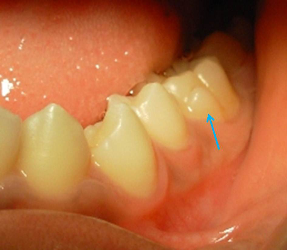
A 14-year-old male patient reported to the private dental clinic for seeking treatment for alignment of his irregular teeth. Patient was physically appeared normal, healthy and well nourished. No other systemic disorders or metabolic problems observed. On intra oral examination, patient was found with complete permanent dentition with all third molars still not erupted. Patient had class II malocclusion with increased proclination of maxillary anterior teeth. On further examination, presence of extra small, rudimentary cusp like structure was observed attached on to the buccal surface of the mesio-buccal cusp of permanent mandibular left first molar was observed (Figures 1 & 2). This cusp was triangular in shape with its base attached to the cervical margin of the buccal surface of first molar and apex was facing occlusally and was extending till 2 mm below the occlusal plane of the first molar. A shallow developmental
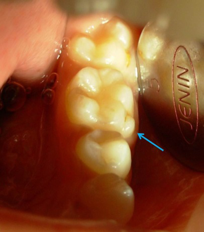
grove was observed with this cusp and buccal surface of the first molar which was discolored because of presence of stains (Figures 1 & 2). No other teeth were found with this type of extra cusp. Finally based on clinical examination and literature evidence this extra cusp was diagnosed as protostylid or Protoconidal Cingulum. As this cusp appeared self-cleansing and patient did not have any discomfort from this extra cusp, no particular treatment was done except scaling and patient was scheduled for orthodontic treatment. Patient was advised to regularly maintain the oral hygiene particularly with respect to this extra cusp like structure.
Discussion
Various nomenclatures have been described for the extra cusp like structure seen on the buccal surface of the mandibular molars in the literature. The first description on this cingular feature was given by Bolk in 1916 [6]. According Bolk, “an additional cusp on the buccal surface of a molar is termed as a “paramolar tublercle” or Bolk’s cusp [6]. de Jonge-Cohen [7] called this structure as “mesiobuccal edge prominencies.” Dahlberg [4] used the same term “paramolar tubercle” for any stylar anomalous cusp, supernumerary inclusion, or eminence occurring on the buccal surface of both upper and lower premolar and molars. Again in 1945 he introduced a new, specific paleontological nomenclature referring to this structure as “parastyle” when it is present in the maxillary molars and as “protostylid” when it is present in the mandibular molars [8]. Broom termed the protostylid structure as “a rudimentary external cingulum” [9]. Dart [10] called it as “a laterally-disposed enamel ridge,” which is separated from the protoconid by a cingular furrow. Recently in 1991, Turner, et al. [2] described it as “a paramolar cusp found on the buccal surface of the protoconid that is normally associated with the buccal groove.” In 2004, Hlusko used the two terms “protostylid” and “Protoconidal Cingulum” which can be used interchangeably [5].
The reported prevalence of the protostylid varies from population of the different countries [3]. In the general population, about 40% of prevalence is reported [11]. Most of the time, it is observed with cusp of carabelli’s trait on the maxillary molars of Arctic population. Hanihara found a high prevalence of protostylid character in Pima Indians. In Mongoloid population, more than 40% of prevalence is recorded and in non-Mongolid people the reported prevalence was less than 20% [12]. In Japanese population, its frequency of occurrence in the mandibular molars showed less frequency as reported by Suzuki and Sakai in 1954 [13]. The prevalence of protostylid is documented more in primary molars including both first and second molars as compared to permanent molars [3]. In Southern Chinese population, the prevalence in the primary dentition was more than 93.7% and in the permanent dentition the reported prevalence was only 37.5% [10, 13]. The detailed description of various prevalence studies carried out in different population across the globe is mentioned in (Table 1) [1, 14].
| Author | Year | Population studied | Number of subjects investigated | Prevalence of Protostylid (%) (only Cusp type) |
|---|---|---|---|---|
| King, et al. [1] | 2010 | Southern Chinese | 725 | 10.1 |
| Ling [14] | 1992 | Southern Chinese | 435 | 5.2 |
| Saunders, et al. [15] | 1982 | Canadian whites | 827 | 0 |
| Liu KL [16] | 1977 | Taiwan Aborigines | 112 | 82 |
| Ooshima, et al. [17] | 1996 | Japanese | 745 | 0.4 |
| Moorrees [18] | 1957 | Aleuts | 60 | 0 |
| Scot, et al. [19] | 1983 | Pima Indians | 532 | 7.4 |
Table 1: Reported Studies Showing the Prevalence of Protoconidal Cingulum/Protostylid Trait on the Permanent Mandibular Molars in
| Grade | Description of Protostylid/Protoconidal Cingulum |
|---|---|
| 0 | The mesiobuccal groove is straight and there is no trace of any irregularity |
| 1 | No evidence of a protostylid, but its presence is suggested by the curvature and branching of the mesio-buccal groove. There may be a small but distint pit at the lower terminus of the mesio-buccal groove separating the protoconid from the hypoconid. The buccal groove is slightly bent distal side at the point of the pit. |
| 2 | The divergence of the mesiobuccal groove is evident. |
| 3 | The two branches of the mesiobuccal groove are more strongly developed than in Grade 2. A small triangular area with its tip downward occurs between the branches of the buccal groove. |
| 4 | A shallow groove appears at the mesial corner of the buccal surface. The area between this groove and the mesial branch of the mesiobuccal groove bulges slightly and gives a triangular shape with its tip upward. |
| 5 | The triangular area is more strongly developed than in Grade 4. |
| 6 | The protostylid is strongly developed so that the tooth seems to have an extra cusp on the buccal surface of mesiobuccal cusp. |
Table 2: Hanihara’s Classification System (1961) for Grading of Protoconidal Cingulum/Protostylid.
Hanihara [20] gave a classification system consisting of seven grades of the protostylid which is elaborated in (Table 2). Based on this grading system, the protostylid in the present case was classified as grade 5 as there was a prominent triangular shaped cusp with its apex facing upwards and attached to the buccal surface (Figures 1 & 2). The base of the triangular eminence had its base attached to the cervical margin of the molar with its apex below the occlusal plane of the first molar. The different prevalence studies reported in the literature shows varied presentation of protostylid based on this grading system [13, 19].
Different theories have been postulated to explain the formation of multi-cusp mammalian teeth such as tri- tubercular theory, differentiation theory and concrescence theory. However, the etiology of extra cusp formation or abnormal shape is multifactorial [1, 12, 19]. In the literature, various articles have been published regarding different forms of protostylid [14, 19, 21]. In a case report published by Desai, et al. [21], the protostylid was attached to the buccal surface of mandibular third molars (bilateral occurrence).
Moreover, protostylid had two distinct cusps resembling a small premolar rather than having a triangular bulge which is a rare presentation of protostylid not mentioned in the classification system given by either Hanihara or Dahlberg. Nagaveni, et al. [22] reported case series of paramolar tubercle in the maxillary primary first molars consisting of varied expressions of paramolar tubercle in Indian children.
Author has also reported occurrence of cusp 7 or metaconulid and accessory roots in the permanent mandibular molars, 6 cusps with central occlusal tubercle in maxillary third molars in Indian population [23, 24, 25]. Reporting of different variations in normal morphological trait or abnormal dental anomalies helps other researchers to come up with more standardized scientific evidence of knowledge regarding these different dental variations.
The rare cusp trait “Protostylid” provides insight into the dental evolution and development of human teeth. The development of protostylid also represents the one of the rarest dental polymorphisms and unique paleontological forms of the human dentition. In addition to this, presence of protostylid can give rise to various clinical implications [21, 23]. It can interfere with orthodontic banding, cementation of brackets and proper alignment of wires during orthodontic treatment. However, some orthodontists preferred to remove the protostylids during orthodontic treatment by enameloplasty procedure or selective removal of enamel by grinding, as protostylids interfere with cementation of the brackets or during banding on molars and during correct alignment of arch wires. But this clinical procedure should be kept as a last option, as it involves the mutilation of an epigenetic variant of the dental morphologic trait. In addition, there are possibility of plaque and food debris accumulation in the groove leading to initiation of dental caries, localized gingivitis and periodontitis. Protostylid can also pose problems during placement of stainless steel crowns or any other type of crowns. If the cusp is sharp or too bulky, it can cause frictional keratosis or ulcerations in the buccal mucosa. This clinical problem has been reported in a case presented by Desai, et al. [21]. In the present case as the cusp was less prominent it did not cause any such problems in the patient.
Conclusion
Though occurrence of protostylid is less common in permanent molars as compared to primary molars, but its presence makes clinical implications. Therefore, knowledge about this rare dental morphological variant is highly essential among practicing clinicians to provide utmost care for the patient.
References
-
King NN, Tsai JSJ, Wong HM (2010) Morphological and numerical characteristics of the southern Chinese definitions. Part II: traits in the permanent dentition. Open Anthropol J 3: 71-84.
-
Turner CG II, Nichol CR, Scott GR (1991) Scoring procedures for key morphological traits of the permanent dentition: the Arizona State University Dental Anthropology System. In: Kelley M, Larsen CS (Eds.), Advances Dent Anthropol. Wiley-Liss, New York, USA: 13-31.
-
Dahlberg AA (1963) Analysis of the American Indian dentition. In: Brothwell DR (Ed) Dent Anthropol. Oxford: Pergamon, pp: 149-177.
-
Dahlberg AA (1950) The evolutionary significance of the protostylid. Am J Phys Anthropol 8(1): 15-25.
-
Hlusko LJ (2004) Protostylid variation in Australopithecus. J Hum Evol 46: 579-594.
-
Bolk L (1916) Problems of human dentition. Am J Anat 19: 91-148.
-
De Jonge Cohen (1923) Some reflections following the researchers of Gottardi. Magazines Dent Imag 8(35): 5-18.
-
Dahlberg AA (1945) The changing dentition of man. J Am Dent Assoc 32: 676-690.
-
Broom R (1937) Discovery of a lower molar of Australopithecus. Nature 140: 681-682.
-
Dart RA (1948) The adolescent mandible of Australo- pithecus Prometheus. Am J Phys Anthropol 6: 391-411.
-
Turner CG (1967) Dental genetics and microevolution in prehistoric and living Koniag Eskimo. J Dent Res 46: 911-917.
-
Hanihara K (1968) Morphological pattern of deciduous dentition-the Japanese American hybrids. J Anthropol Soc Nippon 76: 114-121.
-
Suzuki M, Sakai T (1954) On the “protostylid” of the Japanese. Zinruigaku Zassi 63: 81-84.
-
Ling JYK (1992) A morphometric study of the dentition of 12 year old Chinese children in Hong Kong. 1992, PhD Thesis, The University of Hong Kong.
-
Saunders SR, Mayhall JT (1982) Developmental patterns of human dental morphological traits. Arch Oral Biol 27: 45-49.
-
Liu KL (1977) Dental condition of two tribes of Taiwan aborigines – Ami and Atayal. J Dent Res 56: 117-127.
-
Ooshima T, Ishida R, Mishima K, Sobue S (1996) The prevalence of development anomalies of teeth and their association with tooth size in the primary and permanent dentition of 1650 Japanese children. Int J Pediatr Dent 6: 87-94.
-
Moorress CFA (1957) The Aleut Dentition. Boston: Harvard University Press 36-41.
-
Scott GR, Potter RH, Noss JF, Dahlberg AA, Dahlberg T (1983) The dental morphology of Pima Indians. Am J Phys Anthropol 61: 13-31.
-
Hanihara K (1961) Criteria for classification of crown characters of human deciduous dentition. J Anthropol Soc Nippon 69: 27-45.
-
Desai VD, Sadnani H, Kumar SM, Pratik P (2016) Protostylid: As never reported before! A unique case with variation. J Indian Acad Oral Med Radiol 28: 57-60.
-
Nagaveni NB, Umashankara KV, Radhika NB, Garewal RS (2009) Paramolar Tubercle: case reports and literature review. Inter J Dent Anthropol 14: 12-18.
-
Nagaveni NB (2008) Occurrence of cusp 7 (Metaconulid) in permanent lower first molars – report of 4 cases and review of literature. Inter J Dent Anthropol 13: 22-27.
-
Nagaveni NB, Umashankara KV, Radhika NB (2012) A retrospective analysis of accessory roots in mandibular molars of Indian pediatric patients. Inter J Dent Anthropol 20: 38-46.
-
Nagaveni NB, Umashankara KV (2013) Maxillary molar with dens evaginatus and multiple cusps: Report of a rare case and literature review. Int J Oral Health Sci 3: 92-97.
- Genomic Landscape of Aggressive Penile Squamous Cell Carcinoma including TERT-p and NOTCH1 Mutations – An Institutional Experience
- Establishment of Baseline Haematological Values for Canine Population in North-Central Nigeria: A Cross-Sectional Study in the Federal Capital Territory
- Biochemical Assessment of Uroliths Extracted in Patients with Urolithiasis in a Tertiary Health Institution
- Update on Gastrointestinal Pecomas: Molecular Pathogenesis and Risk Stratification
- A Comparative Study of Serum C-reactive Protein Level Between Pre-eclampsia and Normal Pregnancy in Tertiary Level Hospital
- From Deformity to Alignment: Clinical Outcomes of the Schnepp Osteotomy in Hallux Valgus in 47 Feet