Clinical Utility of Platelet Indices and HbA1c in Type-2 Diabetes Mellitus Patients
Diabetes Mellitus is a chronic metabolic syndrome principally characterized by persistent hyperglycemia. Most of the type 2 DM patient suffers from micro or macrovascular complications. Early diagnosis of these complications can help to treat the conditions successfully. The positive association between platelet indices (MPV, PDW, P-LCR) and glycaemic control parameters (FBS, RBS & HbA1c) indicate the early detection of diabetic complications. The diagnostic use of these biomarkers will provide reliable, accurate, and cost-effective information to predict future thrombotic events, especially in countries with poor socioeconomic status like Bangladesh. This study aimed to evaluate MPV, PDW, and P-LCR along with HbA1c to establish them as potential markers for diabetic complications. A prospective cross-sectional study of platelet indices (MPV, PDW, and P-LCR) was carried out on 75 cases of Type 2 Diabetes Mellitus patients and 75 controls of normal blood glucose levels. Blood was collected in EDTA (ethylene diamine tetraacetic acid) and analyzed in a Sysmex XN-1000 automated hematology analyzer for platelet indices [MPV, platelet distribution width (PDW) and platelet‐large cell ratio]. Statistical evaluation was performed by using the Student’s unpaired t-test for mean and comparison. The age of the patients with type 2 diabetes mellitus was 41 to 70 years. The mean duration of diabetes was 3.6 ± 1.7 years. MPV, PDW, and P-LCR were significantly higher in cases compared to control (11.3 ± 1.0 vs. 9.1 ± 0.6, 14.2 ± 2.5 vs. 10.7 ± 0.7 fl, 32.0 ± 8.1 vs. 21.0 ± 2.4%). which was statistically significant. The higher values of MPV, PDW, and P-LCR indicates that they serve as better risk indicator for the evaluation of initial vascular complications in diabetes mellitus patients.
Introduction
Diabetes Mellitus is a chronic metabolic syndrome principally characterized by persistent hyperglycemia [1]. Diabetes mellitus currently affects more than 171 million people worldwide and will affect an estimated 366 million by 2030 [2]. Type-2 diabetes accounts for over 80% of cases of DM and is a slow onset, a heterogeneous disorder resulting from the interaction between environmental factors and polygenetic inheritance [3]. Diabetes is a growing health problem associated with an increased risk of microvascular and macrovascular complications in Bangladesh. Microvascular complications would go along of diabetes are significant causes of morbidity in diabetes and are usually detected quite late in the course of the disease. With the easy availability of various blood tests such as platelet indices (MPV, PDW, and P-LCR), efforts will made to identify and prove their utility to act as biomarkers for early detection of diabetic complications. The high HbA1c level indicates poor glycemic control. Human platelets are anucleate discoid cells that circulate in the bloodstream and participate in hemostasis. In response to stimuli generated by the endothelium of blood vessels, platelets change shape and adhere to subendothelial surfaces, secret the contents of intracellular organelles, and aggregate to form a thrombus [4]. Diabetes Mellitus has been recognized as a prothrombotic tendency with increased platelet reactivity. This reactivity has been postulated to play a role in the microvascular complications of diabetes [1, 5, 6, 7]. Among platelet indices mean platelet volume changes in either platelet stimulation or the rate of platelet production [8, 9, 10]. Higher MPV in diabetic patients indicates larger platelet size suggesting stimulated thrombopoiesis and augmented platelet activation [11]. Platelet distribution width (PDW) is a measure of platelet heterogeneity which in turn may be due to aging of platelets or heterogenous demarcation of megakaryocytes. The third platelet index Platelet large cell (P-LCR) ratio is the measure of larger platelets [6]. People with diabetes exhibit increased platelet reactivity through direct effects and by promoting glycation of platelet protein. Both insulin resistance and insulin deficiency increased platelet reactivity [1, 7]. The increased values of MPV, PDW, and CRP levels will serve as a confirmatory test in predicting the risk of developing complications. These platelet indices are conveniently obtained from an automated hematology analyzer. It is a simple and cost-effective laboratory test in the follow-up of DM along with HbA1c and thereby helps to reduce morbidity and mortality. An early indication of the presence of one of the complications would go a long way in reducing morbidity and healthcare costs in patients with diabetes. Fewer studies have been found about these newer biomarkers in the literature. Although several measurements of platelet activity have emerged as potential contributors to atherothrombosis. Many of them are time- consuming, expensive, and use a high sample volume. Alternatively, MPV, PDW, and P-LCR can be easily determined on routine automated hemograms available at low cost [11]. Patients with larger platelets can early be identified during routine hematological analysis and timely treatment could be undertaken. Some studies showed that Mean platelet volume is increased in diabetes mellitus and had a positive correlation with HbA1c [1, 12, 13]. This present study will be aimed at evaluating the platelet indices along with HbA1c and CRP in patients with type-2 diabetes for diagnosing initiation or progression of diabetic complications.
Materials and Methods
It was a cross-sectional study. This study took place in BIRDEM General Hospital, Shahbagh, Dhaka, Bangladesh where patients were enrolled in the study from in and out-patient departments. The study used the Probability sampling method. This study included 75 patients with type 2 DM as the ‘study group’ and 75 non‐diabetic patients from the outpatient department of the hospital as a control for comparison. Patients with abnormal hematocrit and/ or abnormal platelet numbers were excluded from the study. Diagnosis of DM was established using the American Diabetes Association criteria of fasting blood glucose level of ⩾7mmol/l and/or 2 hours post‐prandial blood glucose of ⩾11.1 mmol/l on two occasions. Control subjects had a blood glucose level of <6.1 mmol/l. Venous blood (10.0 ml) was taken with aseptic precaution. A drop was used for preparing a blood smear and the remaining samples were collected in two plastic tubes, one tube containing potassium ethylene diamine tetra acetate (K2-EDTA). Blood was also collected in ash tubes for blood sugar and another EDTA tube for HBA1c. Complete blood count analyses were done in Analyzer XN- 1000 including the platelet indices (MPV, PDW, and P-LCR). HbA1c was measured using an autoanalyzer which is based on a specific method. Statistical tests used for handling the data were unpaired t-tests for mean and comparison of various platelet indices with biochemical parameters. A value of P < 0.05 was accepted as level significance (two- tailed). The statistical software used for the study was SPSS version 20 IBM.
Results
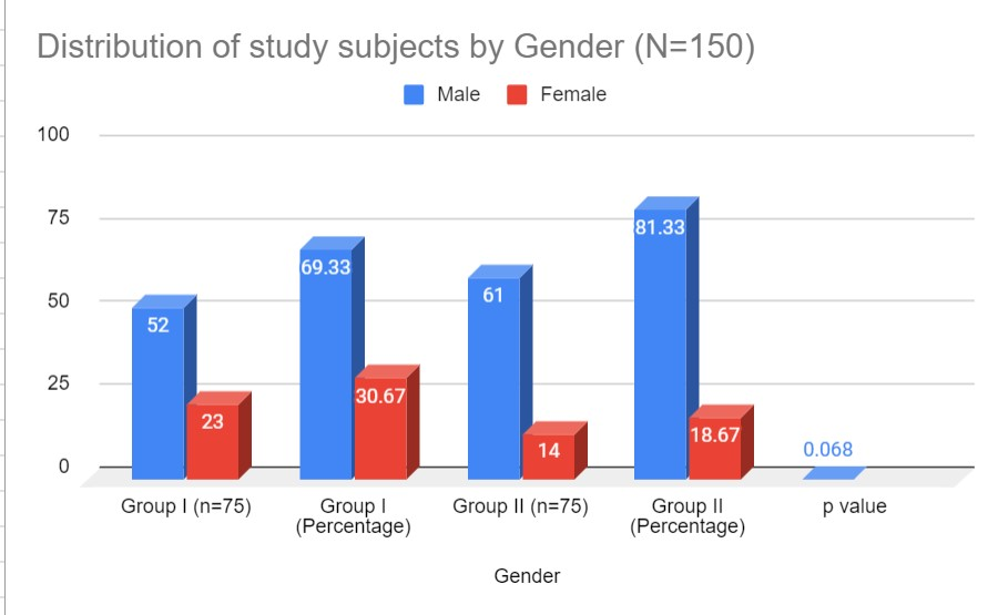
In this study, 75 Type 2 diabetic cases and 75 controls with normal blood glucose parameters were included. In the present study, the age ranged from 41 to 70 years. The mean age of diabetic patients in our study was 50.5±11.71 years and that of the control group was 45.53±9.97 years (Table 1). The majority of the patients diagnosed with Type 2 DM belonged to the 5th decade of life. There was no statistical difference in age between diabetic subjects and controls (p =0·082) The number of males in the diabetic group was 52 (69.33%) compared to 61 (81.33%) in the non-diabetic group & number of females in diabetics was 23 (30.67%) compared to 14 (18.67%) in nondiabetics. There was a male predominance in both groups (male/female: 2∶1·3 in controls and 2∶1·88 in diabetic patients). The mean duration of diabetes was 3.6 ± 1.7 years. The blood glucose parameters (FBS, RBS, and HbA1c) were statistically significantly higher in diabetics compared to non-diabetics (p value<0.001) (Table 2). The mean MPV in diabetic cases was 11.4 ± 1.0 fl compared to 9.0 ± 0.6 fl in nondiabetics with a p-value of 0.003. Mean PDW and P-LCR in diabetic patients were 14.1 ± 2.4 fl and 32.0 ± 8.1 compared to nondiabetics where it was
10.5 ± 0.7 fl and 21.0 ± 2.4% respectively (Table 2). However, no statistical correlation was seen between MPV, PDW, and P-LCR and the duration of DM in the diabetic group (Table 3). Diabetic patients were also divided into two groups according to their HbA1c level. Out of 75 Type 2 DM cases, there were 15 patients (20.0%) in group A (mean HbA1c<6.5%) and 60 patients (80.0%) in group B(mean HbA1c≥6.5%) (Table 3). The mean MPV in group B (11.7 ± 1.0 fl) was significantly higher in group A (11.1 ± 1.2 fl). Significantly higher mean PDW in group B (14.3 ± 2.4 fl) was observed in our study compared to group A (13.5 ± 2.7 fl) (Table 3). Our study also observed a statistically significant difference between mean P-LCR in group B (35.6 ± 7.7%) than group A (32.7 ± 9.1%). Duration of diabetes was not statistically significant between groups A&B (p-value <0.746) (Table 3).
| Age (years) | Group I(n=75) n % | Group II(n=75) n % | p-value |
|---|---|---|---|
| 41-50 | 17 26.7 | 24 36.7 | |
| 51-60 | 35 30.0 | 41 10.0 | |
| 60-70 | 23 20.0 | 1 0 10.0 | |
| Mean±SD Range (min to max) | 50.5±11.71 40 – 70 | 45.53±9.97 40 – 70 | 0.082 ns |
Table 1: Distribution of the Study Subjects by Age (N=150).
| Parameters | Diabetic cases(n = 75) Mean ± SD | Non-diabetic control(n = 75) Mean ± SD | p-value |
|---|---|---|---|
| HbA1c (%) | 7.3 ± 1.1 | 5.3 ± 0.4 | 0.002s |
| FBS (mmol/l) | 10.2 ± 2.6 | 5.7 ± 0.7 | 0.001 s |
| RBS (mmol/l) | 15.2 ± 2.1 | 6.5± 0.4 | 0.002 s |
| MPV | 11.4 ± 1.0 | 9.0 ± 0.6 | 0.003 s |
| PDW | 14.1 ± 2.4 | 10.5 ± 0.7 | 0.002 s |
| P-LCR | 32.0 ± 8.1 | 21.0 ± 2.4 | 0.002 s |
| Parameters | Group A Hba1c<6.5%(n = 15) Mean ± SD | Group B Hba1c ≥6.5%(n = 60) Mean ± SD | t-test p-value |
| Age | 52.1 ± 4.6 | 53.2 ± 5.4 | 0.001 s |
| MPV | 11.1 ± 1.2 | 11.7 ± 1.0 | 0.025 s |
| PDW | 13.5 ± 2.7 | 14.3 ± 2.4 | 0.032 s |
| P-LCR | 32.7 ± 9.1 | 35.6 ± 7.7 | 0.023 s |
| HbA1c (%) | 10.3 ± 1.0 | 8.0 ± 0.6 | 0.004 s |
| FBS (mmol/l) | 10.7 ± 0.7 | 14.2 ± 2.5 | 0.003 s |
| RBS (mmol/l) | 23.0 ± 2.4 | 35.0 ± 8.1 | 0.002 s |
| Duration of Disease | 3.4 ± 1.6 | 3.6 ± 1.7 | 0.643 ns |
Table 2: Comparison of Blood Sugar Level and Platelet Indices among Diabetic Cases and Non-Diabetic Controls.
Discussion
Diabetes mellitus is a major global health problem [15]. It is one of the major causes of morbidity and mortality affecting youth and middle-aged people [14]. The mean age of onset is 42 years. Deficient insulin action is the cardinal factor for the development of DM and clearly contributes to platelet dysfunction [16]. Platelets from patients with type 2 diabetes mellitus have increased reactivity and baseline activation which are likely to play a key role in the development and sustainment of vascular complications [17]. Higher MPV in diabetic patients indicates larger platelet size suggesting stimulated thrombopoiesis and augmented platelet activation [11]. One possible mechanism of increased MPV in DM is osmotic swelling due to raised blood glucose and perhaps due to a shorter life span of platelets in diabetic patients [18, 19]. In our study, the MPV was significantly higher in the diabetic group than the nondiabetic controls which was similar to the studies done by other researchers. An earlier study showed lower MPV in diabetic cases compared to the controls with no statistically significant difference [20, 21]. In addition, another platelet indices PDW was also significantly higher in diabetic subjects compared to controls (p< 0.002 respectively). Similar results were noted in other studies done by Demirtas, et al. [22], Jabeen, et al. [7], and Dalamaga, et al. [23] with significantly higher PDW levels among diabetic cases. The P- LCR is relatively a new platelet volume parameter. It is generated by only a few machines, with the Sysmex analyzer being one of them. Our study concluded that P-LCR was significantly higher (p-value<0.002) in diabetic cases. The study done by Jindal, et al. [19] and Ashraf, et al. [24] concluded significantly higher P-LCR in diabetes compared to non-diabetics. In our study, MPV, PDW, and P-LCR were significantly higher in diabetics with HbA1c levels <6.5% than in diabetics with HbA1c levels ≥6.5%. This is similar to the studies conducted by Kodiatte, et al. [1], Ozder, et al. [21], Ulutas, et al. [12] and Demirtas, et al. [22]. There was a higher number of diabetics with HbA1c levels ≥6.5% which is similar to the observation in the study done by Kodiatte, et al. [1]. This was due to poor dietary practices and a lack of knowledge regarding the diet and exercise regimens that ought to be followed in diabetic patients. Few studies showed HbA1c and MPV tend to decrease when glycemic control improves [21]. Therefore, it may be concluded that glycemic control improves platelet activity and function and may delay possible diabetic vascular complications.
Limitations of the Study
Small sample size. Follow-up of the cases was not possible to find out the prognostic significance of these findings. Platelet function tests could not be conducted on the sample to substantiate the findings of this study.
Conclusion
This study showed that increased platelet volume indices and larger platelets contribute to the prothrombotic state of diabetes mellitus. Because larger platelets are hemostatically more active, therefore its presence probably is a risk factor for developing diabetic vascular complications. Platelets with larger platelets can be easily identified during routine hematological analysis as MPV, PDW, and P-LCR are generated as by-products of the automated blood counts. Hence, MPV, PDW, and P-LCR would be useful prognostic markers of vascular complications in diabetes. Thus, platelet indices MPV, PDW, and P-LCR provide an important, simple, effortless, and cost-effective tool that can be useful in predicting an impending thrombotic state and vascular complications of diabetes.
Acknowledgments
Granted by the Ministry of Science & Technology, Bangladesh.
Conflict of Interest
Author Dr. Mst. Shaila Yesmin and other Author declares that they have no conflict of interest.
References
-
Kodiatyte TA, Manikyam UK, Rao SB, Jagadish TM, Reddy M, et al. (2012) Mean Platelet Volume in Type 2 Diabetes Mellitus. J Lab Physician 4: 5-9.
-
Keating FK, Sobel BE, Schneider DJ (2003) Effects of Increased Concentrations of Glucose on Platelet Reactivity in Healthy Subjects and in Patients with or Without Diabetes. Am J Cardiol 92: 1362-1365.
-
Ostenson CG (2001) The Pathophysiology of Type 2 Diabetes Mellitus: An Overview. Acta Physiol Scand 171: 241-247.
-
Italiano JE, Gresele P, Fuster V, Lopez JA (2008) The Structure and Production of Platelets. In: Gresele P, Fuster V, et al. (Eds.) Platelets in Hematologic and Cardiovascular Disorders. In: 1st (Edn.), Cambridge University Press, New York, USA, pp: 1-20.
-
Koltai K, Feher G, Kesmarky G, Keszthelyi Z, Zopf L, et al. (2006) The Effect of Blood Glucose Levels on Hematological Parameters, Platelet Activation, and Aggregation in Oral Glucose Tolerance Tests. Clin Hemorheol Microcirc 35: 517-525.
-
Borkataky S, Jain R, Gupta R, Singh S, Gupta K, et al. (2009) Role Of Platelet Volume Indices in the Differential Diagnosis of Thrombocytopenia: A Simple and Inexpensive Method. Haematolgy 14: 182-186.
-
Jabeen F, Riz HA (2013) Hyperglycaemic Induced Variations in Haematological Indices in Type 2 Diabetics. Int J Advanc Res 1: 322-334.
-
Fiedel BA (1976) Effect of C-Reactive Protein on Platelet Function. J Immunol 116(5): 1289-1294.
-
Vermeire S, Van Assche G, Rutgeerts P (2004) C-Reactive Protein as a Marker for Inflammatory Bowel Disease. Inflamm Bowel Dis 10(5): 661-665.
-
Saifullah A, Abdulla J, Muralikrishnan G (2013) Association of CRP with Diabetic and Non-Diabetic Individuals. Jordan J Biological Sci 3(1): 7-12.
-
Chang HA, Hwang HS, Park HK, Chun MY, Sung JY (2010) The Role of Mean Platelet Volume as a Predicting Factor of Asymptomatic Coronary Artery Disease. Korean J Fam Med 31: 600-606.
-
Ulutas KT, Dokuyucu R, Sefil F, Yengil E, Sumbul AT, et al. (2014) Evaluation of MVP in Patients with Type 2 Diabetes Mellitus and Blood Glucose Regulation. A Marker of Atherosclerosis. Int J Clin Exp Med 7(4): 955- 961.
-
Lutfullah Cakir, Gulali Aktas (2014) Mean Platelet Volume Increases in Type 2 Diabetes Mellitus Independent of Hba1c Levels. Acta Medica Mediterranea 30: 425.
-
Genuth S, Alberti KG, Bennett P, Buse J, Defronzo R, et al. (2003) Expert Committee on the Diagnosis and Classification of Diabetes Mellitus 2. Follow-Up Report on the Diagnosis of Diabetes Mellitus. Diabetes Care 26: 3160-3167.
-
Hekimsoy Z, Payzin B, Ornek T, Kandogan G (2004) Mean Platelet Volume in Type 2 Diabetic Patients. J Diabet Complicat 18: 173-176.
-
Hers Im (2007) Insulin-Like Growth Factor-1 Potentiates Platelet Activation Via the IRS/PI3Kalpha Pathway. Blood 110: 4243-4252.
-
Stratmann B, Tschoepe D (2005) Pathobiology and Cell Interactions of Platelets in Diabetes. Diab Vasc Dis Res 2: 16-23.
-
Vinik AI, Erbas T, Park TS, Nolan R, Pittenger GL (2001) Platelet Dysfunction in Type 2 Diabetes. Diabetes Care 24: 1476-1485.
-
Jindal S, Gupta S, Gupta R, Kakkar A, Singh HV, et al. (2011) Platelet Indices in Diabetes Mellitus: Indicators of Diabetic Microvascular Complications. Hematology 16: 86-90.
-
Akinsegun A, Olusola D, Sarah J, Olajumoke O, Adewumi A, et al. (2014) Mean Platelet Volume and Platelet Counts in Type 2 Diabetes: Mellitus on Treatment and Non- Diabetic Mellitus Controls in Lagos, Nigeria. Pan Afr Med J 18: 1-5.
-
Ozder A, Eker H (2014) Investigation of Mean Platelet Volume in Patients with Type 2 Diabetes Mellitus and in Subjects with Impaired Fasting Glucose: A Cost-Effective Tool in Primary Health Care. Int J Clin Exp Med 7: 2292- 2297.
-
Demirtas L, Degirmenci H, Akbas E, Ozcicek A, Timuroglu A, et al. (2015) Association of Hematological Indicies with Diabetes, Impaired Glucose Regulation and Microvascular Complications of Diabetes. Int J Clin Exp Med 8: 11420-11427.
-
Dalamaga M, Karmaniolas K, Lekkab A, Antonakosa G, Thrasyvoulides A, et al. (2010) Platelet Markers Correlate with Glycemic Indices in Diabetic, but not Diabetic Myelodysplastic Patients with Normal Platelet Count. Dis Markers 29: 55-61.
-
Ashraf S, Ranjan R, Singh S, Singh H, Kudesia M, et al. (2018) Diabetes Disease Burden by Platelet Indices as Possible Biomarkers in Evaluation of Initial Vascular Risks in Grading Diabetes Mellitus: Correlation of Platelet Dysfunction Indices with Hematopoietic and Biochemical Biomarkers in Diabetes Mellitus. Open J Biochem Press pp: 1-15.
- Genomic Landscape of Aggressive Penile Squamous Cell Carcinoma including TERT-p and NOTCH1 Mutations – An Institutional Experience
- Establishment of Baseline Haematological Values for Canine Population in North-Central Nigeria: A Cross-Sectional Study in the Federal Capital Territory
- Biochemical Assessment of Uroliths Extracted in Patients with Urolithiasis in a Tertiary Health Institution
- Update on Gastrointestinal Pecomas: Molecular Pathogenesis and Risk Stratification
- A Comparative Study of Serum C-reactive Protein Level Between Pre-eclampsia and Normal Pregnancy in Tertiary Level Hospital
- From Deformity to Alignment: Clinical Outcomes of the Schnepp Osteotomy in Hallux Valgus in 47 Feet