‘Inversion’ of Impacted Mandibular Third Molar in Ascending Ramus of the Mandible – Report of Rarest Case
‘Wisdom tooth’ or ‘Third molars’ are very interesting tooth not only for oral and maxillofacial surgeons but also for all other specialists of medicine and dentistry. Mandibular third molars are the most often affected tooth and show variations in eruption, size, shape, development, positions and associated with anomalies. The purpose of this article is to present a rarest case of inverted unilateral impacted mandibular left third molar in the ascending ramus of the mandible and was associated with a cystic lesion which is rarely encountered in clinical practice during impaction procedures. Reporting of such interesting and uncommon presentations of dental structures is highly essential to provide utmost care to the patient as well as to make guidelines and protocols for their management.
Introduction
Mandibular third molars exhibit lot of variations during their development as well as in eruptive stage [1, 2]. An ‘impacted tooth’ refers to a tooth that is absolutely or in part unerupted and is located in opposition to any other teeth, bone or soft tissue so that it eruption is unlikely. Whereas, ‘inversion of tooth’ is the condition where a tooth is mispositioned and reversed in the upside-down position [3, 4]. Impaction of third molars is more commonly observable finding but inversion is a rare phenomenon. The teeth frequently affected by this condition are incisors, canines, premolars and supernumerary tooth [5, 6, 7]. But molars are seldom reported in inverse position. There are publications showing inverted and impacted third molars in the maxillary arch [8, 9, 10]. but their occurrence in mandibular arch is a rare entity. Till date only seven cases have been reported in the dental literature (Table 1) [10, 11, 12, 13, 14, 15]. Clinically such teeth in inversion usually cause headache, loss of adjacent teeth, crowding, late or ectopic eruption, diastema, eruption of teeth into the floor of the nasal cavity (in case of maxilla), or inferior border of the mandible (in mandible), development of serious pathology and worsening of existing conditions [11, 12, 13]. In the maxillary arch, the impacted third molar is knocked out anywhere between the gums and the orbital floor. In mandible, the most common site of getting impacted and inverted is the ascending part of the ramus. This kind of impaction is called as complex, because of the relatively peculiar position of the crown and the root. The exact etiology for development of tooth in the inverse position is not known, however, it is attributed to the peculiar growth of odontogenic epithelium before the formation of the teeth bud. Regarding treatment of inverted third molars there is no definitive treatment protocols exist for the removal of inverted teeth [5, 6, 7, 8, 9, 10, 11, 12, 13, 14, 15].
In the present article, author have presented a rarest occurrence of mandibular left third molar which was impacted and inversed in the ascending part of the ramus of the mandible and also associated with a cystic lesion. The key points learned from this publication is that any impacted third molars should be neglected and must go for detailed investigation including advanced imaging techniques like cone-beam computed tomography.
Case Report
A 55-year-old male Indian patient reported to a private dental clinic complaining of decayed tooth in the upper left region causing pain from past one week. There was no history of medications, or signs and symptoms of systemic or metabolic disorders. On physical examination patient was moderately built and nourished. Intraoral examination showed deep proximal caries involving maxillary left first and second molars which were tender to percussion test. All permanent teeth were erupted including maxillary third molars except mandibular right and left third molars. Severe lower anterior teeth crowding was also observed. Patient did not give any history of pain or removal of teeth from the lower third molars region. To study the status of decayed teeth, patient was subjected to radiographic evaluation. On radiographic examination (orthopantomograph), caries involving pulp was observed in both first and second molars. Further examination of the radiograph revealed impacted mandibular right and left third molars. Mandibular right third molar was in horizontal impaction position. Surprisingly on the left side, the impacted third molar showed some unusual position. It was completely inverted upside-down in the ascending region of the ramus of the mandible with crown facing downwards and roots facing upward (Figure 1). It was also associated with a huge radiolucent cystic lesion surrounding the crown and extending to the inferior border of the mandible (Figure 1). The cystic lesion was also eroded the mandibular canal. But astonishingly this was not led to any signs and symptoms in the patient. It was assumed that due to large size of the teeth, and inadequate arch length, the third molar might have insufficient space to develop and erupt. Therefore, during development of tooth bud itself due to lack of space, the tooth germ is drifted downwards due to the inherent force resulting in ‘inversion’ of the third molar. Patient was explained about the existing condition of the impacted tooth and associated pathology, its consequences and surgical removal of the impacted third molar.
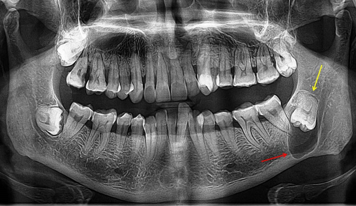
Figure 1: Radiograph Showing Impacted and Inverted Mandibular Left Third Molar in Ascending Ramus of the Mandible. The Crown is Facing Downward with its Roots Upward (Yellow Arrow). It is surrounded by a Radiolucent Cystic Lesion (Red Arrow), Eroding Mandibular Canal and Extending to Inferior Border of the Mandible. Right Side, the Third Molar is in Horizontal Impaction Position.
Discussion
Various authors have given different classification pertaining to impaction of mandibular third molars in the surgical field [12, 14]. But all those mentioned classifications were indicated for the molars with normal position. There is no classification system for inverted and impacted third molars in the mandibular arch. There are reports showing inversion of maxillary third molars in the dental literature [7, 8]. But publications on occurrence of inverted mandibular molars are extremely rare with very few reports are published in the English literature (Table 1). Investigation using radiographic imaging plays a significant role in determining the precise location of inverted, impacted teeth and their interaction with critical anatomic structures [13]. Moreover, prior knowledge of existing position of impacted molars helps in easy removal with minimum surgical trauma [16, 17, 18, 19].
Some of the etiological factors have been suggested in the review articles for the cause of inversion of impacted third molars. They are numerous underlying systemic conditions such as endocrinology, radiation, Down syndrome, cleidocranial dysplasia and other factors like abnormal eruptive pathway, malposed teeth germs, presence of supernumerary tooth, over-retained primary teeth, deficiency of arch length and odontogenic tumors and cyst.
| Author/Ethnicity | Age (in years) & Gender | Laterality | Location | Associated pathology | Treatment provided | |
|---|---|---|---|---|---|---|
| Present case | Nagaveni, et al. India | 55, Male | Unilateral Left side | Ascending ramus of the mandible | Cystic lesion | Surgical removal advised |
| 1 | Talib, et al. [10] UAE | 1.50, Female | Bilateral Right & Left | Ascending ramus of the mandible | None | Surgical removal |
| 1 | Talib, et al. [10] UAE | 2.32, Female | Unilateral Right | - | None | Surgical removal |
| 2 | Sunil, et al. [12] India | 26, Male | Unilateral Right | Ascending ramus of the mandible - | None | Surgical removal |
| 3 | Mohan, et al. [11] India | 40, Male | Unilateral Right | Ascending ramus of the mandible - | None | Surgical removal |
| 4 | Tripathi, et al. [14] India | 35, Male | Bilateral Right & Left | - | None | No treatment |
| 5 | Singh, et al. [13] India | 1.45 Male | Unilateral Right | - | None | No treatment |
| 5 | Singh, et al. [13] India | 2.35 Male | Bilateral Right & Left | - | - | No treatment |
| 5 | Singh, et al. [13] India | 3.26 Female | Unilateral Left | - | - | No treatment |
| 6 | Patel, et al. [16] India | 28, Female | Unilateral Right | - | None | No treatment |
| 7 | Pai, et al. [15] India | 30, female | Unilateral Right | - | None | No treatment |
Table 1: Reported Articles on Inverted and Impacted Mandibular Third Molars.
Investigation using radiographic imaging mainly including orthopantomography plays a significant role in determining the precise location of inverted, impacted teeth and their interaction with critical anatomic structures [13]. Recently with the invention of advanced imaging techniques such as Cone Beam Computed Tomography (CBCT) scan, there is great revolution in medical field including dentistry. Because it is associated with less radiographic exposure compared to conventional plain radiographs, as they require multiple exposure from different angulation to depict any pathology.
In addition to this, CBCT provides three-dimensional picture of the lesion and its associated structures. CBCT play a major role in identification of rare dental phenomenon like inversion of impacted molars. Because it provides detailed information regarding molars depth within the bone, bone details, adjacent anatomic structures like inferior alveolar or mandibular canal or foramen in case of mandibular arch. In maxilla, it provides details on relation with the maxillary structures like maxillary sinuses and infratemporal fossa. Hence, prior knowledge of existing position of impacted molars also helps in easy removal with minimum surgical trauma and no damage to the adjacent important anatomic structures [16, 17, 18, 19, 20, 21].
Knowledge about the level of impaction is very important to plan for type of surgical removal to be selected. The two researchers Pell and Gregory [22] gave a classification system on the level of impaction pertaining to third molars occurring in the mandibular arch. In their classification they have given total six types which is elaborated in Table 2. Based on this classification, the inverted and impacted third molar presented in this paper was classified as Position III, as complete crown of the inverted, impacted third molar was contained within the ramus.
| Types | Description of Third molar Impaction |
|---|---|
| Position A | The impacted tooth’s occlusal level is parallel to that of the surrounding teeth. |
| Position B | The impacted tooth’s occlusal level is midway between the occlusal plane and cervical line of the adjacent tooth. |
| Position C | The impacted tooth’s occlusal level is apical to the cervical line of the adjacent tooth. |
| Position I | The impacted third molar’s crown does not extend into the ramus and the molar is visible in front of the ramus’s anterior border. |
| Position II | When the crown of the tooth is outside of the mandibular ramus. |
| Position III | More than half of the crown must be contained within the ramus. |
Table 2: Pell and Gregory’s Classification to Depict the Level of Impaction in Third Molars (Mandibular Arch).
Surgical science shows no established therapeutic guidelines or protocols for the removal of the molars which are inverted [12, 15]. Most of the authors suggested conservative treatment is the safest and routine treatment modality for these teeth which includes ‘wait and watch method’ until pathological symptoms appear. For this patient has to undergo for repeated and regular routine investigation including radiographic evaluation (either orthopantomography or CBCT scan). But this protocol does not hold right for all cases. The present case is the example where the inverted and impacted third molar was developed with a cystic lesion if it is unnoticed would have resulted in adverse complications. This suggests at the age from 20 to 30, patient’s with unerupted third molars compulsorily undergo for thorough investigation including radiographic evaluation to rule out such asymptomatic third molars. In addition to this, ‘The National Institute for Clinical Excellence’ (NICE) has given therapeutic guidelines for the management of third molars. These NICE guidelines suggests that only patients with pathology, such as unrestorable caries, untreatable pulpal and/or peri-apical pathology, cellulitis, abscess, osteomyelitis, internal or external resorption of the tooth or adjacent teeth, recurrent episodes of pericoronitis, fracture of the tooth, disease of the follicle including cyst/tumor, tooth/teeth impending surgery, or reconstructive dentistry, the surgical removal of third molars is indicated [12, 13, 14, 15, 16, 17, 18, 19, 20, 21, 22]. Other factors that should be considered are age of the patient and deeper positioning of the inverted tooth. In such conditions, its extraction becomes more complex than the extraction of a normally impacted tooth. Because, the teeth are firmly set inside the bone, therefore significant amounts of bone must be removed during surgery. During surgical removal of impacted teeth several methods, including the use of rotary burs, chisel mallets, Lasers, piezosurgery, have been suggested for the easy removal of bone around the impacted tooth [6, 7, 8]. Few authors have suggested that conservative treatment is the treatment of choice for the bilateral occurrence of inverted and impacted third molars if they are not associated with any pathology [6, 7, 8].
In the present case, the inverted and impacted mandibular third molar was diagnosed following radiographic examination taken for some other dental problem. Patient was absolutely normal with no history of any pain, food lodgment or pericoronitis in the third molar region. But following radiographic examination the third molar was totally impacted inside the bone in the inverted position with crown placed below the roots of second molar. Moreover, the tooth was associated with a cystic lesion which was almost encroaching towards inferior border of the mandible and mandibular canal. This fact alarms all dental surgeons about the possibility of abnormalities should expected in case of clinically missing tooth and to for thorough radiographic evaluation including advanced imaging techniques such as CBCT.
Conclusion
Occurrence of inverted mandibular third molars is an extremely rare finding and not only oral surgeon but also all specialties of dentistry should have knowledge in detail about this phenomenon. Moreover, radiographic examination including advanced imaging scans like CBCT should be considered a mandatory procedure in all patients above 18
years of age and also in unerupted cases of mandibular third molars in order to diagnose variations in third molars and also associated pathology.
References
-
Nagaveni NB, Umashankara KV (2013) Maxillary molar with dens evaginatus and multiple cusps: Report of a rare case and literature review. Int J Oral Health Sci 3(2): 92.
-
Nagaveni NB (2023) Inversion of impacted mesiodens: Report of case series with literature review. Global Journal of Research in Dental Sciences 3(5): 7-12.
-
Nagaveni NB (2024) Idiopathic Intracoronal resorption in impacted maxillary tooth – Report of a rare case. EC Dental Science 23(2): 1-4.
-
Yuvaraj, Agarwal GD (2011) Inverted maxillary third molar impaction—a case report. Peoples J Sci Res 4(1): 57-58.
-
Nagaveni NB, Shashikiran N, Reddy VS (2009) Surgical management of palatal placed, inverted, dilacerated and impacted mesiodens. Int J Clin Pediatr Dent 2(1): 30-32.
-
Nagaveni NB (2023) Vertical, Intra-Osseous Impaction of permanent maxillary central incisor in association with multiple anomalies – Report of a rare case. EC Dental Science 22(9): 1-4.
-
Seraj B, Ghadimi S, Mighani G, Zare H, Rabbani M (2012) Inverted Impacted Primary Maxillary Incisors: A Case Report. J Dent (Tehran) 9(2): 174-177.
-
Agarwal P, Kumar S, Jain K, Kiran K (2019) Inverted maxillary third molar impactions. Ann Maxillofac Surg 9(2): 484-488.
-
Khouri C, Aoun G, Khouri C, Saade M, Salameh Z, et al. (2022) Evaluation of Third Molar Impaction Distribution and Patterns in a Sample of Lebanese Population. J Maxillofac Oral Surg 21(2): 599-607.
-
Talib YM, Albalushi NW, Fouad DM, Salloum AM, Kukreja BJ, et al. (2023) Bilateral Inverted and Impacted Mandibular Third Molars: A Rare Case Report. Cureus 15(3): e36573.
-
Mohan A, Mirgan D (2018) An Inverted Impacted Mandibular Third Molar: A Rare Case Report and Literature Review. Journal of Pierre Fauchard Academy (India Section) 32(3-4): 68-70.
-
Sunil V, Partha D, Sahana P, Jacob MS, Koshy J (2020) Inverted impacted mandibular third molar: An unusual eruption. J Adv Clin Res Insights 7(2): 42-44.
-
Singh S, Rahman H, Chandra R, Tripathi S, Jyoti J, et al. (2016) Assessment of inverted mandibular third molar impaction by 3d Reconstruction – A rare case series. IOSR J Dent Med Sci 15(5): 4-7.
-
Tripathi S, Chandra R, Tripathi P, Rahman H, Singh S, et al. (2015) Bipronged inverted impacted third molar-A CBCT analysis. Br J Med Med Res 9(9): 1-5.
-
Pai V, Kundabala M, Sequeira PS, Rao A (2008) Inverted and impacted maxillary and mandibular 3rd molars; a very rare case. Oral Health Comm Dent 2(1): 8-9.
-
Patel R, Patel J, Shah V, Shah R, Bhakhar V (2014) Inverted and impacted third molars-report of two rare cases with literature. Adv Hum Biol 4(2): 74-77.
-
Padhye MN, Dabir AV, Girotra CS, Pandhi VH (2013) Pattern of mandibular third molar impaction in the Indian population: a retrospective clinico-radiographic survey. Oral Surg Oral Med Oral Pathol Oral Radiol 116(3): 161-166.
-
Terauchi M, Akiya S, Kumagai J, Ohyama Y, Yamaguchi S (2019) An Analysis of Dentigerous Cysts Developed around a Mandibular Third Molar by Panoramic Radiographs. Dent J (Basel) 7(1): 13.
-
Sachdeva SK, Jayachandran S, Kayal L, Bakyalakshmi K (2016) Inverted and Impacted Maxillary and Mandibular Third Molar: Unusual Case Reports with Review of the Literature. Saudi J Med Med Sci 4(1): 32-34.
-
Valdec S, Al-Haj Husain A, Winklhofer S, Müller M, Piccirelli M, Stadlinger B (2021) Comparison of Preoperative Cone-Beam Computed Tomography and 3D-Double Echo Steady-State MRI in Third Molar Surgery. J Clin Med 10(20): 4768.
-
Nagaveni NB, Umashankar KV (2023) A giant radicular cyst involving the left maxillary sinus diagnosed on CBCT image-Report of a rare case. J Oral Health Dent 7(1): 591-595.
-
Pell GJ, Gregory BT (1933) Impacted mandibular third molars: classification and modified techniques for removal. Dent Dig 39(9): 330-338.
- Genomic Landscape of Aggressive Penile Squamous Cell Carcinoma including TERT-p and NOTCH1 Mutations – An Institutional Experience
- Establishment of Baseline Haematological Values for Canine Population in North-Central Nigeria: A Cross-Sectional Study in the Federal Capital Territory
- Biochemical Assessment of Uroliths Extracted in Patients with Urolithiasis in a Tertiary Health Institution
- Update on Gastrointestinal Pecomas: Molecular Pathogenesis and Risk Stratification
- A Comparative Study of Serum C-reactive Protein Level Between Pre-eclampsia and Normal Pregnancy in Tertiary Level Hospital
- From Deformity to Alignment: Clinical Outcomes of the Schnepp Osteotomy in Hallux Valgus in 47 Feet