Labial Triple Talon Cusp in a Mesiodens – Report of a New Morphological Variant with Revised New Definition and Classification System
Literature shows various case reports and prevalence studies on mesiodens which is a type of supernumerary tooth occurring in the midline of the maxillary or mandibular arch between two central incisors. It may occur in both primary and permanent dentition and characterized by various morphology. Talon cusp is an anomaly of the tooth shape and numerous reports on different shapes and sizes are evident. The present article shows occurrence of a triple labial talon cusp in the maxillary midline mesiodens which makes a unique expression and not reported so far in the dental anomalies’ literature. This manuscript also provides a new insight pertaining to definition and classification system for talon cusp. Therefore, the present case highlights the pioneer case record of triple labial talon cusp in a mesiodens associated with another dental anomaly.
Introduction
The term ‘Talon cusp’ was suggested by Mellor and Ripa because of its resemblance in shape to an eagle’s claw and represents a rare anomaly but much debated morphological variant [1]. It is a developmental dental anomaly pertaining to shape refers to an accessory cusp-like structure projecting from the cingulum area or cementoenamel junction of the maxillary or mandibular anterior teeth. This may be found unilateral or bilateral, on single or double tooth, and on the palatal/lingual or labial aspects of the affected taloned teeth [1]. Literature shows varied forms of talon cusps [1, 2, 3, 4, 5, 6, 7, 8, 9, 10, 11, 12, 13, 14, 15, 16, 17, 18]. They also vary from sharp and spike-like, or tubercle- like cingulum while others have rounded and smooth tips. Sometimes, it can be horn-like, conical, pyramidal, bifid or tubercle-like cingulum. They can occur in either primary or dentition affecting more frequently permanent maxillary incisors followed by mandibular permanent incisors and primary maxillary incisors. It is documented more in males than females and there is scarcity in epidemiological data pertaining to true prevalence of this anomaly and it varies with age, race and criteria used to define this anomaly [3].
The historical published data suggests that 75% (only 22 cases) of the talon cusp have been encountered in permanent incisors including lateral incisors followed by central incisors and canines and therefore, the occurrence of talon cusp in supernumerary tooth like mesiodens is an uncommon finding [4]. Till date, only 19 cases of talon cusp formation in supernumerary tooth have been documented as evident from the published literature. Moreover, literature shows occurrence of only either palatal or facial/labial talon and with a single or double talon cusp (Table 1) [5, 6, 7, 8, 9, 10, 11, 12, 13, 14, 15, 16, 17, 18, 19]. A publication on occurrence of triple labial talon cusp is an extremely uncommon finding. Therefore, the aim of this manuscript is to present an occurrence of three (triple) labial talon cusp in a mesiodens associated with another developmental anomaly which encountered in Indian 11-year-old male patient.
| Case No. | Author/Year | Patient Age (in years)/ Gender | Mesiodens Type, Shape and Location | Talon Cusp Location | Talon Cusp Type | Associated Anomalies |
|---|---|---|---|---|---|---|
| Present case Nagaveni NB /2024 | 11/Male | Permanent Supplemental Maxillary | Facial | Triple talon Type 6, B (Current classification as proposed by author) | Vertical Impaction of supernumerary tooth | |
| 1 | Nagaveni NB /2023 | 8/Male | Permanent Three-lobed Incisoriform Maxillary | Palatal | Type I | - |
| 2 | Nagaveni NB, et al. /2014 | 14/Male | Permanent Conical Maxillary | Facial | Type III | - |
| 3 | Nagaveni NB, et al. /2014 | 10/Female | Permanent, Conical Maxillary | Facial | Type I | - |
| 4 | Nagaveni NB, et al. /2014 | 11/Male | Permanent Conical Maxillary | Facial | Type I | - |
| 5 | Nagaveni NB, et al. /2014 | 5/Female | Primary, Conical Maxillary | Facial | Type I | - |
| 6 | Hattab, et al. /2014 | 10/female | Permanent Supplemental Maxillary | Palatal Double talon | Type II | - |
| 7 | Nuvvula, et al. /2011 | 14/Male | Permanent Supplemental Maxillary | Palatal | Type III | |
| 8 | Nagaveni, et al. /2010 | 10/Male | Permanent Conical Maxillary | Facial | Type I | - |
| 9 | Rani, et al. /2010 | - | Permanent Conical Maxillary | Palatal Fused with Lateral Incisor | Type I | |
| 10 | Babaji, et al. /2010 | 6/Male | Permanent, Supplemental Maxillary | Palatal | Type I | Erupted supernumerary tooth |
| 11 | Nagaveni NB, et al. /2010 | 11/Male | Permanent Multi-lobed Maxillary | Palatal | Type I | |
| 12 | Topaloglue, et al. /2008 | - | Primary Conical Maxillary | Facial & Palatal | Type I | - |
| 13 | Arora, et al. /2008 | 12/Female | Permanent Maxillary Multi-lobed | Labial | Type I | - |
| 14 | Siraci, et al. /2006 | - | Primary Conical Maxillary | Facial & Palatal | Type II | - |
| 15 | Nadakarni, et al. /2002 | - | Permanent Conical Maxillary | Palatal | Type III | - |
| 16 | Zhu, et al. /1997 | - | Permanent Conical Maxillary | Palatal | Type III | - |
| 17 | Hattab, et al. /1996 | - | Permanent Conical Maxillary | Palatal | Type I | - |
| 18 | Salama, et al. /1990 | - | 1.Primary Conical Maxillary 2.Permanent Conical Maxillary | Palatal Palatal | Type III Type III | - |
Table 1: Published Cases of Supernumerary Tooth (Mesiodens) With Talon Cusp (for Classification of Talon Cusp Type, Hattab, et al
Moreover, literature emphasizes need for the revised formulation of existing both conventional definition and classification system on talon cusp. As a result, taking this into account, author of this paper debated on existing literature addressing talon cusp, to propose a revised definition and new classification system for talon cusp and re-formulated a new definition and classification system (Table 2).
| Talon Cusp Type | Description |
|---|---|
| Type 1 Single Palatal Talon | Extra cusp that prominently projects from the palatal/lingual surface of anterior teeth (incisors & canines) and extends at least half the distance from the cement-enamel junction to the incisal edge (primary or permanent, maxillary or mandibular) |
| Type 2 Single Labial/Facial Talon | Extra cusp that prominently projects from the labial/facial surface of anterior teeth (incisors & canines) and extends at least half the distance from the cement-enamel junction to the incisal edge (primary or permanent, maxillary or mandibular) |
| Type 3 Single Palatal and Labial Talon | Extra cusp that prominently projects from the both palatal/lingual and labial/facial surface of anterior teeth (incisors & canines) and extends at least half the distance from the cement-enamel junction to the incisal edge (primary or permanent, maxillary or mandibular) |
| Type 4 Single Labial/Palatal Talon cusp in Anomalous tooth | Extra cusp that prominently projects from the both palatal/lingual and labial/facial surface of any anomalous tooth (supernumerary/fused/double/geminated tooth) and extends at least half the distance from the cement-enamel junction to the incisal edge (primary or permanent, maxillary or mandibular) |
| Type 5 Double Talon Cusp | Extra two cusps/spikes that prominently projects from either palatal/lingual or labial/ facial surface of anterior teeth or any anomalous tooth (supernumerary/fused/double/ geminated tooth) and extends at least half the distance from the cement-enamel junction to the incisal edge (primary or permanent, maxillary or mandibular) Based on length of the cusps/spikes and presence of developmental grooves, it can be further sub classified into Type 5, A: Equal-sized and separated by a developmental groove Type 5, B: Unequal size and separated by a developmental groove Type 5, C: Equal-sized and fused Type 5, D: Unequal size and fused |
| Type 6 Triple Talon Cusp | Extra three cusps/spikes that prominently projects from either palatal/lingual or labial/ facial surface of anterior teeth or any anomalous tooth (supernumerary/fused/double/ geminated tooth) (primary or permanent, maxillary or mandibular) Type 6, A: Equal-sized and separated by a developmental groove Type 6, B: Unequal size and separated by a developmental groove Type 6, C: Equal-sized and fused Type 6, D: Unequal size and fused |
| Type 7 Single Semi Talon | Extra cusp of 1 mm or more, but extending less than half the distance from the cement- enamel junction to the incisal edge. It may blend with the palatal or labial surface or stand away from the rest of the crown (anterior teeth or anomalous teeth, primary or permanent, maxillary or mandibular). |
| Type 8 Single Trace Talon | En enlarged or prominent cingulam sor its different forms (conical, or tubercle-like) originating from the cervical third of the crown in anteriors (primary or permanent, maxillary or mandibular). |
Table 2: Proposed Revised Classification of Talon cusp.
Case Report
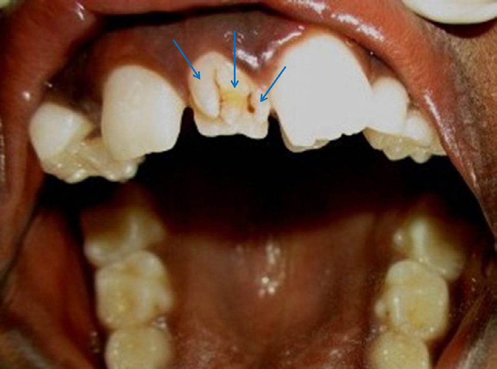
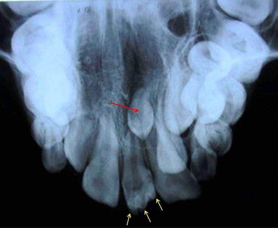
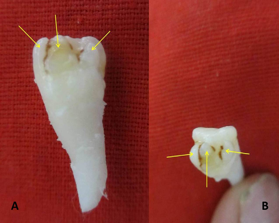
An 11-year-old male patient reported to a private dental clinic complaining of ugly looking tooth in the upper front tooth region. On physical examination, patient appeared apparently normal with no signs and symptoms of any systemic, metabolic or syndromic disorders. On intraoral examination, patient was in mixed dentition stage showing erupted maxillary right and left central and lateral incisors. However, between two central incisors huge gap was present consisting of an extra tooth (supernumerary) in the midline. Careful examination of this extra tooth revealed three spikes like structure extending from cingulum to three fourth level distance of incisal edge. Well developed grooves separated these three spikes and were discolored with stains (Figure 1). As this extra tooth was occupied the central space, both right and left maxillary central incisors were distally drifted, so the left lateral incisor was erupted lingually as there was no space for its eruption. Patient was subjected to a radiographic examination, which showed the presence of three separate V-shaped radiopaque structures extending from cingulum to the incisal edge. Radiograph also showed presence of impacted vertically placed midline another supernumerary tooth (Figure 2). Finally, based on the both clinical, literature search and radiographic examination, this case was diagnosed as mesiodens with triple labial talon cusp associated with vertically impacted supernumerary tooth. According to revised classification as suggested by the current author, the talon cusp in this particular case was diagnosed as Type 6, B (Triple talon cusp, with unequal sized three spikes separated by developmental grooves). Patient was explained about the existing anomaly and extraction was carried out. Following extraction, when the tooth was observed, it showed presence of three well developed talon cusps attached to the labial surface of the supernumerary tooth (Figure 3).
Discussion
Mellor, et al. [1, 2, 3, 4, 5, 6, 7, 8, 9, 10, 11, 12, 13, 14, 15, 16, 17, 18, 19, 20, 21, 22, 23, 24, 25] have researched on this anomaly and debated on their etiology, morphology, prevalence, various definitions and classification systems. After its first description suggested by Mellor, et al. [1] Gorlin, et al. [20] defined it as ‘a high accessory cusp reaching the incisal edge to produce a T-form or a Y-shaped tooth crown.’ Recently it got revised and it defines talon’s cusp as ‘an accessory cusp on the lingual or labial aspect of incisors or canines [3]. According to previous author’s opinion like Nuvvula, et al. [8] Jowharji, et al. [21] Nagaveni, et al. [3, 5, 6, 9, 12] Chin-Ying, et al. [22] Mallineni, et al. [2] and Bommanavar, et al. [4] and based on the current different expression of the talon cusp, author would like to revise the conventional definition and it can be defined as “an accessory cusp/spike/lobe/tubercle/ pyramidal like structure projecting from the cingulum area or cemento-enamel junction ending with smooth/round/ sharp tip in either maxillary or mandibular, primary or permanent, incisors (central and lateral) or canines or any anomalous teeth (supernumerary/fused/double tooth/ geminated tooth) on facial/labial or lingual surface of the affected tooth.” This definition should be stressed upon and needs to be implemented in the text books and in the dental literature constituting this anomaly.
In addition to the definitions, various classification systems have been suggested in the literature due to the difference, location and reason in varied presentation of this dental anomaly and its appearance on different surfaces like labial or lingual surfaces including different teeth. Tracing back to historiography, Hattab, et al. [18] classified talon cusp into three types as true talon, semi-talon and trace talon based on its degree of cusp formation, extension and its morphological characteristics. Chin-Ying, et al. [22] classified them as major, minor and trace talons. Whereas, Maynes [23] studied the dentitions of 301 skeletons of pre-European- contact American Indian races, for the presence or absence of talon cusp, in that five skeletons (2%) had the trait and graded this trait as stage 1, 2 and 3 based on degree of severity from slightest to most extreme form. Mallineni, et al. [2] classified them into three types as facial, lingual and facial and lingual. Recently Bommanavar, et al. [4] suggested a new classification for double facial talon cusps based on their size (equal/unequal) and presence of developmental grooves (separate or fused) as type 1: equal-sized and separated by a developmental groove, type 2: unequal size and separated by a developmental groove, type 3: equal-sized and fused, and type 4 as unequal size and fused. Author of the current paper based on her previous published cases [3, 5, 6, 9, 12] and from the present case made an effort to propose a new classification system by compiling all previous classification and is revised in Table 2.
Occurrence of one anomaly in association with another anomaly is an uncommon finding and needs to be documented for further research output. In the current case, vertical impacted supernumerary tooth was also found following radiographic examination. In maximum cases, talon cusp occurred on lingual surface of anterior teeth. Literature evidence of labial talon cusp is rare and no epidemiological information is available. Therefore, combination of both labial and triple talon cusp is an extremely rare entity in the dental anomalies. Author previously documented ample research work pertaining to such uncommon dental abnormalities to the dental literature. [3, 5, 6, 9, 12, 25] In one survey [3] on talon cusps, author reported a facial talon on mesiodens in the primary dentition which is also a rare finding. In another case, mesiodens was of multi-lobed having palatal talon cusp being another rarity [12]. Recently, Nagaveni [25] explored another unique case three-lobed incisoriform mesiodens constituting type 1 palatal talon cusp. The present case is another addition to such unique cases’ list.
Extensive literature review compiled 21 cases of facial talon cusps in primary and permanent dentition [4]. In these, 19 cases were occurred in the permanent dentition and two cases in the primary dentition. Moreover, those 15 cases exhibited a single talon cusp in primary dentition and remaining 15 cases also showed solitary talon in the permanent dentition [4]. Surprisingly only two cases of double facial talon cusp were reported, one by Pillai, et al. [24] which formed on the facial surface of the permanent left maxillary central and lateral incisor. Another second case was documented recently by Bommanavar, et al. [4] which occurred in permanent maxillary left central incisor. But reports on occurrence of facial talon cusp in a supernumerary tooth like mesiodens are scarce with only countable number of cases published. Till date only 19 cases [5, 6, 7, 8, 9, 10, 11, 12, 13, 14, 15, 16, 17, 18, 19] of labial talon cusp formation in supernumerary tooth have been documented as evident from the published literature (Table 1). Among these, 15 cases occurred in the permanent supernumerary tooth, with remaining 4 cases documented in a mesiodens belonging to primary dentition. In 12 cases, talon was found on palatal surface, and remaining on facial surface. Two cases of both facial and lingual talon cusp were reported by Siraci, et al. [15] and Topaloglu, et al. [13]. In the above reported cases, out of 19, all 18 cases exhibited single talon cusps, with only one case of double talon reported by Hattab [7]. Occurrence of triple talon cusp is not reported till date according to best of best author’s knowledge and also based on extensive literature review performed from PUBMED database. Therefore, it is essential to highlight such unique cases to the dental research for further evaluation which help in formulation of new diagnostic and therapeutic guidelines for implementation into the daily clinical practice.
Conclusion
The current case report sheds light on the proposed reformulation of conventional definition and classification system on talon cusps following overview of the available literature on talon cusps and emphasizing need for the documentation of such unique cases in order to strengthen the current literature evidence.
References
-
Mellor JK, Ripa LW (1970) Talon cusp: a clinically significant anomaly. Oral Surg Oral Med Oral Pathol 29(2): 225-228.
-
Mallineni SK, Panampally GK, Chen Y, Tian T (2014) Mandibular talon cusps: A Systematic review and data analysis. J Clin Exp Dent 6(4): e408-e413.
-
Nagaveni NB, Umashankara KV (2014) A clinical and radiographic retrospective analysis of talon cusps in ethnic Indian children. J Craniomax Dis 3(2): 79-84.
-
Bommanavar S, Rajhans N, Rajput DV, Pawar VR (2022) Double facial talons on maxillary incisor—A rare case report and new proposed classification system. J Oral Maxillofac Pathol 26(3): 423.
-
Nagaveni NB (2023) Three lobed (multi-lobed) incisoriform mesiodens with type 1 talon cusp – Report of a unique dental anomaly. Global J Res Dent Sci 3(6): 7-10.
-
Nagaveni NB, Shah R, Poornima, Roshan NM (2014) An unusual presentation of mesiodens tooth with talon cusp – Report of four cases and literature review. J Res Pract Dent 2014: 1-7.
-
Hattab FN (2014) Double talon cusps on supernumerary tooth fused to maxillary central incisor: Review of literature and report of case. J Clin Exp Dent 6(4): e400-e407.
-
Nuvvula S, Pavuluri C, Mohapatra A, Nirmala SV (2011) Atypical presentation of bilateral supplemental maxillary central incisors with unusual talon cusp. J Indian Soc Pedodo Preven Dent 29(2): 149-154.
-
Nagaveni NB, Sreedevi B, Praveen BS, Praveen Reddy B, Vidyullatha BG, et al. (2010) Survey of mesiodens and its characteristics in 2500 children of Davangere city, India. Eur J Paediatr Dent 11(4): 185-188.
-
Rani AK, Metgud S, Yakub SS, Pai U, Toshniwal NG, et al. (2010) Endodontic and esthetic management of maxillary lateral incisor fused to a supernumerary tooth associated with a talon cusp by using spiral computed tomography as a diagnostic aid: a case report. J Endod 36(2): 345-349.
-
Babaji P, Sanadi F, Melkundi M (2010) Unusual case of a talon cusp on a supernumerary tooth in association with a mesiodens. J Dent Research Dent Clin Dent Prospec 4(2): 60-63.
-
Nagaveni NB, Umashankara KV, Sreedevi, Reddy BP, Radhika NB, et al. (2010) Multi-lobed mesiodens with a palatal talon cusp – A rare case report. Braz Dent J 21(4): 375-378.
-
Topaloglu AKA, Eden E, Ertugrul F, Sutekin E (2008) Supernumerary primary tooth with facial and palatal talon cusps: A case report. J Dent Child 75(3): 309-312.
-
Arora R, Marwah N, Goel M, Dutta S (2008) Multilobed supernumerary teeth with a talon: Pioneer case report. J Oral Health Comm Dent 2(1): 13-15.
-
Siraci E, Gungor HC, Taner B, Cehreli ZC (2006) Buccal and palatal talon cusps with pulp extensions on a supernumerary primary tooth. Dentomaxillofac Radiol 35: 469-472.
-
Nadakarni UM, Munshi A, Damle SG (2002) Unusual presentation of talon cusp: two case reports. Int J Pediatr Dent 12(5): 332-335.
-
Zhu JF, King DL, Henry RJ (1997) Talon cusp associated with adjacent supernumerary tooth. Gen Dent 45(2): 178-181.
-
Hattab FN, Yassin OM, Al-Nimri KS (1996) Talon cusp in permanent dentition associated with other dental anomalies: Review of literature and report of seven cases. J Dent Child 63: 368-376.
-
Salama FS, Hanes CM, Hanes PJ, Ready MA (1990) Talon cusp: a review and two case reports on supernumerary primary and permanent teeth. J Dent Child 57(2): 147- 149.
-
Gorlin RJ, Goldman HM (1970) Thoma’s Oral Pathology. 6 th (Edn.), CV Mosby Co, St. Louis, USA, pp: 1-96.
-
Jowharji N, Noonan RG, Tylka JA (1992) An unusual case of dental anomaly: A facial talon cusp. J Dent Child 59(2): 156-158.
-
Chin-Ying SH, Girija V, Fei YJ (2001) Bilateral talon cusps in primary teeth: clinical significance and treatment. ASDC J Dent Child 68(4): 239-243
-
Mayes AT (2007) Labial talon cusp: a case study of pre- European-contact American Indians. J Am Dent Assoc 138(4): 515-518.
-
Pillai DS, Babu SG, Castelino RL, Ram GS, Bhat S, et al. (2017) A rare cooccurrence of facial talon’s cusp on fused maxillary permanent central and lateral incisor. J Turgut Ozal Med Cent 24: 494496.
-
Nagaveni NB (2023) Talon Cusp, Tooth Transposition, Taurodontism-Occurrence of “T Anomalies” Together in a Non-Syndromic Child-A Rarest Case Report. Clin Pathol 7(1): 1-7.
- Genomic Landscape of Aggressive Penile Squamous Cell Carcinoma including TERT-p and NOTCH1 Mutations – An Institutional Experience
- Establishment of Baseline Haematological Values for Canine Population in North-Central Nigeria: A Cross-Sectional Study in the Federal Capital Territory
- Biochemical Assessment of Uroliths Extracted in Patients with Urolithiasis in a Tertiary Health Institution
- Update on Gastrointestinal Pecomas: Molecular Pathogenesis and Risk Stratification
- A Comparative Study of Serum C-reactive Protein Level Between Pre-eclampsia and Normal Pregnancy in Tertiary Level Hospital
- From Deformity to Alignment: Clinical Outcomes of the Schnepp Osteotomy in Hallux Valgus in 47 Feet