Assessment of Hematological Recovery by Reticulocyte Parameters and Platelet Count after Chemotherapy in Remission Induction Phase
Reticulocyte count performed on a supravitally stained blood film is an initial and common laboratory method of determining early hematological recovery. This method is rapidly replaced by automated flow cytometry methods which have the advantage of much greater precision before other tests become positive after chemotherapy. A prospective observational study was carried out in the Department of Laboratory Medicine with the Department of Paediatric Haematology and Oncology, Bangabandhu Sheikh Mujib Medical University, Dhaka, Bangladesh during a period of one year to evaluate the bone marrow recovery in children with acute lymphoblastic leukemia automated reticulocyte analysis over platelet count. A total of fifty patients were enrolled in this study in the remission induction phase. All patients were taken up to 18 years age range with a mean age of 5.5 ± 3.2 years. At the end of the study out of 50 cases, Ret% and Abs ret count showed early recovery in a median of 6 days before the platelet count recovery and concluded that the reticulocyte parameter showed earlier hematopoietic recovery than the platelet count recovery.
Introduction
Acute lymphoblastic leukemia (ALL) is the most common malignancy diagnosed in children aged 2-5 years, representing nearly one-third of all pediatric cancers [1]. Chemotherapy is the mainstay of treatment in ALL. ALL is a highly curable disease due to chemotherapy responsiveness. The cure rate in Western countries lies between 70-80% [2]. Chemotherapy uses anti-cancer (cytotoxic) drugs to destroy the leukemic cells. In ALL patients, aplasia occurred after chemotherapy and leaves the patient with little or no red cell, white cell, or platelet production. As a result, infection may occur which increases the time of aplasia and prolonged the period of hospital stay after chemotherapy [3]. After chemotherapy, blood counts generally fall within a week of treatment and may take some time to recover [4, 6]. At this period, extensive monitoring of bone marrow recovery is needed. Among the hematological parameters reticulocyte parameters, and platelet count can predict bone marrow recovery over others. So, serial measurement of reticulocyte parameters & platelet count is useful to monitor bone marrow recovery [7].
Reticulocytes are immature red blood cells. Reticulocytes typically compose less than 1% of the red cells in the human body [8]. They are released in the peripheral blood after a period of maturation in the bone marrow and undergo further differentiation into mature RBC [8]. The number of reticulocytes in the peripheral blood is a fairly accurate reflection of erythropoietic activity assuming that the reticulocytes are released normally from the bone marrow and they remain in circulation for the normal time period. An increase in the reticulocyte percentage>1% is used as an indicator of erythroid regeneration. Spanish Multicentric Study Group for Haematopoietic recovery defined Absolute reticulocyte count>50x109 /L as hematopoietic recovery [5, 6]. Platelet count >20x109/l is defined as a predictor of bone marrow recovery [6, 7].
Platelet recovery acts as a conventional indicator of bone marrow regeneration. But it is a delayed parameter over other hematological parameters [8]. Platelet count may be influenced by frequent platelet transfusions. For platelet recovery, a minimum of 4 weeks’ time interval is needed [9]. So, Platelet counts are less frequently monitored as a useful predictor [6, 7]. The aim of this study is to compare the earliest indicator of marrow recovery among the reticulocyte parameter and platelet count in children with ALL.
Materials and Methods
This study was carried out in the Department of Laboratory Medicine and Department of Paediatric Haematology and Oncology, Bangabandhu Sheikh Mujib Medical University (BSMMU), Dhaka, Bangladesh after taking ethical clearance from the institutional review board of BSMMU. 50 Children up to 18 years of age irrespective of sex with acute lymphoblastic leukemia attended in Paediatric Hemato-Oncology outpatients and inpatients department were included in this study on the basis of inclusion and exclusion criteria. Blood sample (2 ml) was collected in an EDTA tube for complete blood count (CBC), total platelet count, reticulocyte profile (both manual and automated methods), and peripheral blood film (PBF) examination. The count was done preferably within 2 hours of collection. Supravital staining of unfixed RBCs was done with new methylene blue (NMB) in 50 patients. Mixing of 100 µl whole blood with 100 µl of 1% NMB solution was done. After incubation at 37° for 15-20 minutes, the dilution was remixed and a wedge smear was performed [10, 11]. The number of reticulocytes per 1000 RBCs was determined microscopically on the x100 objective. The percentage of reticulocytes was done. A reticulocyte was defined as an RBC containing at least 2 granules of reticulum (Figure 1). Absolute reticulocyte count was calculated from the RBC count obtained from an automated hematology analyzer. Automated reticulocyte count was done in 50 patients in a Sysmex xt-2000i hematology analyzer. The measuring principle of the system is based on flow cytometry combined with hydrodynamic focusing. Platelet count was performed by examining 10 high-power fields under a light microscope. An average of platelets per high power field was calculated. The platelet was multiplied by 10000. The calculated number was the total platelet count [8]. Serial hemogram with reticulocyte count, Abs ret and platelet count measurements were done in every 4 days interval up to 32 days of the therapy. All necessary and relevant data were processed. Data were evaluated by standard statistical methods. Analysis was done by Statistical Package for social science (SPSS) 16 by applying the appropriate formula. Hematopoietic recovery was evaluated by Mean, Median, Mode, and ANOVA tests.
Results
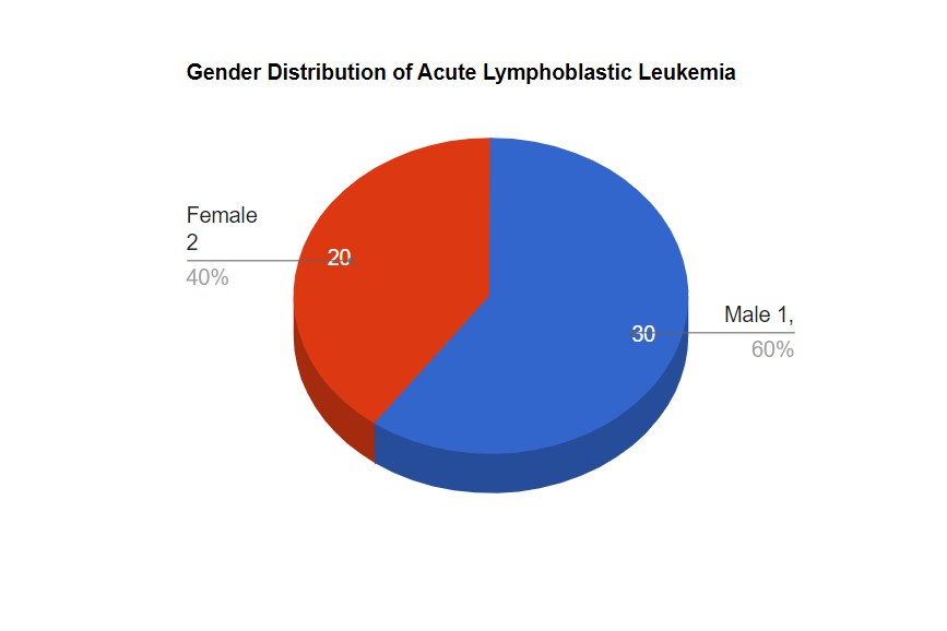
In this study, fifty (50) children with ALL were enrolled. The age limit was between 8 months to 15 years of age and the mean age of the patients was 5.5±3.2 years (Table 1). Maximum patients were male. The male and female ratio was 1.5:1(Fig-1). In this study, during the induction remission phase hemoglobin (Hb) levels were gradually decreased up to the 12th day (p>0.05) (Paired t-test). At diagnosis mean total count of WBC was 14.74±25.47 (x109 /L). During induction remission, the total count of WBC sharply declined on day 8 and continued up to the last follow-up. Statistically significant differences were observed between different follow-ups day. This study found the mean distribution of Ret %, Ret abs, and platelet count according to the recovery of the study patients. In this study, Ret % significantly declined up to day 4 and then gradually increased or remain the same with diagnosis day in both automated and manual methods. Ret % recovery occurred on the first day of a persistent 3 days rise of the reticulocyte from the 18th to 28 days. It recovered at a median of 18 days and the mean ±SD Ret % was 18.5 ±6.8 days, 28. ±7.4 days and 24.3±6.7 in early, late, and same recovery respectively (Table 2). The absolute reticulocyte concentration significantly declined up to day 4 then it remains the same up to the last follow-up (p<0.05). The mean ±SD Ret abs were 18.3±4.7 days in early recovery,
24.4±5.3 days in late recovery, and 20.7±3.5 days in same recovery (Table 2). The platelet count declined significantly (p<0.05) up to 16 days and then remain the same with the baseline status. The mean platelet count was 24.1±6.5 days in early recovery, 28 ±6.4 days in late recovery, and 20.5±5.7 days in the same recovery (Table 2). Ret %, Ret abs, and platelet recovery were statistically significant (p<0.05) in different statuses. Recovery of Ret % at a median of 20 days, Ret abs 18 days, and platelet counts occurred at a median of 24 days, which was 4 days delayed than Ret %, 6 days delayed than Ret abs(Table 2).
| Age group years | Number of patients | Percentage |
|---|---|---|
| ≤ 5 | 29 | 58 |
| 6 – 10 | 18 | 36 |
| 11 – 15 | 3 | 6 |
| Mean ±SD | 5.5±3.2 | |
| Range (min-max) | (8 months | -15 years) |
Table 1: Age distribution of the acute lymphoblastic leukemia patients up to 18 years (N=50).
| Early recovery | Late recovery | Same recovery | P value | |
|---|---|---|---|---|
| Ret | ||||
| Mean+- SD | 18.5+-6.8 | 28.4+-7.4 | 24.3 6.7 | 0.013s |
| Median | 20 | 28 | 24 | |
| Mode | 18 | 24 | 20 | |
| Range(Min-Max) | 18-26 | 20-32 | 18-32 | |
| Ret abs | ||||
| Mean+- SD | 18.3 4.7 | 24.4 5.3 | 0.884085648 | 0.001s |
| Median | 18 | 24 | 20 | |
| Mode | 20 | 10 | 20 | |
| Range(Min-Max) | 16-32 | 20-32 | 14-32 | |
| Platelet | ||||
| Mean+- SD | 24.1+-6.5 | 28+-6.4 | 20.5+-5.7 | 0.001s |
| Median | 24 | 28 | 22 | |
| Mode | 24 | 24 | 20 | |
| Range(Min-Max) | 16-32 | 20-32 | 16-32 |
Table 2: Comparison of Ret %, Ret abs, and Platelet count with recovery in days in acute lymphoblastic leukemia patients (N=50).
P value reached from ANOVA test Level of significance (p<0.05). Table 2: Comparison of Ret %, Ret abs, and Platelet count with recovery in days in acute lymphoblastic leukemia patients (N=50).
Discussion
Few studies were performed to evaluate the utility of using flow cytometric analysis of reticulocytes to predict bone marrow recovery after chemotherapy. Though the high cost of using flow cytometry or third-generation automated blood cell counters for automated reticulocyte counts is presently a limiting factor in developing countries such as Bangladesh. But despite the fact, ret%, ret abs can effectively serve as an additional index to indicate bone marrow recovery other than platelet count. Ret %, ret abs and platelet recovery were found statistically significant (p<0.05) in different statuses in this study. The mean ±SD Ret % was 18.5 ±6.8 days,28. ±7.4 days and 24.3±6.7 in early, late, and same recovery respectively. Ret% recovered at a median of 20 days. Dekoninck, et al. [12] found Ret % recovery from 5 to 33 days (median 19), which was 1 day earlier than this study. But this study found the recovery of ret abs was in 18 days and the mean ±SD Ret abs were 18.3±4.7 days in early recovery,24.4±5.3 days in late recovery, and 20.7±3.5 days in the same recovery. Kuse, et al. [13] confirmed that ARC recovered at a median of 10 days in acute leukemia which was 8 days earlier than our study. The mean platelet was 24.1±6.5 days in early recovery, 28. ±6.4 days in late recovery and 20.5±5.7 days in the same recovery. Dalal, et al. [14] found platelet recovery was a median of 29 days in a study that was 5 days delayed recovery from our study. Dekononick, et al. [12] showed platelet recovery occurred in a median of 17.2 days compared to ret on 8.7 days which was earlier than our outcome. Bhatnagar, et al. [15] in a study found, platelet recovery 1 day was delayed than Ret %which was nearly consistent with our study. 85.7% percent of them had thrombocytopenia where platelet was < 100x 109/l. In this study, recovery of Ret% at a median of 20 days, ret abs in 18 days and platelet counts occurred at a median of 24 days; platelet recovery was 4 days delayed than Ret %, 6 days delayed than Ret abs. So, our study confirmed, Ret abs were the earlier marker than Ret % and platelet count [16, 17, 18, 19, 20].
This study concluded that the reticulocyte parameter showed earlier hematopoietic recovery than the platelet recovery for monitoring in children with acute lymphoblastic leukemia after chemotherapy. This early laboratory indicator will guide the clinicians to make important therapeutic decisions, which will be economic savings as well as life savings. Nowadays, reticulocyte is offered in most third-generation hematology analyzers. Moreover, this test is a simple, quick, cost-effective, reproducible, and reliable tool on the automated hematology analyzer. Thus its potential use as a routine test to see bone marrow recovery is important.
Conclusion
This study concluded that absolute reticulocyte count was the earlier marker than reticulocyte percentage and platelet count for hematopoietic recovery in acute lymphoblastic leukemia patients in the remission induction phase. Thus, it could facilitate clinical decision-making about hematopoietic recovery.
Recommendations
Further study may be done on other phases of chemotherapy on a daily basis.
Acknowledgment
I would like to thank all the consultants, doctors, and staff of the Department of Laboratory Medicine, BSMMU, and Dhaka for their sincere help in conducting this study.
Declaration
This study was completed self-funded by the first author.
References
-
Satake N, Yoon JM (2009) Acute Lymphoblastic Leukaemia’. E medicine Pediatric: medscape.com. 12: 1-26.
-
Veerman AJP (2003) Acute lymphoblastic leukemia in children: Experiences in West and East. Bangladseh’J Child Health 27: 16-17.
-
Torres A, Sanchez J, Lakomsky D, Serrano J, Alvarez AM, et al. (2001) Assessment of hematologic progenitor engraftment by complete reticulocyte maturation parameters after autologous and allogenic hematopoietic stem cell transplantation. Haematologica 86(1): 24-29.
-
Kuse R (1993) The appearance of reticulocytes with medium or high RNA content is a sensitive indicator of beginning granulocyte recovery after aplasiogenic cytostatic drug therapy in patients with AML. Ann Hematol 66(4): 213-214.
-
Davis BH (1996) Immature reticulocyte fraction (IFR): by any name, a useful clinical parameter of erythropoietic activity. Lab Hematol 2: 2-8.
-
Remacha (1994) Flow cytometric reticulocyte quantification in the evaluation of hematologic recovery. Spanish Multicentric Study Group for Hematopoietic Recovery. Eur J Haematol 53(5): 293-297.
-
Hellgriel K (1993) The clinical significance of reticulocyte determination. In: Basting T, 5th (Edn.), The Emerging Importance of Accurate Reticulocyte Counting. Miles, Inc. Diagnostic Division, Tarrytown: pp: 7-10.
-
Norhana A, De Souza CA, Vigorito AC, Aranha FJP, Zulli R, et al. (2003) Immature reticulocytes as an early predictor of engraftment in autologous and allogenic bone marrow transplantation. Clin Lab Haematol 25: 47-54.
-
Khalil F, Cauling H Cogburn J, Miles L (2007) The criteria or Bone Marrow Recovery Post Myelosuppressive Therapy or Acute Myelogenous Leukaemia: cA Quantitative Study. Archives of Pathology and Laboratory Medicine 131(8): 1281-1289.
-
Robert V (2002) Reticulocytes: Their usefulness and measurement in peripheral blood. Clinics in Laboratory Medicine 22(1): 63-79.
-
Imazu M (2002) General Description of the Automated Hematology Analyzer, XT-2000i. Sysmex J Int. 12: 13-17.
-
Dekoninck A, Brusselmans C, Goossens W (2002) Indicators for hematopoietic recovery in patients after bone marrow transplantation or intensive chemotherapy. Department of Laboratory Medicine, University Hospital Leuven, Leuven, Belgium pp: 39.
-
Kuse R, Foures C, Jou JM, d’Onofrio G, Paterakis G (1996) Automated reticulocyte counting for monitoring patients on chemotherapy for acute leukemias and malignant lymphomas. Clin Lab Haematol 18(S1): 39-43.
-
Dalal BI, Stockford GR, Naiman SC, Spinelli JJ, Phillips GL (1996) Criteria for marrow engraftment: comparison of reticulocyte maturity index with conventional parameters. Bone Marrow Transplant 17(1): 91-92.
-
Bhatnagor S, Chandra J, Naayanb S (2002) Hematological Changes and Predictors of Bone Marrow Recovery in Patients with Neutropenic Episodes in Acute Lymphoblastic Leukemia. Journal of Tropical Pediatrics 48(4): 200-203.
-
Davis BH, Bigelow NC (1989) Flow cytometric reticulocyte quantification using thiazole orange provides clinically useful reticulocyte maturity index. Arch Pathol Lab Med 113(6): 684-689.
-
Riley S, Jonathon M, Ezra B, Goel R, Tidwell A (2001) Reticulocytes and Reticulocyte Enumeration. Journal of Clinical Laboratory Analysis 15(5): 267-294.
-
Bain BJ, Lewis SM, Bates I (2008) Basic hematological techniques. In: Lewis SM, Bain Bf/.; Bates I, Dacie and Lewis Practical Haematology, 10th (Edn.), Philadelphia: Churchill living stone pp: 25-57.
-
Greinix HT, Linkesch W, Keil F, Kalhs P, Schwarzinger I, et al. (1994) Early detection of hematopoietic engraftment after bone marrow and peripheral blood stem cell transplantation by highly fluorescent reticulocyte counts. Bone Marrow Transplant 14(2): 307-313.
-
Praful B, Godkar G, Darshan D, Godkar P (2003) routine hematological tests, Textbook o Medical Technology. In: 2nd (Edn.), pp: 750-751.
- Genomic Landscape of Aggressive Penile Squamous Cell Carcinoma including TERT-p and NOTCH1 Mutations – An Institutional Experience
- Establishment of Baseline Haematological Values for Canine Population in North-Central Nigeria: A Cross-Sectional Study in the Federal Capital Territory
- Biochemical Assessment of Uroliths Extracted in Patients with Urolithiasis in a Tertiary Health Institution
- Update on Gastrointestinal Pecomas: Molecular Pathogenesis and Risk Stratification
- A Comparative Study of Serum C-reactive Protein Level Between Pre-eclampsia and Normal Pregnancy in Tertiary Level Hospital
- From Deformity to Alignment: Clinical Outcomes of the Schnepp Osteotomy in Hallux Valgus in 47 Feet