Hepatoprotective and Antioxidative Activity of Avicennia marina Forsk
Avicennia marina Forssk. is a common mangrove tree found around estuarine coasts of India. The literature survey on this plant so far has reported the presence of several novel compounds with prospective medicinal value. Avicennia marina has been reported to posses flavones and flavonoids along with triterpenoids like betulin, lupeol etc. and has been reported for antiviral activity invitro on hepatits B virus. But scientific data on its hepatoprotective potential is unavailable. Thus the present study was aimed to evaluate hepatoprotective activity of standardized ethyl acetate extract of the leaves of Avicennia marina. The extract was standardized using validated HPTLC method for betulinic acid, betulin, lupeol, ursolic acid and β-sitosterol content. The antioxidant activity of Avicennia marina leaves was assessed by DPPH and Galvinoxyl free radical scavenging activity. Hepatoprotective activity was assessed against CCl4 induced liver intoxication. CCl4 induced albino Wistar rats were treated with three different doses of the standardized extract for three days. The results were compared with the established positive control, Silymarin. The extract provided concentration dependent percent protection which was evident by the changes in the levels of biochemical parameters (SGOT, SGPT, ALP, TPN, TG, CHO, HDL, LDL, direct and total bilirubin and liver glycogen content) in albino Wistar rats. The observations of biochemical parameters, antioxidant assays and histopathology, endorse an overall promising effect against liver disorders.
Introduction
Liver damage is a common complication. Liver has to detoxify many toxic substances. Most of the hepatotoxic chemicals damage liver cells by producing reactive species which form covalent bond with the lipids of the tissue. Due to excessive exposure to hazardous chemicals like CCl4, alcohol, etc., the number of free radicals generated is so high that they overpower the natural defense system of the body causing hepatic damage and lead to jaundice, cirrhosis and fatty liver etc. [1, 2, 3]. Till date, the synthetic hepatoprotective agents used in clinical practices are therapeutically non-promising and may they lead to hepatotoxicity. Herbal medicines and their bioactives are considered to be relatively safe and have been used in the treatment of liver diseases for a long time [4]. Modern medicines have little to offer for alleviation of hepatic disorders and diseases, and only limited number of drugs like Ribavarin, alpha interferon; Zeffix, etc are available for the treatment of the same [5]. The studies based on the knowledge of minority ethnic groups and literature search revealed that the mangroves have been neglected for their scientific investigation [6]. Mangrove species have been reported to be efficacious in the treatment of toothache, sore throat, constipation, fungal infections, fever, kidney stone, rheumatism, dysentery and malaria [7]. Traditionally, many of the folk remedies of plant origin have long been used for treatment of liver diseases [8, 9]. Mangroves and related species have also been reported to have hepatoprotective activity [10]. Hepatoprotective activity of Rhizophora mucronata [5], Lumnitzera racemosa [11], Acanthus ilicifolius [12] etc has been analyzed on CCl4 intoxicated rats. Hepatoprotective activity of Suaeda maritama has been reported against concalavalin-A induced hepatotoxicity in albino Wistar rats [13]. Methanolic extract of Avicennia marina has also been reported to possess antiviral properties against hepatitis B virus [14]. The current research work focuses on, Avicennia marina Forssk., a common mangrove which has not been thoroughly and scientifically evaluated so far. It has been reported to possess phytoconstituents like betulinic acid, lupeol etc [15]. The study aims to evaluate the efficacy of the ethyl acetate extract of the leaves of Avicennia marina against CCl4 induced hepatic damage in albino Wistar rats.
Materials and methods
Plant material
The leaves of Avicennia marina Forssk. Were collected from Airoli, Mumbai and authenticated at Agharkar Research Institute, Pune, (Voucher no. Auth. 13-017). The leaves were shade dried for a week and further oven dried at 37°C ± 2°C to constant weight, powdered in a mixer grinder, sieved (85 mm mesh – BSS Sieve) and stored in airtight plastic containers.
Apparatus and chemicals
CCl4 (GR grade, Batch no.: IG8G580178, Merck Specialties Pvt. Ltd) and Silybon tablets (Silymarin as silybin 70 mg, Batch no. SIAD0025, Micro Labs Limited) were procured from market. DPPH, and Galvinoxyl radicals were procured from Sigma-Aldrich Chemical Company, (Steinheim, Germany). All other chemicals used were of analytical grade.
Extract preparation
Ethyl acetate extract of the leaves of Avicennia marina was prepared by adding 1000 mL ethyl acetate to 10 g of plant powder using soxhlet extraction method (yeild = 3.04%). The extract was standardized using a validated HPTLC method and content of reported hepatoprotective triterpenoid markers like; Ursolic acid [16], β-sitosterol [17], Lupeol [18] Betulin [19], betulinic acid [20], was estimated for the standardization of the extract. The content was found to be 17.73 %, 5.33 %, 13.59 %, 3.28 % and 4.97 % respectively. The extract was further evaporated to dryness in a water-bath preset at 65°C to remove all traces of ethyl acetate from the extract prior to animal studies. Animals were dosed by weighing this dried extract accurately as per the weight of the animal and suspending it in distilled water (total volume > 1 mL/ 100 g).
Animals
Adult albino Wistar rats (females, 180-220 g) and Swiss Albino mice (females, 18-22 g) were procured from Haffkines Biopharmaceuticals Pvt. Ltd., Mumbai. The animals were maintained under standard laboratory conditions at an ambient temperature of 25 ± 2º C, relative humidity of 50 – 55 % and with 12-h light and dark cycle in an animal house with standard facilities as per CPCSEA guidelines (CPCSEA/315). They were fed with commercially available standard pellet diet (AMRUT feed) and filtered drinking water was provided ad libitum. After one week of acclimatization, the animals were used for experimentation. The approval from institutional animal ethics committee of Ramnarain Ruia College, Matunga, for the usage of animals in the experiment was obtained as per Indian CPCSEA guidelines (Approval number: MJ – 130624 – 02).
Safety evaluation
Safety study of standardized extract of Avicennia marina was conducted on albino Swiss mice as per OECD guidelines (No. 420, fixed dose procedure). The mice were fasted overnight for 10-14 hours and administered with standardized extract of the leaves of Avicennia marina (2.0 g/kg) orally. The animals were observed individually during the first 30 min for all reflexes, periodically during the first 48 hours with special attention given during the first 4 hours (short-term toxicity) and daily thereafter for a total of 14 days (long-term toxicity) for alteration from general behavior and clinical symptoms like alteration of skin and fur texture, ptosis, diarrhea etc. Daily body weight, food and water intake record was also maintained. The results were compared with control group (orally administered with D/W).
Assessment of hepatoprotective activity
For the study, albino Wistar rats were randomly divided into seven groups with six animals in each. The group details are as follows: Group I: Normal control (NC) Group II: CCl4 control (0.7 ml/kg body weight) (IC) Group III: CCl4 control as natural recovery group (NR) Group IV: CCl4 + Silymarin (70 mg /kg body weight) (POS) Group V: CCl4 + Extract of Avicennia marina leaves (50 mg/kg body weight) (AM 1) Group VI: CCl4 + Extract of Avicennia marina leaves (150 mg/kg body weight) (AM 2) Group VII: CCl4 + Extract of Avicennia marina leaves (300 mg/kg body weight) (AM 3) The animals were fasted overnight before the initiation of the study. Animals from Group I received an intraperitoneal injection of 0.5 ml liquid paraffin/animal on the zeroth day of the study and were treated as normal control. Animals from groups II, III, IV, V, VI and VII received an intraperitoneal injection of 0.7 ml/kg body weight of CCl4 [21] in 0.5 ml liquid paraffin/animal on the zeroth day of the study. The animals from Groups I, II and III received an oral dose of 2 ml of distilled water (D/W) once daily. A dose of 70 mg / kg Silymarin [22] (in the form of Silybon tablets) suspended in 2 ml of D/W was administered orally to each animal of Group IV once daily, starting 24 h post induction on the first day. The animals from Group V, VI and VII received an oral dose of extract of leaves of Avicennia marina suspended in distilled water (50 mg/kg, 150 mg/Kg and 300 mg/kg body weight respectively) / animal, daily and 24 h post induction on the first day of the study. The animals from Groups I, II, IV, V, VI and VII were sacrificed on the fourth day (72 hr after first dosing) and those from Group III were sacrificed on seventh day of the study for comparative evaluation of the natural recovery in the study. Daily record of body weight and food and water intake was maintained. Prior to the sacrifice, blood was collected from retro orbital plexus in heparinized vials and plasma was separated. Biochemical parameters like Serum Glutamate Oxaloacetate Transaminase (SGOT), Serum Glutamate Pyruvate Transaminase (SGPT), Alkaline phosphatase (ALP), direct (D. Bil) and total Bilirubin (T. Bil), Cholesterol (CHO) and Triglycerides (TG), Total Proteins (TPN), Low-density lipoprotein (LDL) High- density lipoprotein (HDL) content, and Liver glycogen content was analyzed from the plasma samples obtained. During autopsy, liver was excised, rinsed in saline, blotted and weighed. A small piece of liver tissue from the largest lobe was cut and fixed into bouin’s fixative. Fixed tissues were processed for routine haematoxylin and eosin staining and evaluated. The extent of liver recovery was compared with positive control. Percent protection in individual biochemical parameters was calculated as 100 × (values of CCl4 Control - values of sample) / (values of CCl4 control - values of vehicle) [23].
Assessment of antioxidant activity
The antioxidant activity of the extracts, based on the scavenging activity of the stable 1,1 – diphenyl – 2 – picryl hydrazyl (DPPH) free radical, was determined [24] Also, a similar method with a different stable radical (Galvinoxyl) was carried out [25]. Percent inhibition (I) was calculated by the following equation:
I = (A control – A sample) / A control X 100 % Where Acontrol is the absorbance of the ethanol containing control, and Asample is the absorbance of the reaction mixture with the tested sample. EC50 values were determined to be the concentration at which DPPH radical and Galvinoxyl radical are scavenging by 50 %. Ascorbic acid was used as reference standard.
Statistical analysis
The results were expressed as mean ± SE. The statistical analysis of the values was carried out by using Microsoft Excel and GraphPad Prism 5.0 software for one- way analysis of Variance (ANOVA) followed by Dunnett’s _t-_test. P value of < 0.001 was considered to be significant.
Results
The standardization of the ethyl acetate extract Avicennia marina using HPTLC showed that the extract is rich in markers like ursolic acid, lupeol, β – sitosterol, betulinic acid and betulin, all of which have been reported to possess hepatoprotective activity. Before the animal experimentation, the extract was evaporated to dryness to remove all the traces of ethyl acetate. This dried extract was administered to the animals during the experimentation using distilled water as the vehicle. The extract was first screened for its safety profile in Albino
Wistar rats following OECD Guidelines No. 420 and was found to be safe up to the maximum dose of 2.0 g/ Kg body weight. No mortality was observed during the study [26]. The antioxidant activity of the extract was assessed in DPPH and Galvinoxyl radical scavenging models. In order to quantify the antioxidant activity, the IC50 value, i.e., the concentration of sample required to decrease the absorbance of specific free radical at specific λmax by 50%, was calculated [27]. The extract showed concentration dependant antiradical activity by inhibiting DPPH and Galvinoxyl radicals with an IC50 value of 519.24 µg/mL and 540.09 µg/mL for both the radicals respectively (Table 1).
| A. marina IC50 | Ascorbic acid | |||||||
|---|---|---|---|---|---|---|---|---|
| Parameters | ||||||||
| (µg/mL) | IC50 (µg/mL) | |||||||
| DPPH radical scavenging activity | 519.24 ± 1.17 | 2.28 ± 0.98 | ||||||
| Galvinoxyl radical Scavenging activity | 540.09 ± 1.41 | 1.54 ± 0.77 |
Table 1: Antioxidant activity of the standardized extract expressed in IC50 (µg/mL) value.
Hepatic toxicity caused by the induction of CCl4 (0.7 mL/ Kg body weight) caused acute liver injury which was indicated by the sudden increase in the concentration of biomarkers like SGOT, SGPT, direct and total bilirubin, ALP, CHO, TG, LDL and liver glycogen along with decrease in the concentration of HDL and TPN in the induction control (IC) group of animals when compared with the animals of normal control (NC). The fluxed levels of the biomarkers were significantly brought back to normal by the treatment of standardized extract of the leaves of Avicennia marina (AM 1) (AM 2) (AM 3) and in the animals treated as positive control (POS). The results are tabulated in table 2 and 3. The percent protection offered by the extract shows a dose dependant increase with the highest dose at 300 mg/ Kg body weight showing significant recovery (P< 0.001 using Dunnett’s test). Thus indicating the ability of the extract to reverse the damage caused by CCl4 intoxication and significantly enhance the recovery process as indicated by the comparison of the treated group of animals (AM 1) (AM 2) (AM 3) (POS) with the animals of the natural recovery group (NR).
| SGOT (Ul/ L) | SGPT (Ul/ L) | ALP (Ul/ L) | T. Bil (mg/ dl) | D. Bil (mg/ dl) | |||||||||||
| NC | 41.38 ± 0.289 | 33.42 ± 0.183 | 25.17 ± 0.165 | 0.56 ± 0.070 | 0.08 ± 0.100 | ||||||||||
| IC | 117.58 ± 0.451@ | 132.00 ± 0.126@ | 89.15 ± 0.269@ | 4.36 ± 0.254@ | 1.68 ± 0.960@ | ||||||||||
| NR | 105.05 ± 0.472 | 112.74 ± 0.204 | 70.58 ± 0.248 | 3.26 ± 1.332 | 1.42 ± 0.700 | ||||||||||
| % protection | 18.92 | 20.18 | 29.32 | 28.86 | 15.96 | ||||||||||
| POS | 40.53 ± 0.249* | 31.93 ± 0.190* | 26.73 ± 0.125* | 0.87 ± 0.078* | 0.19 ± 0.110* | ||||||||||
| % protection | 101.54 | 103.1 | 97.62 | 91.87 | 93.39 | ||||||||||
| AM 1 | 79.55 ± 0.104 | 63.29 ± 0.445 | 55.16 ± 0.171 | 1.95 ± 1.059 | 0.84 ± 0.480 | ||||||||||
| % protection | 50.59 | 70.93 | 53.33 | 63.47 | 52.13 | ||||||||||
| AM 2 | 64.87 ± 0.326 | 60.11 ± 0.240 | 50.11 ± 0.301 | 1.56 ± 0.770 | 0.44 ± 0.377* | ||||||||||
| % protection | 69.31 | 74.19 | 61.21 | 73.63 | 77.71 | ||||||||||
| AM 3 | 51.81 ± 0.402* | 34.86 ± 0.222* | 41.29 ± 0.207* | 1.46 ± 0.102* | 0.40 ± 0.232* | ||||||||||
| % protection | 86.75 | 100.11 | 74.95 | 76.32 | 79.91 |
Table 2: Effect of standardized extract on biochemical parameters. (Values are expressed in mean ± S.E, n=6 rats in each group) V
HDL (mg/ dl) LDL (mg/ dl) CHO (mg/ dl) TG (mg/ dl) TPN (mg/ dl)L.Gly (mg/ dl) NC 10.11 ± 0.078 30.29 ± 0.278 52.27 ± 0.264 56.22 ± 0.443 6.48 ± 0.077 8.41 ± 0.079 IC 1.92 ± 0.164@ 79.20 ± 0.473@ 117.00 ± 0.526@177.33 ± 0.300@2.48 ± 0.254@4.33 ± 0.342@ NR 3.65 ± 0.411 73.55 ± 0.252 104.52 ± 0.490 130.29 ± 0.304 3.01 ± 0.133 4.87 ± 0.074 % protection 16.95 12.34 19.64 38.84 13.83 13.24 POS 11.52 ± 0.724* 26.88 ± 0.237* 52.03 ± 0.168* 63.13 ± 0.322* 6.98 ± 0.780* 7.55± 0.412* % protection 112.1 106.46 99.71 94.29 110.98 78.92 AM 1 8.00 ± 1.263 54.00 ± 0.336 81.61 ± 0.220 100.54 ± 0.300 3.82 ± 0.105 5.23 ± 0.363 % protection 69.51 51.77 54.59 63.4 33.67 22.06 AM 2 8.30 ± 1.899 50.11 ± 0.404 71.71 ± 0.303 54.95 ± 0.288* 4.31 ± 0.077 5.66 ± 0.304 % protection 73.18 59.61 69.7 101.05 45.54 32.6 AM 3 8.79 ± 0.456* 38.06 ± 0.354* 58.00 ± 0.294* 53.32 ± 0.373* 4.82 ± 0.102 5.87 ± 0.386 % protection 79.06 83.91 90.62 102.4 58.13 37.75 Table 3: Effect of standardized extract on biochemical parameters. (Values are expressed in mean ± S.E, n = 6 rats in each group) Values are statistically analyzed for significance using dunnetts test *= p<0.001 when compared with values of CCl4 control and @ = p<0.001 against NC The hepatoprotective effect of the standardized extracts of the leaves of Avicennia marina was further confirmed by histopathological observations of the excised liver tissue. The histoarchitecture of extract treated and POS group animals showed significant recovery to normalcy in comparison to IC group (Figure: 1-7). The liver tissue of the animals of NR group did not show significant improvement when compared to IC. The current study thus demonstrates the hepatoprotective potential of standardized extract of the leaves of Avicennia marina.
Discussion
Carbon tetrachloride is one of the most commonly used hepatotoxins. CCl4 is biotransformed under the action of cytochrome P450 enzyme in the microsomal compartment of liver to trichloro methyl radical (CCl3) which readily reacts with molecular oxygen to form trichloromethylperoxy radical [28]. Both the radicals can bind covalently to the macromolecules and induce peroxidative degradation of the membrane lipids of endoplasmic reticulum rich in polyunsaturated fatty acids. This leads to the formation of lipid peroxides [29]. This lipid peroxidation leads to a cascade of reactions leading to liver necrosis [30]. This liver damage can be detected by measuring the content of liver function marker enzymes which are released into blood from damaged cells and liver tissues for example, transamines, which are cytoplasmic enzymes in nature and are released into circulation after cellular damage [31]. Thus, these markers also form the basic indicators of liver damage. The hepatoprotective activity of doses of Avicennia marina was monitored by estimating the transamines, ALP, Bilirubin, etc biomarkers, which gave a good idea about the functional state of the liver [32]. The increase in the levels of both direct and total bilirubin reflected the depth of jaundice and increase of transamines and alkaline phosphatase was a clear indication of cellular leakage and loss of functional integrity of cell membrane [33]. The normalization of these marker enzymes (Table 2 and 3) after the administration of standardized ethyl acetate extract of the leaves of Avicennia marina establishes its hepatoprotective potential which may also be due to the ability of the extracts to accelerate regeneration of liver cells, thus reducing the leakage of marker enzymes into the rat blood. The investigation also statistically reveals that the protection offered by the extracts is comparable to the positive control used i.e silymarin. The recovery towards normalcy of enzyme markers content and that of tissue histoachitecture caused by the extracts was found to be similar to that caused by silymarin (POS) in the present study. Among the two transmarine, 300 mg dose of the extract showed better result in the SGPT levels with a recovery of 100.11%. GPT is a better index of liver injury as liver GPT represents 90% of the total enzyme in the body and it catalyses the conversion of alanine to pyruvate and glutamate and is released in a similar manner [34].
The reduction in total protein content (TPN) post induction can be attributed to the initial damage produced and localized in the endoplasmic reticulum which leads to functional failure with a decrease in protein synthesis and accumulation of triglycerides leading to fatty liver [35]. The plant treatment groups show increased content of total protein post treatment indicating enhanced synthesis of proteins which corresponds to acceleration in the regeneration process. This is also supported by the significant reduction in the triglyceride content in serum. High dose of the extract has shown better protection in case of TG (102.4 %) when compared with the positive control (94.29 %). Inhibition of the bile acids synthesis from cholesterol leads to the increase in cholesterol levels, which is also a result of CCl4 intoxication [36]. Significant suppression of cholesterol levels (90.62 %) by the test groups suggests that the bile acids synthesis inhibition was also successfully reversed by the standardized extracts of Avicennia marina.
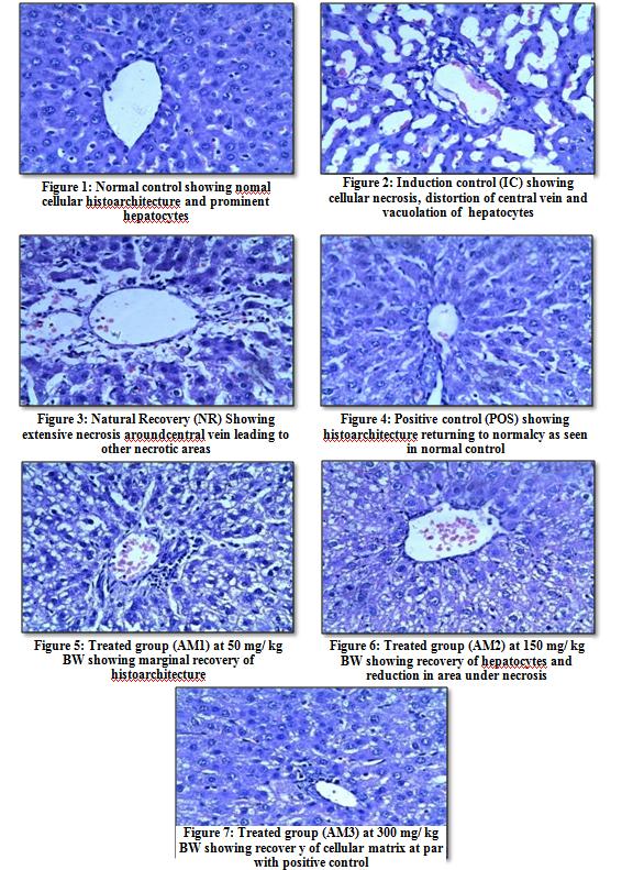
Figure 1-7: Effect of standardized extract on CCl4 induced rat liver (H & E, X400).
The histopathological profiles of CCl4 induced liver (Figure 1-7) revealed extensive necrosis near the central veins leading to other necrotic areas. The sections also show distorted hepatocytes along with vacuolated sinusoids in the liver tissue. The liver tissues of the animals of POS group (Figure 4) show significant recovery of all the above observations. Albino Wistar rats treated with plant extract (Figure: 5, 6, 7) also exhibit significant improvement of hepatocellular architecture over IC group (Figure 2) as evident from considerable reduction observed in the areas under necrosis and distortion caused by CCl4. The liver tissue of the animals of NR group (Figure 3) did not show significant improvement when compared to IC, which also seconds the hypothesis that the treatment groups and silymarin control showed enhanced recovery from the hepatic damage in comparison to the natural rate of liver recovery. The extract of leaves of Avicennia marina also showed potential antioxidant property as observed from DPPH and Galvinoxyl radical scavenging activity assay (Table 1). Hence, this antioxidant property of the extract in turn might have prevented formation of trichloromethyl peroxy radical thereby reducing tissue damage. Therefore, the hepatoprotective activity shown by the extract can also be attributed to its antioxidant potential. Furthermore, the presence of phytoconstituents such as betulinic acid, betulin, ursolic acid, β-sitosterol and lupeol, which have been reported to possess hepatoprotective potential can also partially bear claim of the triterpenoid rich standardized extract’s hepatoprotective activity.
Conclusion
From the above results it can be stated that the standardized ethyl acetate extract of the leaves of Avicennia marina shows dose dependant hepatoprotective activity which at the dose of 300 mg/ kg body weight showed results at par with established positive control, silymarin. The extract lowered the levels of biochemical markers and showed significant recovery in histopathological analysis. Though the exact mode of action is not yet clear, the presence of favorable phytochemical markers and antioxidant potential of the extract justify the hepatoprotective activity. Further studies can be undertaken to understand the specific pharmacokinetic profile of these markers and to understand in detail the mechanism of action of the standardized extract.
References
-
Maity T, Maity S, Pahari N, Kar DR, Ganguli S (2015) A review on hepatic diseases and development of herbal drugs for the treatment of liver complications. World Journal of Pharmaceutical research 4(6): 677- 691.
-
Gupta Amartya K, Ganguly Partha, Majumder Upal K, Ghosal Shibnath (2009) Hepatoprotective & antioxidant effect & stereoidal saponins of solanum of _Solanum xanthocarpum_ & _Solanum nigrum_ in paracetomol induce hepatotoxicity in rats. Pharmacologyonline 1: 757-768.
-
Areefa S, Elumalai A, Chinna Eswaraiah M, Usha (2012) An Updated Review On Hepatoprotective Medicinal Plants. J Drug Del Therap 2(2): 1-3.
-
Alvari A, Mehrnaz SO, Ahmad FJ, Abdin MZ (2012) Contemporary Overview on Clinical Trials and Future Prospects of Hepatoprotective Herbal Medicines. Review on recent clinical trials. 7(3): 214-223.
-
Sundaram R, Gyanadesign M (2012) Hepatoprotective and antioxidant properties of _Rhizophora mucronata_ plant in CCl4 Intoxicated rats. Journal of Experimental and Clinical medicine 4(1): 66-72.
-
Chanyong S (2009) Plant species in mangrove and beach forest. Neopoint, Songkhla 1-11.
-
Bandaranayake WM (1998) Traditional and medicinal uses of mangroves. Mangroves and Salt Marshes 2(3): 133-148.
-
Bandaranayke WM (2002) Bioactivities, bioactive compounds and chemical constituents of Mangrove plants. Wetlands Ecology and Management 10(6): 421-422.
-
Ashok SK, Somayaji SN, Bairy KL (2001) Hepatoprotective effect of _Ginkyo biloba_ against CCl4 induced hepatic injury in Rats. Indian J Pharmacol 33 (2): 260-266.
-
Rouf R, Uddin SJ, Shilpi JA, Alamgir M, (2007) Assessment of antidiarrhoeal activity of the methanol extract of _Xylocorpus granatum_ bark in mice model. Journal of Ethnopharmacology, 109(3): 539-542.
-
Sundaram R, Gnanadesigan M (2011) Hepatoprotective and antioxidant activity of a mangrove plant _Lumnitzera racemosa._ Asian Pacific Journal of Tropical Biomedicine 348-352.
-
Babu BH, Shylesh BS, Padikkala J, (2001) Antioxidant and hepatoprotective effect of _Acanthus ilicifolius_. Fitoterapia 72(3): 272-277.
-
Ravikumar S, Gyanadesigan M, Inbaneson JS, Kalaiarasi A (2011) Hepatoprotective and antioxidant properties of _Suaeda maritime_ L. Dumortethanolic extract on concanavalin-A induced hepatotoxicity in rats. Indian journal of experimental biology, 49(6): 455-460.
-
Namazi R, Zabihollahi R, Behbahani M, RezaeicA (2013) Inhibitory Activity of Avicennia marina, a Medicinal Plant in Persian Folk Medicine, against HIV and HSV. Iranian Journal of pharmaceuticalresearch. 12(2): 435-443
-
Mehera SA, Ahmad VU, Saifullah SM, Mohammad FV, Ambreen K (2011) Steroids and triterpenoids from gray mangrove _Avicennia marina_. Pakistan Journal of Botany, 43(2): 1417-1422.
-
Tamanna S, Abdur R, Ahad A, Samsuddin FM (2010) Hepatoprotective and Antibacterial Activity of Ursolic Acid Extracted from _Hedyotis_ _corymbos_a L. Bangladesh Journal of Scientific and Industrial Research 45(1)**:** 27-34.
-
Gawade SP, Chandrashekar Rao MV (2012) Antihepatotoxic activities of Ci Compound: β- sitosterol isolated from fruits and leaves of _Coccinia_ _indica_. Indian Journal of Pharmaceutical Education and Research 46(1): 7-11.
-
Prasad S, Kara N, Shukla Y (2007) Hepatoprotective effects of lupeol and mango pulp extract of carcinogen induced alteration in Swiss albino mice. Molecular Nutrition & Food Research. 51(3): 352-359.
-
Agnieszka SC, Karolina P, Martyna KS (2010) Protective effect of betulin and betulinic acid on acetaminophen and ethanol-induced cytotoxicity and reactive oxygen species production in HepG2 cells. Journal of Pre-Clinical and Clinical Research 4(2): 096-100.
-
Moghaddam MG, Ahmad FBH, Kirmani AS (2012) Biological activity of Betulinic acid: A Review. Pharmacology and Pharmacy 3: 119-123.
-
Frank PR, Suresh V, Arunachalam G, Kanthlal SK, Ziaudheen VM (2012) Evaluation of hepatoprotective effect of _Adiantum incisum_ Forsk. Leaf extract against CCl4 induced hepatoptoxicity in rats. International Research Journal of Pharmacy 3(3): 230-234.
-
Silymarin Clinical Update, _Profile_ _on_ _the_ _liver_ _herb,_ (Scientific Communications International Limited, Hongkong) 1995.
-
Ganga Rao B, Venkateswara Rao Y, Mallikarjuna Rao T (2012) Hepatoprotective activity of _Spillanthes_ _acmella_ Extracts against CCl 4-induced liver toxicity in rats. Asian Pacific Journal of Tropical Disease 208- S211
-
Narendhirakannan RT, Rajeshwari K (2010) In vitro antioxidant properties of three varieties of _Allium_ _sativum_ L. extracts. E-J Chem 7(1): 573-579.
-
Ling LT, Yap SA, Radhakrishnan AK, Subramaniam T, Cheng HM, et al. (2009) Standardised _Mangifera_ _indica_ extract is an ideal antioxidant. Food Chemistry 113: 1154- 1159
-
Shailajan S, Joshi M, Tiwari B (2014) Hepatoprotective activity of Parmelia perlata (Huds.) Ach. against CCl4 induced liver toxicity in Albino Wistar rats. Journal of Applied Pharmaceutical Science 4(2): 070-074.
-
Acharya SR, Acharya NS, Bhnagale JO, Shah SK, Pandya SS (2012) Antioxidant and hepatoprotective action of _Asparagus racemosus_ Willd. root extracts. Indian Journal of Experimental Biology. 50: 795-801.
-
Raucy JL, Kraner JC, Lasker J (1993) Bioactivation of halogenated hydrocarbons by cytochrome P 450 E1. Critical Reviews in toxicology 23(1): 1-20.
-
Nayak DP, Dinda SC, Swain PK, Kar B, Patro VJ (2012) Hepatoprotective activity against CCl4 induced hepatotoxicity in rats of _Chenopodium album_ aerial parts. Journal of Phytotherapy and Pharmacology 1(2): 33-41.
-
Meera R, Devi P, Kameshwari B, Madhumita B, Merlin NJ (2009) Antioxidant and hepatoprotective activities of _Ocimum basilicum_ Linn. And _Trigonella foenum-_ _graecum_ linn. Against H2O2 and CCl4 induced hepatotoxicity in goat liver. Indian Journal of Experimental Biology 47(7): 584-590.
-
Freitag AF, Cardia GFE, Rocha BA, Aguiar RP, Silva- Comar FM, et al. (2015) Hepatoprotective Effect of Silymarin (Silybum marianum) on Hepatotoxicity Induced by Acetaminophen in Spontaneously Hypertensive Rats. Evidence based Complementary and Alternate Medicine 1-8.
-
Rao KS, Mishra SH (1997) Anti-inflammatory and hepatoprotective activities of _Sida rhombifolia_ Linn. Indian Journal of Pharmacology 29(2): 110-116.
-
Saraswat B, Visen PK, Patnaik GK, Dhawan BN (1993) Anticholeststic effect of Picroliv, active hepatoprotective principle of _Picrorhiza kurroa_ against CCl4 induced cholestasis. Indian J. Expt. Biol 31: 316-318.
-
Hegde K, Joshi AB (2009) Hepatoprotective effect of _Carissa carandas_ Linn root extract against CCl4 and paracetamol induced hepatic oxidative stress. Indian Journal of Experimental Biology 47(8): 660-667.
-
Suresh Kumar SV, Sujatha C, Syamala J, Nagasudha B, Mishra SH (2007) Hepatoprotective activity of extracts from _Pergularia daemia Forsk_. against Carbon tetrachloride induced toxicity in rats. Pharmacognosy magazine 3(11): 187-191.
-
Anusha M, Venkateswarlu M, Prabhakaran V, Shareen Taj , Pushpa Kumari B, et al. (2011) Hepatoprotective activity of aqueous extract of _Portulaca oleracea_ in combination with lycopene in rats. Indian Journal of Pharmacology. 43(5): 563–567.
- Management of Gallbladder Perforations: A Review
- From The Mouth to the Gut: The Oral Microbiome's Role in Promoting Gastrointestinal Disease
- Case Report: Intraductal Papillary Mucinous Neoplasm (IPMN) Complicated by Portal Vein Plaquing and Biliary Obstruction Mimicking Pancreatic Metastatic Malignancy
- Management of Non-Cirrhotic Portal Hypertension during Pregnancy: A Review
- Effectiveness of Omeprazole versus Pantoprazole for Symptomatic Relief of Gastro-Esophageal Reflux Disease (GERD)/ Acid Peptic Disease (APD): A Real-World Evidence (RWE) Study
- Case of Splenic Infarction; A Rare Presentation of Complicated Enteric Fever in a Pediatric Patient