Study of VDR Expression after Treatment with the Biofield Energy Healing-Based Proprietary Test Formulation Using Cellular Models
The present study was designed to evaluate the VDR (vitamin D receptor) expression after treatment with the Trivedi Effect®- Biofield Energy Treated/Blessed Test formulation (TI) composed of minerals (magnesium, zinc, copper, calcium, selenium, and iron), vitamins (ascorbic acid, pyridoxine HCl, alpha tocopherol, cyanocobalamin, and cholecalciferol), Panax ginseng extract, CBD isolates, and β-carotene on various cell lines in DMEM medium using values of relative quantification (RQ) by RTPCR among different groups. The test formulation constituents were divided into two parts; one part defined as the untreated test formulation (UT), another part received Biofield Energy Healing Treatment (BT)/Blessing by a renowned Biofield Energy Healer, Mr. Mahendra Kumar Trivedi. The test formulation was treated with Biofield Energy Healing Treatment/Blessing and was divided as Biofield Energy Treated/Blessed (BT) and untreated (UT) test items. MTT data showed that all the cell lines after treatment with the test formulation in various tested concentrations were found as safe and nontoxic with viability range 72% to 144% up to 1 μg/mL test formulation concentration. In addition, RQ values were calculated in all the cell lines and were compared with the untreated test formulation, which showed significant improved values. The MG-63 cell line showed significant increased RQ values by 140.2% (UT- DMEM + BT-TI) to 817.7% (BT-DMEM + BT-TI) among the experimental groups as compared with the untreated test formulation group. The MDA-MB-231 cell lines showed a significant increased RQ value in all the test formulation groups by 55.3% (BT-DMEM + UT-TI group) to 2308.8% (UT- DMEM + BT-TI group) among the tested groups as compared with the untreated test group. The SH-SY5Y cells were used for the VDR expression in the showed a significant increased RQ value by 15.7% (BT- DMEM + UT-TI group) to 457.7% (BT-DMEM + UT-TI group), while HEK-293 cells significant increased RQ value by 14.6% to 695.8% among the tested group in the UT- DMEM + BT-TI group as compared with the untreated test group. The HaCaT cells showed a significant increased RQ value by 41.9% (UT- DMEM + BT-TI group) to 2123.5% (BT-DMEM + BT-TI group) among the tested groups as compared with the untreated test group. Hence, it can be concluded that the Biofield Energy Treatment may have the potential to modulate vitamin D3 metabolism via VDR expression. Overall, the results showed the significant increased RQ values in all the tested cell line. VDR expression in different cell lines were significantly modulated in MG-63 (Bone), MDA-MB-231 (Breast), SH-SY5Y (Brain), HEK293 (Kidney), and HaCaT (Skin) cell lines. Thus, it can be concluded that the Biofield Energy Treatment/Blessing may have the excellent potential to upregulate the VDR expression in multiple cell-lines, and that could help calcium and bone homeostasis, and slowdown of the disease progression rate related to vitamin D3 deficiency.
Introduction
Vitamin D (cholecalciferol) and its associated metabolites are the steroidal hormones, which are increasingly renowned as a vital physiological regulator with many pleiotropic functions [1]. 3β-hydroxyl-secosteroid supplied from diet as well as produced in the skin considered as an inert compound, that showed significant biological activity via two successive hydroxylations to provide 1α, 25-dihydroxyvitamin D3 (calcitriol) [2]. However, 1,25-dihydroxyvitamin D3 [1,25-(OH)2D3], a hormone action was mediated by ligand- inducible transcription factor known as vitamin D receptor (VDR). This hormone is associated with calcium and bone homeostasis along with other cellular processes, such as proliferation and differentiation. The expression of VDR depends on the binding with specific DNA elements of target genes and inducing or repressing various transcriptional activities [3, 4]. VDR levels are regulated by many factors such as hormones, growth factors, and calcium [5, 6, 7]. Some internal factors increased (cAMP-activated protein kinase A), while other (protein kinase C) may reduced the expression of VDR [8]. MG-63 (Human Osteosarcoma like cells), MDA- MB-231 (Human Breast adenocarcinoma), SH-SY5Y (Human Neuroblastoma), HEK-293 (Human Embryonic Kidney Cells), and HaCaT (Human Keratinocytes) cell lines were used in order to detect the expression of VDR after treatment with the novel test formulation. The novel test formulation was designed and was composed of essential minerals (Ca, Zn, Mg, Se, Fe, Cu), vitamins (B12, E, D3, C, B6), and some biological active plant-based extracts such as β-carotene, Ginseng, and cannabidiol isolate (CBD). All the vitamins and minerals included in the novel formulation have reported with significant physiological action [9, 10, 11]. In addition, the cannabidiol itself has wide range of biological action [12, 13], while ginseng extract is regarded as the one of the best immune booster for overall biological activity [14]. Vitamins are immunity builder and works through various pathways for bone health. Vitamin D is itself used and reported for improved strength, skin elasticity [15, 16], improve arterial stiffness, neuronal plasticity, and many more [17, 18, 19]. The effect of Biofield Energy Treatment on media (DMEM) and a novel test formulation was evaluated for its VDR expression using MG-63 (Human Osteosarcoma like cells), SH-SY5Y (Human Neuroblastoma cells), HaCaT (Human Keratinocytes), HEK-293 (Human Embryonic Kidney Cells), and MDA-MB-231 (Human Breast adenocarcinoma) cell lines through the values of relative quantification (RQ) in RT-PCR.
Biofield Energy Healing Treatment approach is among the successful Complementary and Alternative Medicine (CAM) one of the emerging CAM treatment that aimed in building a scientific network with respect to the complex homeodynamic regulation of living systems. Biofield Energy Therapy is highly effective with respect to the physical, mental, and emotional human wellness [20], which improved the endogenous energy flows. CAM therapies have been accepted by the National Centre of Complementary and Integrative Health (NCCIH) along with the Biofield Energy Healing, such as deep breathing, Tai Chi, yoga, therapeutic touch, Reiki, chiropractic/ osteopathic manipulation, relaxation techniques, pranic healing, meditation, homeopathy, Ayurvedic medicine, movement therapy, mindfulness, traditional Chinese herbs and medicines in biological systems, etc. [21]. However, the Trivedi Effect®-Consciousness Energy Healing Treatment have significant clinical, preclinical, and scientific studies in different scientific disciplines such as materials science [22, 23], agriculture science [24], antiaging [25], gut health [26], nutraceuticals [27], pharmaceuticals [28], and overall human health and wellness. The present study focused on the impact of the Biofield Energy Treatment/Blessing on the given novel test formulation in order to detect the expression of VDR after treatment with the novel test formulation.
Material and Methods
Chemicals and Reagents
Pyridoxine hydrochloride (vitamin B6), calcitriol, zinc chloride, magnesium (II) gluconate, and β-carotene (retinol, provit A) were purchased from TCI, Japan. Copper chloride, cyanocobalamin (vitamin B12), calcium chloride, vitamin E (Alpha-Tocopherol), cholecalciferol (vitamin D3), iron (II) sulfate, and sodiumcarboxymethylcellulose (Na-CMC) were procured from Sigma-Aldrich, USA. Ascorbic acid (vitamin C) and sodium selenate were obtained from Alfa Aesar, India. Cannabidiol isolate and Panax ginseng extract were obtained from Panacea Phytoextracts, India and Standard Hemp Company, USA, respectively. Resveratrol was purchased from Acros Organics, and DMEM was purchased from Lonza, USA. MG-63 (Human Osteosarcoma like cells), SH-SY5Y (Human Neuroblastoma cells), HaCaT (Human Keratinocytes), HEK- 293 (Human Embryonic Kidney Cells), and MDA-MB-231 (Human Breast adenocarcinoma) cell line were procured from ATCC, USA. However, cell line medium such as DMSO, FBS, EDTA, and MTT were procured from Genexlife, Protaq Biomedical, Genexlife, and Parshuram and Parshuram traders, respectively.
Cell Culture
All the cell lines were used as test system, which were maintained in specific growth medium such as DMEM for routine culture supplemented with 10% FBS. Growth conditions were maintained at 37°C, 5%CO2, and sub-cultured by trypsinization followed by splitting the cell suspension into fresh flasks and supplementing with fresh cell growth medium. Three days before the start of the experiment (i.e., day -3), the growth medium of near-confluent cells was replaced with fresh phenol-free medium, supplemented with 10% charcoal-dextran stripped FBS (CD-FBS) and 1% penicillin-streptomycin [19].
Experimental Design
The experimental groups consisted of cells in baseline control, vehicle control groups (0.05% DMSO with Biofield Energy Treated/Blessed and untreated DMEM), positive control group (resveratrol) and four different experimental test groups. The experimental groups included the combination of the Biofield Energy Treated/Blessed and untreated test formulation/Medium (DMEM). It consisted of four major treatment groups on specified cells with Untreated-DMEM+ Untreated-Test item (UT-TI), UT- DMEM + Biofield Energy Treated test item (BT-TI), BT- DMEM + UT-TI, and BT- DMEM + BT-TI.
Consciousness Energy Healing Strategies
Consciousness Energy Healing treatment was performed in the novel test formulation, which was consisted of zinc chloride, iron (II) sulfate, copper chloride, vitamin B6, vitamin B12, vitamin D3, sodium selenate, calcium chloride, ascorbic acid, vitamin E, beta carotene, Panax ginseng extract, cannabidiol and magnesium (II) gluconate. Each ingredient of the novel test formulation was divided into two parts, one part of the test compound was not received any sort of treatment and were defined as the untreated or control sample. The second part of the test formulation was treated with the Trivedi Effect® - Energy of Consciousness Healing Treatment (Blessing/Biofield Energy Treatment) by a renowned Biofield Energy Healer, Mr. Mahendra Kumar Trivedi under laboratory conditions for ~3 minutes through Trivedi’s unique Energy Transmission process in the laboratory of Dabur Research Foundation, near New Delhi, India without touching. After that, the Biofield Energy Treated/Blessed samples was kept in the similar sealed condition and used as per the study plan. In the same manner, the control test formulation group was subjected to “sham” healer for ~3 minutes energy treatment, under the same laboratory conditions. The “sham” healer not has any knowledge about the Biofield Energy Treatment. The Biofield Energy Treated/Blessed test medium was also taken back to experimental room for further culture methods.
Determination of Non-cytotoxic Concentration
All the cells were used in the assay with cellular density 10000 cells/ well contained 25000 cells/ well. The single cell suspension of all the cells were prepared with 10% FBS. The cells were counted on a hemocytometer, while the cells were seeded with specific cell density in 96-well plates. The cells were incubated in a CO2 incubator for 24 hours. After 24 hours, medium was removed, and following treatments were given in medium along with the 10% FBS in various experimental groups. After incubation for 24 hours, the effect of the test formulation on cell viability was assessed by 3-(4, 5-dimethylthiazol-2-yl)-2,5-diphenyl tetrazolium bromide (MTT) assay. About 20 µL of 5 mg/mL of MTT was added to all the wells and incubated at 37°C for 3 hours. The cells were centrifuged to obtain the pellet. The supernatant was removed and 150 µL of DMSO was added to all wells to dissolve formazan crystals. Further, all the wells were reported using optical density (OD) values at 540 nm using Synergy HT microplate reader. The effect of the test formulation on viability of cells was determined using equation 1.
% Cell viability = 100 - % Cytotoxicity----------------------------------------------- (1) where; % Cytotoxicity = {(O.D. of Control cells – O.D. of cells treated with test formulation)/ OD of Control cells} *100
Estimation of VDR expression
The single cell suspension of all the cells was prepared in 10% FBS along with its specific medium using a hemocytometer. The cells were counted using a hemocytometer and plated in 35-mm culture dishes. The cells were incubated overnight under growth conditions to allow cell recovery and exponential growth. Following overnight incubation, the above cells were subjected to serum starvation in DMEM+ 10%FBS. The cells were then treated with different concentrations of test formulation obtained by serial dilution of main stock (i.e., 19.94 mg/mL in DMSO stock) in DMEM. Following respective treatments, each set was incubated in a CO2 incubator at 37°C, 5% CO2, and 95% humidity and incubated for 24 hours. The cells were harvested by scrapping and washed with PBS. The cell pellets obtained were analyzed for VDR gene expression using human VDR specific primers such as forward: 5’-GCTGACCTGGTCAGTTACAGCA-3’, and reverse: 5’-CACGTCACTGACGCGGTACTT-3’. The VDR gene expression was normalized using Internal Control (IC) reference. Relative quantification (RQ) of VDR gene in treated cells was calculated with respect to the untreated cells using following formula: RQ = 2^-N Where, N is the relative Threshold Cycle (CT) value of treated sample with respect to the untreated sample.
Statistical Analysis
The data were represented as mean ± standard error of mean (SEM) and subjected to statistical analysis using Sigma-Plot statistical software (Version 11.0). For multiple comparison One-way analysis of variance (ANOVA) followed by post-hoc analysis by Dunnett’s test and for between two groups comparison Student’s t-test was performed. The p≤0.05 was considered as statistically significant.
Results and Discussion
MTT Assay- Non-cytotoxic Effect of the Test Formulation
The cytotoxic effect of the test formulation was evaluated on MG-63 (Human Osteosarcoma like cells), MDA- MB-231 (Human Breast adenocarcinoma), SH-SY5Y (Human Neuroblastoma), HEK-293 (Human Embryonic Kidney Cells), and HaCaT (Human Keratinocytes) cells using MTT assay. The results were compared with respect to the defined positive control, calcitriol (nM). The cells were treated with the test formulation for 24 hours. The effect on viability of cells was determined after 24 hours of treatment by MTT assay. The cells were treated with the test formulation and in various experimental test groups. Calcitriol at 100, 500, and 1000 nM in MG-63 cell line showed upto 103% cell viability, while in experimental test groups showed cell viability range from 76% to 144% upto 25 µg/mL test concentration. Similarly, calcitriol upto 1000 nM in MDA-MB-231 cell line showed upto 171.6% cell viability, while in experimental test groups showed cell viability range from 77% to 132% upto 25 µg/mL test concentration. SH-SY5Y cell line showed cell viability upto 113% in positive control, calcitriol, while upto 130% in experimental test group with upto 25µg/mL test formulation concentration. Cell viability in HEK-293 cells showed upto 106.7% in calcitriol group, while cell viability range is from 72% to 131% in experimental test group with concentration upto 10µg/mL. HaCaT cell line showed upto 91% cell viability in calcitriol group, while experimental test groups showed cell viability range from 85% to 120% upto 25 µg/mL test concentration. Overall, the MTT data of all the 5 tested cell lines suggested that the test formulation along with test media groups were found safe at all the tested concentrations range up to maximum 25 µg/mL in the tested cell line.
Assessment of the test formulation on VDR expression
The VDR expression in all the cells were counted using a hemocytometer and plated in 35-mm culture dishes. The cells were treated with the test formulation at different combinations and effect on VDR expression was determined using quantitative-PCR amplification. VDR-CT values were obtained from PCR amplification. Relative quantification (RQ) was calculated from the VDR-CT and IC-CT values for the different cell lines treated with the test formulation.
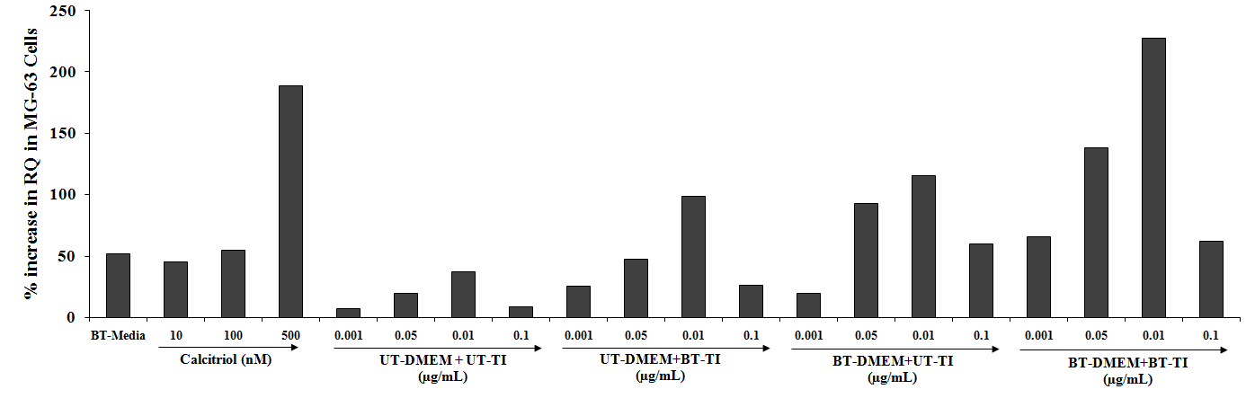
VDR expression of MG-63 cell line The effect of the test formulation on VDR expression in the MG-63 cells showed a significant increased value in test formulation groups. The positive control, calcitriol showed 45.4%, 54.8%, and 188.8% increased value of RQ as compared with the normal control group at 10, 100, and 500 nM concentrations, respectively. However, the experimental treated groups such as UT- DMEM + Biofield Energy Treated test item (BT-TI) group showed improved value of RQ by 140.2%, 162.6%, and 206.4% at 0.001, 0.05, 0.01, and 0.1 µg/mL respectively, as compared with the untreated test formulation, which suggest VDR expression was significantly modulated. Similarly, 175.1%, 371.9%, 208.5%, and 594.2% increased RQ value at 0.001, 0.05, 0.01, and 0.1 µg/mL respectively as compared with the untreated test formulation in the BT-DMEM + UT-TI group. However, BT-DMEM + BT-TI group showed improved RQ value by 817.7%, 598%, 504.9%, and 620% at 0.001, 0.05, 0.01, and 0.1 µg/mL respectively as compared with the untreated test formulation.
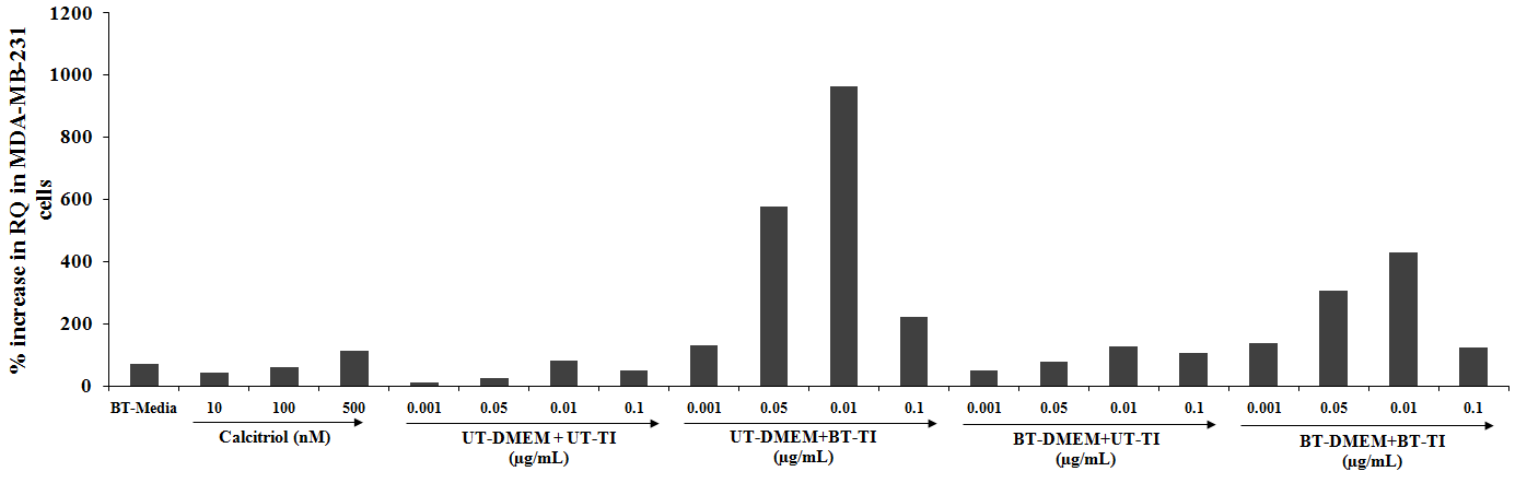
VDR expression of MDA-MB-231 cell line The VDR expression in the MDA-MB-231 cells showed a significant increased RQ value in all the test formulation groups. The positive control, calcitriol showed 41.42%, 61.33%, and 111.40% increased value of RQ as compared with the normal control group at 10, 100, and 500 nM concentrations, respectively. However, the experimental treated groups such as UT- DMEM + Biofield Energy Treated test item (BT-TI) group showed improved value of RQ by 1098.7%, 2308.8%, 1081.5%, and 354.9% at 0.001, 0.05,
0.01, and 0.1 µg/mL respectively as compared with the untreated test formulation. Similarly, 342.2%, 229.6%, 55.3%, and 115.1% increased RQ value at 0.001, 0.05, 0.01, and 0.1 µg/mL respectively, as compared with the untreated test formulation in the BT-DMEM + UT-TI group. However, the BT-DMEM + BT-TI group showed an improved RQ value by 1143%, 1174.8%, 424.9%, and 151.6% at 0.001, 0.05, 0.01, and 0.1 µg/mL respectively, as compared with the untreated test formulation.
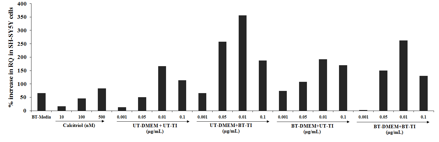
VDR expression of SH-SY5Y (Human Neuroblastoma) cell line The SH-SY5Y cells were used for the VDR expression in the showed a significant increased RQ value in all the test formulation groups. The positive control, calcitriol showed 16.5%, 45.4%, and 82.8% increased value of RQ as compared with the normal control group at 10, 100, and 500 nM concentrations, respectively. Similarly, the experimental treated groups such as UT- DMEM + Biofield Energy Treated test item (BT-TI) group showed improved value of RQ by
395.7%, 410.7%, 114.4%, and 64.7% at 0.001, 0.05, 0.01, and 0.1 µg/mL respectively as compared with the untreated test formulation. Similarly, BT-DMEM + UT-TI group showed improved RQ value by 457.7%, 114.7%, 15.7%, and 49.5% at 0.001, 0.05, 0.01, and 0.1 µg/mL respectively, as compared with the untreated test formulation. However, the BT-DMEM + BT-TI group showed an improved RQ value by 196.2%, 58%, and 14.9% at 0.05, 0.01, and 0.1 µg/mL respectively, as compared with the untreated test formulation.
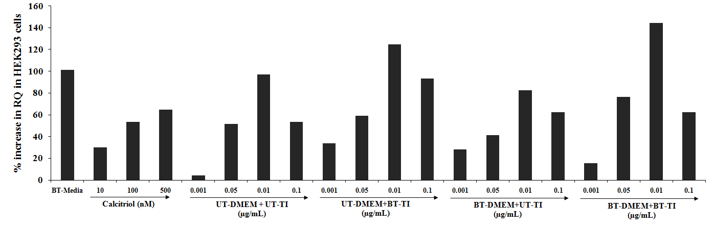
VDR expression of HEK-293 (Human Embryonic Kidney Cells) cell line The HEK-293 cells also showed significant results with respect to the VDR expression with increased RQ value in all the test formulation groups. The positive control, calcitriol showed 30.1%, 53.7%, and 64.7% increased value of RQ as compared with the normal control group at 10, 100, and 500 nM concentrations, respectively. Similarly, the experimental treated groups such as UT- DMEM + Biofield Energy Treated test item (BT-TI) group showed improved value of RQ by
695.8%, 14.6%, 28.6%, and 73.6% at 0.001, 0.05, 0.01, and 0.1 µg/mL respectively as compared with the untreated test formulation. Similarly, BT-DMEM + UT-TI group showed improved RQ value by 567.4% and 16.3% at 0.001 and 0.1 µg/mL respectively, as compared with the untreated test formulation. However, the BT-DMEM + BT-TI group showed an improved RQ value by 269%, 48.4%, 48.6%, and 16.3% at 0.05, 0.01, and 0.1 µg/mL respectively, as compared with the untreated test formulation.
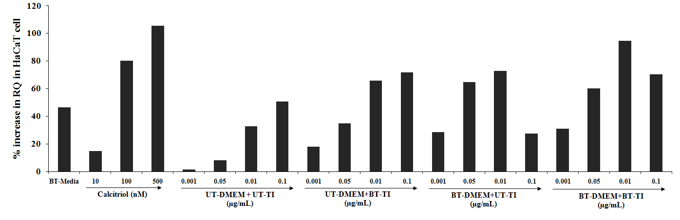
VDR expression of HaCaT (Human Keratinocytes) cell line The HaCaT cells also showed significant results with respect to the VDR expression with increased RQ value in all the test formulation groups. The positive control, calcitriol showed 14.9%, 80.3%, and 105.6% increased value of RQ as compared with the normal control group at 10, 100, and 500 nM concentrations, respectively. Similarly, the experimental treated groups such as UT- DMEM + Biofield Energy Treated test item (BT-TI) group showed improved value of RQ by
1196.6%, 338.3%, 100.4%, and 41.9% at 0.001, 0.05, 0.01, and 0.1 µg/mL respectively as compared with the untreated test formulation. Similarly, BT-DMEM + UT-TI group showed improved RQ value by 1930.3%, 716.9%, and 121.8% at 0.001, 0.05, and 0.01 µg/mL respectively, as compared with the untreated test formulation. However, the BT-DMEM + BT-TI group showed an improved RQ value by 2123.5%, 660%, 187.6%, and 39.6% at 0.001, 0.05, 0.01, and 0.1 µg/mL respectively, as compared with the untreated test formulation.
The active form of vitamin D, 1,25-dihydroxyvitamin D (Calcitriol), regulates the calcium homeostasis, immunity, and cellular growth and differentiation [19]. Calcitriol has been demonstrated to be able to inhibit the proliferation and/ or to induce the differentiation of various types of malignant cells, including breast, prostate, colon, skin, and brain cancer cells, as well as myeloid leukemia cells in vitro [29]. Calcitriol, mediates its action through Vitamin D Receptor (VDR). The VDR regulates the rates of transcription of key target genes involved directly or indirectly in calcium and phosphate regulation [30]. Apart from calcium homeostasis, VDR has been detected in multiple tissues and cell types like skin, muscle, breast, colon and hematopoietic cells [31]. Based on the localization of VDR in multiple tissues, VDR has been reported to have implications in hyper-proliferative skin diseases, hyperparathyroidism, osteoporosis. cancer, CVD, infections, and autoimmune diseases [32].
In the present study, the possible effect of the Biofield Energy Treatment/Blessing on VDR expression was determined in different cell lines of different origin.
In this research plan, the results showed the significant VDR expression in all the tested cell lines, which helps in slowdown of the disease progression, disease-related all other symptoms/complications and also reduced the chances of disease susceptibility. This improved cellular differentiation, contractile functions, exonal extensions, and skin elasticity and firmness of the cell lines used in the study after treatment was very significant. Based on the overall data, it suggests that the Biofield Energy Healing Therapy/ Blessing was found to be most effective and benefited in order to prevent and protect from the occurrence of any type of diseases and can be used as significant way for energy boosting in various disease states that will ultimately improve the overall health and quality of life in human.
Conclusions
The VDR expression in different cell lines were tested and the effect of test formulation was evaluated at different concentrations that was compared with respect to the positive control and untreated test formulation. MTT analysis for evaluation the non-toxic test formulation concentrations were evaluated and data showed that the test formulation in various tested concentrations was found as safe and nontoxic with viability range 72% to 144% among the all five tested cell lines up to maximum 1 µg/mL test formulation concentration. Relative quantification (RQ) was calculated from the VDR-CT and IC-CT values for the different cell lines treated with the test formulation. RQ values in MG-63 cell line showed significant increased values by 140% to 817.7% among the experimental groups as compared with the untreated test formulation group. Similarly, MDA-MB-231 cells showed a significant increased RQ value in all the test formulation groups by 55.3% to 2308.8% among the tested groups as compared with the untreated test group. SH-SY5Y cells were used for the VDR expression in the showed a significant increased RQ value by 64.7% to 457.7%, while HEK-293 cells significant increased RQ value by 14.6% to 695.8% among the tested groups as compared with the untreated test group. HaCaT cells showed a significant increased RQ value by 41.9% to 2123.5% among the tested groups as compared with the untreated test group. Hence, it can be concluded that the Biofield Energy Treatment may have the potential to modulate Vitamin D3 metabolism via VDR expression. Overall, the Biofield Energy Treated (the Trivedi Effect®) test formulation showed a significant improved VDR expression in the tested cell lines, which play a vital role in maintaining various immune and life style related disorders, increase alertness, energy, attention, allergy, Alzheimer, cardiovascular, cancer, diabetes, eye, immune, inflammatory, or Parkinson. Therefore, the Consciousness Energy Healing based test formulation might be suitable alternative nutritional supplement, which could be useful for the management of various immune related disorders. Thus, in conclusion this therapy might also reduce the severity of many acute/chronic diseases and its progression rate.
Acknowledgements
The authors are grateful to Dabur Research Foundation, Trivedi Science, Trivedi Global, Inc., and Trivedi Master Wellness for the assistance and support during the work.
References
-
Dusso AS, Brown AJ, Slatopolsky E (2005) Vitamin D. Am J Physiol Renal Physiol 289: F8-F28
-
Nagpal S, Rathnachalam R (2005) Noncalcemic actions of vitamin D receptor ligands. Endocr Rev 26(5): 662– 687.
-
Aranda A, Pascual A (2001) Nuclear hormone receptors and gene expression. Physiol Rev 81(3): 1269-1304.
-
Dowd DR, Sutton AL, Zhang C, MacDonald P (2005) Comodulators of VDR-mediated gene expression. In: Feldman D, Pike JW, Glorieux FH (Eds.), Vitamin D. Burlington, MA: Elsevier Academic; pp: 291-304.
-
Petkovich PM, Heersche JN, Tinker DO, Jones G (1984) Retinoic acid stimulates 1,25-dihydroxyvitamin D3 binding in rat osteosarcoma cells. J Biol Chem 259(13): 8274-8280.
-
Walters MR (1981) An estrogen-stimulated 1,25-dihydroxyvitamin D3 receptor in rat uterus. Biochem Biophys Res Commun 103(2): 721-726.
-
Uhland-Smith A, DeLuca HF (1993) The necessity for calcium for increased renal vitamin D receptor in response to 1,25-dihydroxyvitamin D. Biochem Biophys Acta 1176(3): 321-326.
-
Krishnan AV, Feldman D (1991) Activation of protein kinase-C inhibits vitamin D receptor gene expression. Mol Endocrinol 5(4): 605-612.
-
Byrne JH, Voogt M, Turner KM, Eyles DW, McGrath JJ, et al. (2013) The impact of adult vitamin D deficiency on behaviour and brain function in male Sprague-Dawley rats. PLoS One 8(8): e71593.
-
Rayman MP (2000) The importance of selenium to human health. Lancet 356: 233-241.
-
Beard JL, Connor JR (2003) Iron status and neural functioning. Ann Rev Nutr 23: 41-58.
-
Peres FF, Lima AC, Hallak JEC, Crippa JA, Silva RH, Abílio VC (2018) Cannabidiol as a Promising Strategy to Treat and Prevent Movement Disorders? Front Pharmacol 9: 482.
-
Nagarkatti P, Pandey R, Rieder SA, Hegde VL, Nagarkatti M (2009) Cannabinoids as novel anti-inflammatory drugs. Future Med Chem 1(7): 1333-1349.
-
Kang S, Min H (2012) Ginseng, the ‘Immunity Boost’: The Effects of Panax ginseng on Immune System. J Ginseng Res 36(4): 354-368.
-
Schagen SK, Zampeli VA, Makrantonaki E, Zouboulis CC (2012) Discovering the link between nutrition and skin aging. Dermatoendocrinol 4(3): 298-307.
-
Mostafa WZ, Hegazy RA (2015) Vitamin D and the skin: Focus on a complex relationship: A review. J Adv Res 6(6): 793-804.
-
Dong Y, Stallmann-Jorgensen IS, Pollock NK, Harris RA, Keeton D, et al. (2010) A 16-week randomized clinical trial of 2000 international units daily vitamin D3 supplementation in black youth: 25-hydroxyvitamin D, adiposity, and arterial stiffness. J Clin Endocrinol Metab 95(10): 4584-4591.
-
Hwang E, Park SY, Yin CS, Kim HT, Kim YM, et al. (2017) Antiaging effects of the mixture of Panax ginseng and Crataegus pinnatifida in human dermal fibroblasts and healthy human skin. J Ginseng Res 41(1): 69-77.
-
Top 15 Health Benefits Of CBD Oil. Redstorm Scientific.
-
Maizes V, Rakel D, Niemiec C (2009) Integrative medicine and patient-centered care. Explore (NY) 5(5): 277-289.
-
Bischof M, Del Giudice E (2013) Communication and the emergence of collective behavior in living organisms: a quantum approach. Mol Biol Int 2013: 987549.
-
Trivedi MK, Branton A, Trivedi D, Jana S (2021) Effect of consciousness energy healing treatment on the metal profile and properties of tellurium. Eng Technol Open Acc 3(5): 555623.
-
Mahendra KT, Alice B, Dahryn T, Snehasis J (2021) Consciousness energy healing treatment impacted the isotopic abundance ratio of 6-Mercaptopurine (6-MP). Nov Appro Drug Des Dev 5(5): 555673.
-
Trivedi MK, Branton A, Trivedi D, Nayak G, Mondal SC, et al. (2015) Morphological characterization, quality, yield and DNA fingerprinting of biofield energy treated alphonso mango (Mangifera indica L.). Journal of Food and Nutrition Sciences 3: 245-250.
-
Trivedi MK, Jana S (2021) Anti-aging activity of biofield energy treated novel proprietary test formulation by assessment of vital biomarkers in cerebrospinal fluid (CSF) in Sprague Dawley rats. On J Neur & Br Disord 5(2): 463-470.
-
Trivedi MK, Jana S (2021) Evaluation of biofield energy healing treatment based proprietary test formulation on gut health potential in colon cancer cell line (HT-29). J Pharmacol Clin Res 8(4): 555743.
-
Trivedi MK, Branton A, Trivedi D & Jana S (2021) Isotopic abundance ratio analysis of consciousness energy healing treated folic acid. Food Nutr Current Res 4(2): 290-295.
-
Trivedi MK, Branton A, Trivedi D, Jana S (2020) The consciousness energy healing treatment and its impact on the isotopic abundance ratio analysis of flutamide. Drug Des Int Prop Int J 3(5): 427-439.
-
Pirotta S, Kidgell DJ, Daly RM (2015) Effects of vitamin D supplementation on neuroplasticity in older adults: a double-blinded, placebo-controlled randomised trial. Osteoporos Int 26(1): 131-140.
-
Daly R (2013) Oral Poster Presentations: Clinical #FR0195. In: The American Society for Bone and Mineral Research 2013 Annual Meeting; Baltimore.
-
Kogan NM, Mechoulam R (2007) Cannabinoids in health and disease. Dialogues Clin Neurosci 9(4): 413‐430.
-
Wee JJ, Mee Park K, Chung AS (2011) Biological Activities of Ginseng and Its Application to Human Health. In: Benzie IFF, Wachtel-Galor S, editors. Herbal Medicine: Biomolecular and Clinical Aspects. 2nd edition. Boca Raton (FL): CRC Press/Taylor & Francis.
- Superposition of Cryo-EM and AlphaFold Predictions of Dengue Antigen-Antibody Complexes
- Jugular-Applied Coherent Low-Level Laser Therapy Enhances Systemic Mitochondrial Metabolic Function and Antioxidant Response
- Role of OMC32 Polypeptide in Acrosin-Mediated Exocytosis during the Bovine Sperm Acrosome Reaction
- Association of Galectin-3 but not Laminin in Tamoxifen-Induced Growth Suppression in Breast Cancer MCF-7 Cells
- Effect of Different Wavelengths of Light on the Rate of Photosynthesis
- Nutritional, Therapeutic, and Environmental Effect of Oyster Mushrooms: An Editorial