Comparative Study of Femur Bone in Human and Non- Human Species
Understanding skeletal remains found at crime scenes or archaeological sites requires a multidisciplinary approach involving anthropology, zoology, and wildlife forensics. Anthropology focuses on analyzing measurements, size, shape, and structure of bones to determine the cause of death, whether it involves humans or animals. Wildlife forensics investigates animal deaths, particularly for illegal activities like poaching and smuggling, by examining remains such as bones, skins, and horns. Zoology contributes by studying the behavior and characteristics of living creatures, as well as preserving and studying extinct species for future research in zoological museums. Examining human remains yields insights beyond individual data, shedding light on broader societal dynamics. Through classification by sex and age, valuable information emerges regarding mortality patterns, demography, and even migration within the community or society where the individual lives. This interdisciplinary approach allows for a comprehensive understanding of past populations, contributing to our knowledge of cultural practices, social structures, and historical contexts. In simpler terms, when bones are discovered at a crime scene, anthropological methods help identify the type of bone. If it's determined to be from an animal, wildlife forensics is employed to investigate the circumstances of its death. If the bone belongs to a rare species, zoology is used to preserve the specimen for further study and display in a zoological museum. This interdisciplinary approach ensures a comprehensive understanding of skeletal remains and their significance in forensic investigations and scientific research.
Introduction
Bones are the structure with minerals, calcium, and phosphorous. It supports and shapes the body protecting its delicate organs. Depending on the environment and time, bones can grow up to a size or shrink. They are responsible for maintaining the body’s structure, preventing critical organs from being damaged, and enabling body movement. Additionally, the body develops bone marrow and blood cells in the bones. Bones support the human body and skeleton. There are 28 bones in the skull, 27 bones in the hand, 26 bones in the feet, and 33 small bones in the spine. Every bone in the skeleton is growing at all times and consists of several thin layers, which are connected by various types of bone tissue. Human bones based on tissues: The skeleton makes up about 15% of the body weight. Humans have approximately 270 soft bones at birth. As they get bigger, some of them fuse. At the age of adulthood, people have approximately
206 to 213 bones. Variations in the number of bones in the ribs, vertebrae, fingers, and toes between individuals can be attributed to these differences. There are significant differences in the bones of man and animal species, although common features such as shape and composition help to distinguish them. Human bones are typically characterized by certain distinctive features, including specific shapes and proportions that are characteristic of the human skeleton. For example, in terms of length and curvature, the proportions of human long bones, such as the femur or humerus, may differ from those of animals. In addition, given the unique biomechanics and musculature of the human body, human bones often show more pronounced muscle attachment sites and surface markings [1, 2, 3, 4, 5].
- There are collar bones, clavicles, and scapulae in humans which makes us more stable than mammals. In animals, there are unique shapes and sizes of some bones, like the femur which is bent in an animal’s body. Other bones that tend to be connected with their sides. Animals have a higher density of bones than humans.
- The human body has large upper limbs, different bones for the radius and ulna, and a wide pelvis. The femur is the most extensive bone of all. Animals have strong upper limbs, their ulna and radiuses are fused, their pelvis is long and slender, and they have a femur just like humans.
- Animals have a skeleton that is different from human skeletons, including hydrostatic and exoskeletons as well as endoskeletons.
- There are 206 bones in the human adult. The amount of muscle and bone in an animal is different. The human skeleton consists of several cartilages and bones, with some animals making up the whole system. Like marine cartilaginous fish (Chondrichthyes), Scoliodon, and Pristis—have an endoskeleton composed entirely of cartilage.
- To facilitate flight, birds’ long bones are pneumatic or hollow with air pockets. There areno human pneumatic bones.
- Exoskeleton-bearing creatures occasionally molt, shedding their exoskeleton several times as they grow. The human skeleton grows to maturity and stabilizes at that time.
- The comparison of femur bone measurements between humans and various animal species reveals interesting similarities and divergences. In addition to the femur bones examined, measurements of buffalo and cow are also found in several areas that closely resemble those of humans [5].
Firstly, the height of the patellar surface in buffalo and cow femur bones closely resembles that of humans, indicating a similar anatomical structure in this region. In terms of knee joint mechanics and muscle attachment points, this similarity seems to imply that there may be a potential equivalence in function [6].
In addition, in buffalo and cow femurs the distance between fovea and lateral epicondyle is comparable to human measurements but has minor variations. In the lower limb anatomy of such species, this similarity in length can result in comparable proportions and biomechanical properties.
Furthermore, measurements such as the intertrochanteric line to the abductee tubercle and the intertrochanter line to the lateral epicondyle exhibit close correspondence between humans and buffalo/cow femur bones. The potential functional similarities between hip joint mechanics and femoral shaft anatomy are apparent from these similarities in length. However, it is important to note that there are differences in some measurements as well. For instance, the diameter of buffalo and cow femur bones is larger than that of humans, indicating potential variations in bone density and mechanical strength. Overall, a potential functional similarity between the anatomy and biomechanics of the lower limbs can be observed concerning certain femoral bone measurements in humans as well as buffalo cows. These findings may have implications for comparative anatomical studies, biomechanical research, and even surgical interventions involving the lower limb in different species [7, 8, 9, 10].
Methodology
Collection of Sample
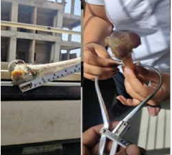
We conducted a differentiation study between human and animal bones focusing on the femur. The samples of non- human species including goat, dog, pig, cow, and buffalo bones were procured from “Kamla Nehru Prani Sangharalaya Indore” on February 20, 2024. This collection was facilitated with the assistance of the zookeeper, “Gabbar sir,” and conducted under the guidance of Dr. Mahajan. Prior permission and consent were obtained from the director and a doctor at the Indore Zoo [11, 12, 13, 14, 15].
For the human bone specimen, a replica of a female femur was acquired from the esteemed faculty of R.D. Gardi Medical College for research purposes. The replica was modeled after the femur of an unidentified individual, aged approximately 12-13 years, whose charred remains were discovered in a jungle. This replica was crafted using plaster of Paris (POP) and dental stone, specifically for research and study purposes [16, 17].
Methodology
- Initially, the bones were received in a wet state, still enveloped in muscular and tissue remnants as obtained from the scene.
- All muscular tissue and residual dried blood were meticulously removed from the bones using a damp cloth and cotton to ensure minimal damage to the bone structure.
- The bones were then wrapped in two layers of newspaper and exposed to sunlight for two days to eliminate any bacterial presence and unpleasant odors.
- Following the sunlight treatment, the bones were carefully placed in a polyethylene bag with three to four layers to prevent direct contact between the dried bone samples and potential natural predators like dogs and cats.
- After the six-day process, the bones were thoroughly washed with distilled water and dried meticulously with the aid of cotton.
- To facilitate the measurement and documentation process, essential tools such as pens, paper, a measuring tape, a scale, and a camera were prepared to record measurements and capture images of the samples.
Result
| S.No | Measurement | Human | Goat | Buffalo | Pig | Cow | Dog |
|---|---|---|---|---|---|---|---|
| 1 | Height of patellar surface | 15.9 cm | 7 cm | 10 cm | 6 cm | 9 cm | 4 cm |
| 2 | Fqvea to Laeral epicondyle | ± 5cm | 7.1 cm | ±9.8 cm | ±7 cm | ±10cm | ±8 cm |
| 3 | Interlochanter line to abductee tubercle | 15 cm | 1.2 cm | ± 9 cm | ±2 cm | ± 8 cm | 3cm |
| 4 | Abductle tubercle to lateral condyle | 14.4 cm | 1.4 cm | 3 cm | 2 cm | 2 cm | 3 cm |
| 5 | Interlochanter line to lateral epicondyle | 15.5 cm | 7.5 cm | 8.8 cm | ±6 cm | ±8 cm | ±6 cm |
| 6 | Patellar surface | 2 cm | 0.5 cm | 9 cm | 0.5 cm | 8 cm | 1 cm |
| 7 | Angle of head | Acute angle | Acute angle | Acute angle | Acute angle | Acute angle | Acute angle |
| 8 | Diameter | 3 cm | 2cm | 4 cm | 2 cm | 4 cm | cm |
Table 1: Measurements for Human, Cow, Goat, Buffalo, Pig, Cow and Dog.
Conclusion
In conclusion, the comparison of femur bone measurements between humans and various animal species, including buffalo and cows, reveals both striking similarities and notable differences. While there are resemblances in certain measurements, such as the height of the patellar surface and distances to specific anatomical landmarks, differences like bone diameter also exist. Overall, these findings suggest potential functional similarities in lower limb anatomy and biomechanics across species. These insights may inform comparative anatomical studies, biomechanical research, and surgical interventions involving the lower limb in diverse species, highlighting the importance of understanding cross-species anatomical variations and functional adaptations.
References
-
Saulsman B, Oxnard CE, Franklin D (2010) Long bone morphometrics for human from non-human discrimination. Forensic Sci Int 202(1-3): 110.e1-5.
-
Canine Hindlimb Anatomy.
-
Frisbie DD, Cross MW, McIlwraith CW (2006) A comparative study of articular cartilage thickness in the stifle of animal species used in human pre-clinical studies compared to articular cartilage thickness in the human knee. Vet Comp Orthop Traumatol 19(3): 142- 146.
-
Brits D, Steyn M, L’Abbé EN (2014) A histomorphological analysis of human and non-human femora. Int J Legal Med 128(2): 369-77.
-
Ubelaker DH, DeGaglia CM (2020) Chapter 17 - Factors of population variation in sex estimation methodology. Sex Estimation of the Human Skeleton pp: 281-293.
-
Evans FG, Lebow M (1951) Regional Differences in Some of the Physical Properties of the human femur. J Appl Physiol 3(9): 563-572.
-
Goat Skeleton Anatomy-Skull Forelimb and Hindlimb Bones.
-
Lavelle CL (1974) An Analysis of the Human Femur. Am J Anat 141(3): 415-426.
-
Hillier ML, Bell LS (2007) Differentiating Human Bone from Animal Bone: A Review of Histological Methods. J Forensic Sci 52(2): 249-263.
-
Morales JP, Roa I, Matamala DZ, Suazo I, Morales G (2012) Determination of the Species from Skeletal Remains Through Histomorphometric Evaluation and Discriminant Analysis. International Journal of Morphology 30(3): 1035-1041.
-
Mulhern DM (2009) Differentiating Human from Nonhuman Skeletal Remains. In Handbook Of Forenisc Anthropology.
-
Oghenemavwe LE, Orupabo CD, Amos II (2022) Comparative study of the histomorphometry of the femur in humans and canis lupus familiars (Dog): Implication for forensic investigations. Journal of Dental and Medical Sciences 21(3): 1-7.
-
Tatare NE, Sciulli PW (2005) Anthropological Analysis of the Lower Extremity. Forensic Medicine of the Lower Extremity pp: 69-98.
-
Hodgskinson R, Currey JD (1990) The effect of variation in structure on the Young’s modulus of cancellous bone: a comparison of human and non- human material. Proc Inst Mech Eng H 204(2): 115-121.
-
Sundari RK, Damaraju PK, Gudepu P, Mekala L, Chandrashekhar EL (2022) Age Related Microanatomical Changes in the Femoral Head Articular Cartilage of Buffalo (Bubalusbubalis). Indian Journal of Veterinary Anatomy 34(2): 101-105.
-
Johnson V, Beckett S, Márquez-Grant N (2017) Differentiating human versus non-human bone by exploring the nutrient foramen: implications for forensic anthropology. Int J Legal Med 131(6):1757-1763.
-
Babosova MZ A scaling study on macroscopic and histomorphometric differences between cow (Bos taurus) and sheep (Ovis aries).
- Narcotics and Digital Forensics: Bridging Crimes in the Digital Age
- Ethics in Forensic Psychiatry: Principles, Dilemmas, and Human Rights
- Impact of Acute Stress on Attentional Orienting to Social Cues
- Head Injury and Intracranial Hemorrhage in Western Region of Libya
- A Forensic Study on Handedness: Examination of Handwriting Features in Right and Left Handed Writers
- Techniques for Latent Fingerprint Development Using Natural and Synthetic Powders: A Review