Pathogenicity and Antibiotic Susceptibility Pattern of Enterococcus faecalis in Clarias gariepinus Juvenile
Background: Disease is the most significant obstacle in aquaculture causing harm to farmers’ livelihoods, reducing their income, and resulting in unemployment and food insecurity. This study was conducted to determine pathogenicity and antibiotic susceptibility pattern of Enterococcus faecalis in Clarias gariepinus juvenile. Methodology: Antibiotic susceptibility pattern of Enterococcus faecalis was carried out using agar disc diffusion method. Clarias gariepinus juvenile (n = 100) with an average weight of 19.0 ± 0.81 g, and length of 11.7 ± 0.6 cm, were randomly divided into five groups, and four groups were infected with 1ml of TSA broth culture containing 2.1 X 10⁶ CFU Enterococcus faecalis OgNG1 by immersion at the rate of 5ml/L of water. The infected and control groups were monitored daily for 14 days. The clinical signs, gross, skin lesions, and mortalities were observed. Haematology, blood chemistry, and histopathological changes in the skin and gill were investigated. An Independent T-test was used for statistical analysis and p < 0.05 was considered significant. Results: The cumulative mortality in infected juvenile was 77.5%, and the gross lesion indicated atrophied barbel, skin ulceration, and necrosis of the gill. Histopathological analysis revealed erosion of secondary lamellae, epidermal and dermal layer. The isolates were multi-drug resistant with MAR index of 0.64, indicating resistance to Aminoglycosides, Quinolones, Tetracyclines, Cephalosporins, and Phenicols classes of antibiotic drugs, though sensitive to penicillin; ampicillin, amoxicillin, and ampicillin-cloxacillin. Clinicopathological investigation showed significantly (p ≤ 0.05) lower packed cell volume, hemoglobin, and red blood cells in infected groups compared to the control group. Meanwhile, white blood cells were significantly (p ≤ 0.05) higher in the infected groups compared with the control group. Moreover, there was a significant increase (p ≤ 0.05) in the activity of serum AST, ALT, and creatinine but a significant decrease (p ≤ 0.05) in total protein, albumin, and globulin concentrations of infected groups compared to the control group. The results of this study indicated that Enterococcus faecalis isolates were highly pathogenic to Clarias gariepinus juveniles. Conclusions: Enterococcus faecalis isolates were multidrug-resistant with a MAR index of 0.64, sensitive to ampicillin, amoxicillin, and ampicillin-cloxacillin, and highly virulent, and pathogenic to Clarias gariepinus juveniles.
Introduction
African catfish also known as Clarias gariepinus (Burchell), is an economically important and most farmed fish species in Nigeria. This supremacy of African catfish production is associated with their aquaculture qualities which include the ability to bear handling stress, disease resistance, high proliferation, fast growth rate, high fertility, and acceptability. Bacterial diseases in fish cause outbreaks that could lead to high economic losses. Economic losses due to disease are likely to increase as a result of rapid growth in aquaculture [1].
Disease is the most significant obstacle in aquaculture causing harm to farmers’ livelihoods, reducing their income, and resulting in unemployment and food insecurity. Worldwide, bacteria are the principal causative agents of diseases in freshwater fishes [2]. Fish bacteria pathogens such as Flavobacterium columnare, Aeromonas hydrophila and many more have been reported earlier. Recently, some opportunistic bacterial pathogens have been characterised as the causal agents of severe diseases outbreak in fish farm facilities [3]. One of these opportunistic bacteria is Enterococcus faecalis, important emerging fish pathogens which adversely affects aquaculture production all over the world [4].
Enterococcus faecalis are bacteria located in the gastrointestinal tract of animals, humans, and they are also present in the environment. The bacteria have been isolated from infected fish. They are classified as evolving pathogens of fish and they are good indicators of antibiotic resistance. They have ability to obtain resistance gene from other bacteria and transfer of antimicrobial resistance. The transmission may be horizontal involving transfer of mobile genetic elements to other pathogenic bacteria [5, 6].
Antibiotic resistance in aquaculture is a global concern for control of bacterial diseases. Moreover, there is dearth of information on pathogenicity and antibiotic susceptibility profile of Enterococcus faecalis isolated from the infected farmed fish in Nigeria. It is a necessity to continue assessing resistance profile of emerging and opportunistic bacteria causing diseases in aquaculture farms. Assessing the tendencies of antimicrobial resistance in Enterococcus faecalis isolated from infected fish is crucial. This will provide important information and knowledge concerning the level of resistance and empirical selection of antimicrobial agents to treat infected fish. Pathogenicity study through experimental infection of animals has been a crucial method in establishing aetiology of disease conditions as well as having information on their pathophysiology and pathogenesis. Hence, pathogenicity and antibiotic susceptibility pattern of Enterococcus faecalis in Clarias gariepinus juvenile.
Materials and Methods
Experimental Infection
Enterococcus faecalis isolates were characterized in an earlier study, using species-specific primers [7]. Moreover, 1ml of TSA broth culture determined by McFarland 0.5 standard opacity tube contained 2.1 x 10⁶ CFU of Enterococcus faecalis. Eighty Clarias gariepinus juveniles were submerged in water containing isolates of Enterococcus faecalis at a rate of 5ml/L [1]. The control group (n=20) was uninfected and the infected fish (n=80) were divided into four tanks and exposed to the bacterial isolates. Feeding began 96 hours post-infection at a rate of three percent body weight once daily. Fifty percent of tank water was changed daily to prevent contamination [2]. Physico- chemical parameters of water in the experimental tank were monitored daily. Dissolved Oxygen, pH, temperature, nitrite, and ammonia were determined. Experimentally infected fish were monitored daily for clinical symptoms, gross, skin lesions, and mortalities for 14 days [8]. The collection of blood samples for haematology and serum biochemistry was performed at seven-day intervals. Organs such as skin and gills were submitted for histopathology using H & E stain.
Antibiotic Susceptibility Testing
A total of ten Enterococcus faecalis isolates were analyzed for antibiotic susceptibility. By using the agar-disc diffusion method, which was carried out by CLSI standards Mo2 [9]. Pure isolates of Enterococcus faecalis isolates were evaluated for their sensitivity and resistance to antibiotics. The susceptibility was evaluated against 12 antibiotics. The drugs belong to seven classes: penicillin (amoxicillin– clavulanic acid 30 μg (AC); ampicillin 10 μg (AP), amoxicillin 25 μg (AM)), cephalosporin (third-generation) ceftriaxone 30 μg (CEF), tetracycline (doxycycline 30 μg (D), tetracycline 30 μg (T), chloramphenicol 30 μg (CH), aminoglycoside (streptomycin 10 μg (S), gentamicin 10 μg (G)), fluoroquinolone (ciprofloxacin 5 μg (CIP), Enrofloxacin 5 μg (ENR)), Polymyxin (Colistin sulphate 10 μg (CS) [9].
Preparation of Antibiotic Disk
The disc was constructed by the CLSI Mo2 disk diffusion guide; six 6-mm sterilized Whatman filter paper disks were placed on a 100-mm plate, and the disks were impregnated with the known antimicrobial concentrations stated above.
Each isolate on a sterile Mueller-Hinton agar plate was made and appropriately labeled with an indelible marker [9].
Preparation of Inoculums
By adjusting the suspension of the Enterococcus faecalis colony in sterile normal saline to the density of a McFarland 0.5 turbidity standard, the direct colony suspension method was employed. Enterococcus faecalis was grown overnight on Tryptic Soy agar (HiMedia®), and pure cultures were harvested using a sterile loop. The colonies were suspended in sterile normal saline that had been blended to consistent turbidity. Saline or additional bacteria were added to get the density of the Enterococcus faecalis suspension to McFarland 0.5. After that, Muller-Hinton agar (HiMedia®), was streaked with the suspended Enterococcus faecalis colonies. Quality control was performed using the Pseudomonas aeruginosa strain [9].
Inoculation of agar plates
The antibiotic disc was picked using sterile pointed forceps and applied to the surface of Mueller-Hinton agar plates. This allowed the discs to effectively touch the medium. Afterward, the plates were incubated for 18 hours at 37°C [9]. To identify which antibiotics the isolate was sensitive to or resistant to, the diameter of the visible zone of growth inhibition surrounding the several discs was measured in millimeters. Each isolate’s resistance pattern was created using the antibiogram.
Statistical Analysis
The significant differences between the control and infected groups were determined using an Independent T-test. A “p” value of less than 0.05 (p < 0.05) was regarded as significant to determine the level of significance between haematology, serum biochemistry, and mortality rate of control and infected groups.
Result
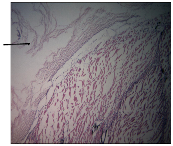
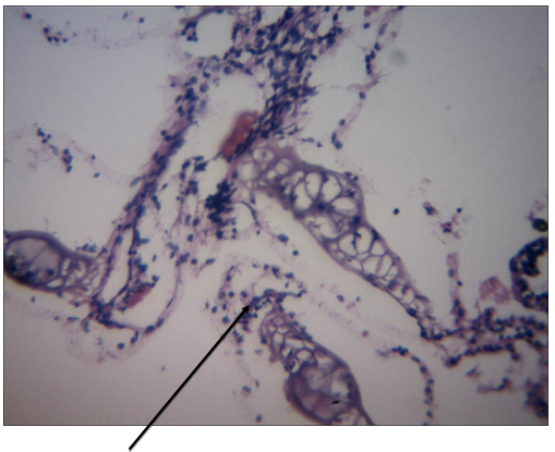
The clinical signs observed in infected juvenile groups were atrophied or reduced barbels, desquamation, or abrasion of the skin. The gross lesion observed in infected juvenile groups were necrosis of the gills (Figure 1). The histopathological changes observed in the organs of infected juvenile groups infected with Enterococcus faecalis isolates included erosion of the entire skin and secondary lamellae of the gill (Figures 2 & 3). The mortality in the first week of the infected group (32) was significantly higher (α0.05) than the control group (0). This progressively increased in the second week, the infected group was thirty while the control group was zero (Table 1).
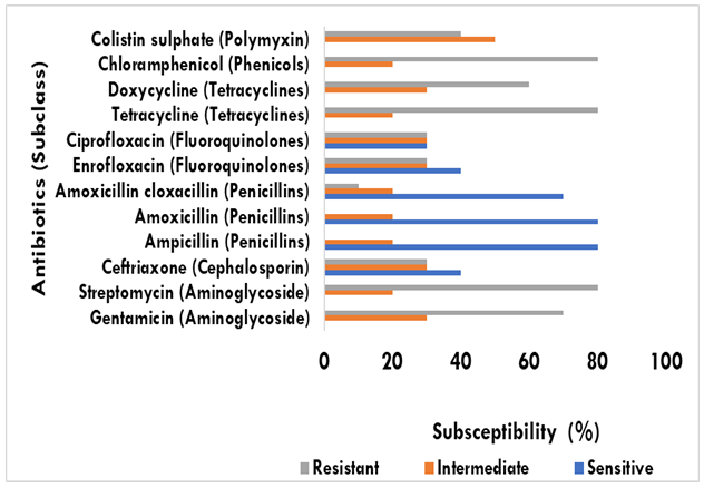
Anaemia was observed in the infected group with reduced packed cell volume (PCV), haemoglobin, and total red blood cells. Leucopaenia was observed with the total white blood cell counts of the infected group significantly (α0.05) lowered in comparison to the control group. The lymphocyte counts of the infected group were significantly (α0.05) lower compared to the control group. The heterophil count of the infected group was significantly higher (α0.05) compared to the control group. The monocyte counts of the infected group were significantly higher (α0.05) compared to the control group. The eosinophil count of the infected group was significantly higher (α0.05) compared to the control group. The basophil counts of the infected group were significantly lower (α0.05) compared to the control group. The platelets count of the infected groups was significantly lower (α0.05) compared to the control group (Table 2). Moreover, serum hypoproteinemia, hypoalbuminemia, and reduced globulin were observed in the infected groups compared to the control group. The serum creatinine, aspartate aminotransferase (AST) and Alanine aminotransferase (ALT) levels of the infected groups were significantly higher compared to the control group (Table 3) Enterococcus faecalis isolates (80%) were sensitive to ampicillin, amoxicillin, and ampicillin-cloxacillin. The bacteria isolates (30%) were resistant to ceftriaxone, enrofloxacin, and ciprofloxacin. The bacteria isolates (70%) were highly resistant to doxycycline and highly resistant to gentamycin (70%). Enterococcus faecalis isolates (80%) were resistant to tetracycline, streptomycin, and chloramphenicol (Figure 4). The MAR index was 0.64
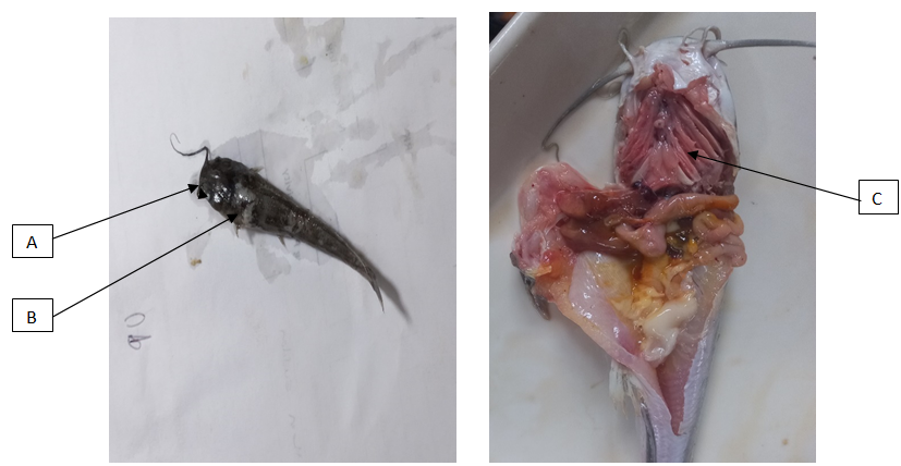
A – Atrophied barbel, B – Skin ulceration, C – Necrosis of gill
| Control Juvenile | Infected Juvenile | |
|---|---|---|
| Week 1 | 0a | 32b |
| Week 2 | 0a | 30b |
| Total | 0 | 62 |
| Percentage | 0 | 77.5 |
Table 1: Mortality rate of Clarias gariepinus juveniles experimentally infected with Enterococcus faecalis isolates.
Values with different superscript along the rows indicate significance (α0.05) according to the Independent T test
| Control Juvenile | Infected Juvenile | |
|---|---|---|
| PCV (%) | 23.80±0.41a | 16.67±0.28b |
| Hb (dL) | 6.29±0.40a | 5.18±0.70b |
| RBC (x10^6/μL) | 2.33±0.20a | 1.24±0.19b |
| MCV (fL) | 102.15±1.75a | 134.4±3.24b |
| MCH (pg) | 27.00±0.71a | 41.8±1.77b |
| MCHC (%) | 26.43±0.90a | 31.1±0.76b |
| WBC (x10^3/μL) | 12.69±0.14a | 15.38±0.11b |
| Lymphocytes (%) | 69.38±0.98a | 61.67±0.76b |
| Heterophils (%) | 24.71±0.81a | 30.00±0.71b |
| Monocytes (%) | 2.28±0.18a | 4.00±0.29b |
| Eosinophils (%) | 2.67±0.14a | 3.78±0.32b |
| Basophils (%) | 0.98±0.02a | 0.78±0.22b |
| Platelets (x10^5/μL) | 2.82±0.01a | 1.13±0.26b |
Table 2: Haematological parameters of juveniles Clarias gariepinus infected with Enterococcus faecalis isolates.
Values with the different superscript along the rows indicate significance (α0.05) according to Independent T-test PCV – Packed cell volume, Hb – Haemoglobin, RBC – Red blood cells, WBC – White blood cells, MCV – Mean corpuscular volume, MCH – Mean Corpuscular hemoglobin, MCHC – Mean corpuscular hemoglobin concentration
| Control Juvenile | Infected Juvenile | |
|---|---|---|
| Total Protein (%) | 6.10±0.30a | 4.62±0.45a |
| Albumin (dL) | 1.94±0.05a | 1.06±0.16a |
| Globulin (dL) | 4.16±0.03a | 3.01±0.13a |
| AST (μL) | 189.80±3.83a | 207.4±4.06a |
| ALT (μL) | 42.60±0.74a | 73.33±4.03a |
| Creatinine (mg/dL) | 0.48±0.02a | 1.03±0.14a |
Table 3: Serum biochemistry parameters of juvenile and adult Clarias gariepinus infected with Enterococcus faecalis isolate.
Values with the same superscript along the rows indicate significance (α0.05) according to the Independent T-testAST – Aspartate aminotransferase, ALT – Alanine transaminase
Discussion
Enterococcus faecalis strains have been reported as antibiotic-resistant bacteria in many countries [10]. In the present study, all of the E. faecalis isolates showed resistance to multiple antibiotics. Enterococci can acquire transposons, resistance plasmids, and sex pheromone plasmids from a wide range of recipients, which facilitate them to act as a reservoir of resistance genes [11]. Resistance of Enterococci to various antibiotics such as chloramphenicol, clindamycin, erythromycin, tetracycline, aminoglycosides, beta-lactamases, and vancomycin has been reported Boccella M, et al. [12]. Various antibiotics and growth-promoting agents are widely used in aquaculture, livestock, and poultry- rearing facilities in Nigeria without proper awareness of their application. This may lead to the resistance of the fish pathogen E. faecalis to multiple antibiotics shown in this study. Resistance of E. faecalis to various cell wall-degrading antibiotics such as penicillin-G, ampicillin, and vancomycin has been reported Li G, et al. [13]. In this study, all E. faecalis isolates were found to be resistant to tetracycline, aminoglycosides, fluoroquinolones, and chloramphenicol but sensitive to ampicillin, amoxicillin, and ampicillin cloxacillin. The antibiotic susceptibility profile of a fish pathogenic E. faecalis demonstrated in this article has not previously been reported.
The severe anemia observed in the infected group in this study may be a result of renal damage thereby hindering erythropoiesis. The monocytosis and heterophilia may be due to systemic bacterial infection. The results are consistent with Aly SM, et al. [14] who observed severe anemia, moderate hypothermia, mild neutrophilia, mild lymphocytopenia, moderate to severe monocytosis, moderate to severe eosinophilia, and severe basophilia with Proteus vulgaris isolates. Hypoalbuminemia and Hyperglobulinemia may be due to the increased production of antibodies caused by bacterial infection. Increased serum AST, ALT, and creatinine were caused by liver and kidney damage [14].
The mortality rate of 77.5% observed in the infected groups was similar to the report of Zahran E, et al. [15] who observed 80% in infected farmed Nile tilapia, and Rahman M, et al. [16] observed 90% in infected tilapia. The high mortality rate may be due to the pathogenic effect of Enterococcus faecalis.
Elgohary I, et al. [17] reported postmortem examination of infected Oreochromis niloticus included loose scales, ulceration in the dorsal part of the head, petechial haemorrhages on the body surfaces, unilateral exophthalmia, and corneal opacity together with congested blood vessels of nearly all fleshy portions of the body fins. Internal examination of the viscera showed congestion of the blood vessels in the kidneys and brain, a dark enlarged spleen, and a liver with a marbled appearance. The gross lesions observed in this study were similar to previous studies, atrophied or reduced barbels, desquamation, or abrasion of the skin, and necrosis of the gills. Rahman M, et al. [16] reported erosion of the caudal fin, haemorrhages at the base of the pelvic fin, and bilateral opacity in infected tilapia. Meanwhile, Zahran E, et al. [15] reported haemorrhages, dark pigmentation, skin erosions, ulcers, corneal opacity, hemorrhagic liver, pale kidney, and splenomegaly.
The histopathological organ changes observed in this study were erosion of the entire epidermis and secondary lamellae. This was similar to previous studies, Zahran E, et al. [15] observation in infected tilapia revealed degenerative and necrotic changes in the heart and liver. Moreover El-nobi G, et al. [18] reported vascular congestion and infiltration with inflammatory cells in examined organ tissues (brain, liver, kidney, spleen, and heart) in infected tilapia.
Conclusion
In conclusion, Enterococcus faecalis isolates were multidrug-resistant with a MAR index of 0.64, sensitive to ampicillin, amoxicillin, and ampicillin-cloxacillin, highly virulent, and pathogenic to Clarias gariepinus juveniles. The enterococcal isolates exhibited resistance to several antimicrobials, including those considered critical for humans. The occurrence of a high percentage of multidrug- resistant E. faecalis in food animals is alarming, especially given the fact that very few antimicrobial agents can be used to control enterococcal infection. Such resistance is likely to be passed from fish consumption to humans through the food chain. Therefore, the prudent use of antimicrobials in aquaculture will be crucial in limiting the public health hazards of Enterococcus in Nigeria.
Declarations
Ethics approval and consent to participate
Ethical approval was received from the University of Ibadan, Animal Care and Use Research Ethics Committee (UI-ACUREC) with the assigned number UI- ACUREC/066-0622/10
Consent for publication
Not applicable.
Availability of Data and Material
The datasets used and or analysed during the current study are available from the corresponding author on reasonable request.
Competing interest
There are no competing interests to declare concerning the actualization of this work.
Funding
This study was completely funded by the personal funds of the authors.
Authors’ contributions
OR and BO Conceptualized the study, OR, BO, and AO generated data and carried out fieldwork, OR, BO analyzed data and made the manuscript draft, OR and AO edited the manuscript, OR, BO, and AO approved the final manuscript.
Acknowledgments
We want to acknowledge Mr Segun for laboratory assistance.
References
-
Anifowose OR, Oladosu GA, Oladele OO (2021) Causal factors of mass mortality of hatchery-reared Clarias gariepinus fry during exogenous feeding. International Journal of Fisheries and Aquatic Studies 9(1): 235-239.
-
Tiamiyu OS, Oladosu GA, Anifowose OR, Ajayi OL (2020) Pathogenicity and Antibiotics Sensitivity Profile of Aeromonas Bestiarum used in Experimental Infection of Different Developmental Stages of Clarias gariepinus. Journal of Aquaculture Marine Biology and Ecology JAMBE: 103.
-
Zorrilla I, Chabrillón M, Arijo S, Dı́az-Rosales P, Balebona MC, et al. (2003) Bacteria recovered from diseased cultured gilthead sea bream (Sparusaurata L.) in southwestern Spain. Aquaculture 218(1-4): 11-20.
-
Hammerum AM (2012) Enterococci of animal origin and their significance for public health. Clin Microbiol Infect 18(7): 619-625.
-
Nilsson O (2012) Vancomycin-resistant enterococci in farm animals–occurrence and importance. Infect Ecol Epidemiol 2: 16959.
-
Torres C, Alonso CA, Ruiz-Ripa L, León-Sampedro R, del Campo R (2018) Antimicrobial resistance in Enterococcus spp. of animal origin. Microbiol Spectrum 6(4).
-
Anifowose OR, Akinniyi O.O, Banwo O.G, Oladosu GA (2023) Occurrence and characterization of Enterococcus faecalis from infected farmed African catfish in Ogun State. International Journal of Oceanography & Aquaculture 7(3): 1-7.
-
Anagor TA (2017) Clinical and Treatment Outcomes in Clarias gariepinus Experimentally Infected with Single and Mixed Escherichia Coli and Salmonella gallinarum strains (Doctoral dissertation).
-
CLSI (2020) CLSI Performance Standards for Antimicrobial Susceptibility Testing. 30th (Edn.), CLSI Supplement M100, Wayne, PA, USA.
-
Miller WR, Murray BE, Rice LB, Arias CA (2020) Resistance in vancomycin-resistant enterococci. Infectious Disease Clinics 34(4): 751-771.
-
Ezeh GC, Ogugua AJ, Nwanta JA (2023) Occurrence, antimicrobial resistance and pathogenic factors of Enterococci. Animal Research International 20(1): 4791-4816.
-
Boccella M, Santella B, Pagliano P, De Filippis A, Casolaro V, et al. (2021) Prevalence and antimicrobial resistance of Enterococcus species: a retrospective cohort study in Italy. Antibiotics, 10(12): 1552.
-
Li G, Walker MJ, De Oliveira DM (2022) Vancomycin resistance in Enterococcus and Staphylococcus aureus. Microorganisms 11(1): 24.
-
Aly SM, Taha R, Dessouki AA (2012) Experimental pathological, hematological and biochemical studies on streptococcosis in Nile tilapia (Oreochromis niloticus).
-
Zahran E, Mahgoub HA, Abdelhamid F, Sadeyen JR, Risha E (2019) Experimental pathogenesis and host immune responses of Enterococcus faecalis infection in Nile tilapia (Oreochromis niloticus). Aquaculture 512: 734319.
-
Rahman M, Rahman MM, Deb SC, Alam MS, Alam MJ, Islam MT (2017) Molecular identification of multiple antibiotic resistant fish pathogenic Enterococcus faecalis and their control by medicinal herbs. Scientific reports 7(1): 3747.
-
Elgohary I, Eissa AE, Fadel NG, Ibrahim Abd Elatief J, Mahmoud MA (2021) Bacteriological, molecular, and pathological studies on the Gram‐positive bacteria Aerococcus viridans and Enterococcus faecalis and their effects on Oreochromis niloticus in Egyptian fish farms. Aquaculture Research 52(5): 2220-2232.
-
El-Nobi G, Hassanin M, El-Hady M, Aboshabana S (2021) Isolation, identification, molecular, and histopathological investigations of two pathogenic Enterococcus species from tilapia in Egyptian farms. Slov Vet Res 58(24): 33-43.
- Genetic Improvement of Nile Tilapia (Oreochromis niloticus): Advances in Selective Breeding and Genomic Approaches for Sustainable Aquaculture
- Microplastics, Contaminants, and Waste Hotspots: Divergences and Faults in Prioritizing Control Efforts
- Creating a Healthier, More Vibrant Open and Closed Aquatic Environment. A Submersible, Centrifugal Magnetically Affixed Current Changing Aquarium Pump
- An Attempt to Assess Alpha Diversity and Sample Size: Using the Ostracod Assemblages off Kumamoto Port, Japan
- Assessment of the Efficiency of Common Fishing Gears and Crafts Used at Mohananda River of Chapai Nawabganj, Bangladesh
- Fish Productivity and Biodiversity Status of Sundarban Mangrove in Bangladesh