Intra-Cranial Hematoma as a First Manifestation of Brain Tumours, a Meta-Analysis
Introduction: Intracerebral Hematoma (ICH), conventionally associated with vascular causes, is increasingly recognized as the initial presentation of underlying brain tumors. This Narrative review aims to synthesize current literature on the incidence, clinical features, diagnostic challenges, and outcomes associated with ICH as the primary manifestation of brain tumors. Methods: A systematic search across PubMed, Embase, and Cochrane Library databases yielded studies meeting inclusion criteria. These encompassed prospective and retrospective observational studies, case-control investigations, and case series reporting instances where ICH was the initial sign of an underlying brain tumor. Results: Analysis of the selected studies revealed variable incidences of ICH as the first manifestation of brain tumors, with differences across tumor types and patient demographics. Clinical presentations mimicked primary vascular causes, posing diagnostic challenges. Common tumor types included with varying intracranial locations. Treatment strategies ranged from surgery to adjuvant therapies, and outcomes were heterogeneous, influenced by tumor histology, location, and patient characteristics. Discussion: This review highlights the significance of recognizing ICH as an initial sign of brain tumors, emphasizing the need for refined diagnostic approaches and enhanced imaging modalities. Challenges in timely identification underscore the importance of heightened clinical suspicion. The variable outcomes suggest the necessity of tailoring treatment strategies based on tumor characteristics and patient factors, paving the way for future research to optimize interventions in ICH associated with brain tumors.
Introduction
Brain tumors and spontaneous intracerebral hemorrhage (ICH) are two distinct neurological conditions with overlapping clinical features and significant implications for patient management [1]. ICH is a type of stroke that results from the rupture of blood vessels within the brain parenchyma, leading to the formation of a hematoma and subsequent edema, mass effect, and neurologic dysfunction. Spontaneous ICH refers to bleeding that occurs in the absence of a clear precipitating factor, such as head trauma, vascular malformation, or anticoagulant use. Brain tumors are neoplastic growths arising from the brain parenchyma or surrounding structures, with varying histologic and molecular characteristics and potential for malignant transformation [2]. As these tumors grow, they can cause elevated intracranial pressure, cerebral edema, and local tissue damage, leading to a range of neurologic signs and symptoms [3]. The relationship between brain tumors and spontaneous ICH is complex and bidirectional [4]. On one hand, patients with brain tumors are at increased risk of developing spontaneous ICH, likely due to the presence of fragile and leaky blood vessels within the tumor microenvironment, as well as the use of anticoagulant or antiplatelet medications to manage symptoms such as seizures or thromboembolism [5]. In addition, the disruption of the blood-brain barrier and the activation of coagulation pathways by tumor cells or inflammatory mediators can further increase the risk of bleeding events [6]. On the other hand, spontaneous ICH can also be a presenting sign of an underlying brain tumor, particularly in cases of deep or lobar hemorrhage, younger age, and absence of vascular risk factors [7]. In these situations, neuroimaging studies such as computed tomography (CT) or magnetic resonance imaging (MRI) may reveal a mass lesion or tumor-related vasculopathy as the underlying cause of the bleeding [8]. The recognition of the relationship between brain tumors and spontaneous ICH is critical for accurate diagnosis, appropriate treatment, and improved outcomes. The management of these conditions may involve a multidisciplinary approach, including neurosurgery, radiation therapy, chemotherapy, and supportive care, depending on the tumor type, location, and grade, as well as the severity and evolution of the ICH. Early detection, risk stratification, and individualized treatment planning are essential to minimize morbidity and mortality in these complex and challenging scenarios.
Methods and Materials
Search Strategy
We conducted a comprehensive literature search on PubMed, Embase, and the Cochrane Library from their inception until September 2021. The search was limited to studies published in English and used the following keywords: “intracerebral hemorrhage,” “brain tumor,” “neoplasms,” “cancer,” “astrocytoma,” “glioblastoma,” “meningioma,” “pituitary adenoma,” “metastasis,” and “secondary neoplasms.” We also reviewed the reference lists of relevant articles to identify additional studies. Search Strategy is followed in appendix-1.
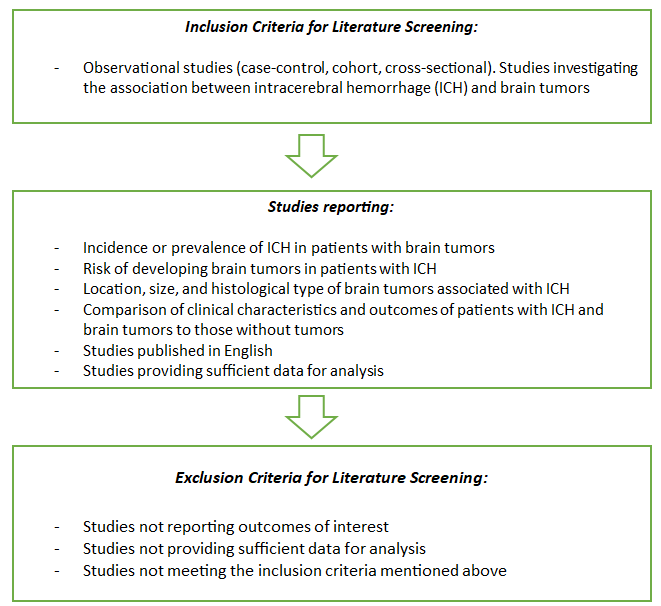
Flowchart of inclusion and exclusion criteria.
Review Process
Two independent reviewers screened titles and abstracts of retrieved studies and full-text articles were retrieved for potentially eligible studies. Disagreements were resolved by consensus between reviewers. This criteria and process were employed to ensure the selection of relevant studies and maintain methodological rigor in the meta-analysis.
Study Selection
We included observational studies (case-control, cohort, cross-sectional) that investigated the association between ICH and brain tumors. Studies that did not report the outcomes of interest or did not provide sufficient data for analysis were excluded. The inclusion criteria were as follows:
- Studies that reported the incidence or prevalence of ICH in patients with brain tumors or the risk of developing brain tumors in patients with ICH.
- Studies that reported the location, size, and histological type of brain tumors associated with ICH.
- Studies that compared the clinical characteristics and outcomes of patients with ICH and brain tumors to those without tumors.
- Two reviewers independently screened the titles and abstracts of all retrieved studies, and full-text articles were retrieved for potentially eligible studies. Disagreements were resolved by consensus.
Data Extraction
Data were extracted from each included study by two reviewers independently. The following information was extracted: study design, sample size, participant characteristics (age, sex, comorbidities), ICH and brain tumours characteristics (location, size, histological type), and outcomes (incidence, prevalence, risk, mortality, morbidity). Any discrepancies were resolved by consensus.
Quality Assessment
We assessed the quality of the included studies using the Cochrane Risk of Bias tool for randomized controlled trials and the Newcastle-Ottawa Scale for observational studies. Two reviewers independently evaluated each study, and any disagreements were resolved by consensus.
Data Analysis
We performed a narrative synthesis of the included studies due to the heterogeneity of the study designs, populations, and outcomes. We summarized the findings of each study and identified the factors that contributed to the association between ICH and brain tumours. We also conducted a subgroup analysis according to the type and location of brain tumours and the risk factors for ICH.
Discussion
ICH has been found in about 10% of cases with BTs, while different clinicopathologic series have indicated that 0.8%–7.4% of BTs are associated with spontaneous ICH [9]. The occurrence of a hemorrhagic presentation in a previously silent BT is even more uncommon, and there is limited current evidence on this topic.
According to Burth s, findings, 33% of intratumoral bleeding originates from GBMs, with metastases, anaplastic gliomas, low-grade gliomas, meningiomas, adenomas, hemangioblastomas, melanomas, neuroblastomas, and pinealoblastomas contributing to varying extents [10]. In a study conducted by Kondziolka et al., it was observed that among 264 GBM cases, about 6.4% displayed macroscopic hemorrhage, while 12.9% exhibited microscopic hemorrhage [11]. An interesting case involves a GBM mimicking acute cerebral hemorrhage. In this particular instance, the hemorrhage took on a mixed type, primarily presenting as macroscopic hemorrhage [12]. In scientific literature, instances of hemorrhaging tumors with varying histologies have been documented. The most commonly observed bleeding tumors are metastases, particularly from melanoma, and malignant gliomas [13]. While glioblastoma demonstrates the highest overall incidence of bleeding tumors, primary melanoma and neuroblastoma, though comparatively rarer, exhibit a significantly higher bleeding ratio [14]. The frequency of bleeding escalates with the malignancy level of the tumor [15]. Noteworthy bleeding rates have been emphasized by some authors in anaplastic astrocytomas, oligodendrogliomas, and oligoastrocytomas, particularly in the context of intratumoral microhemorrhages [16]. These microhemorrhages are reported with comparable incidences in WHO grade IV astrocytomas (56%) and WHO grade III oligodendrogliomas(53%) [13]. Moreover, intratumoral bleeding within glioblastoma multiforme (GBM) may manifest when the tumor infiltrates sizable blood vessels, leading to the debilitation and disintegration of their walls [17]. Low-grade gliomas constitute approximately 15% of adult brain tumors. Despite a comparatively favorable prognosis compared to higher- grade glial tumors, 50% to 75% of patients with low-grade gliomas eventually succumb to their condition [18].
The current research indicates that 2.9% of surgical patients had previously undetected brain tumors causing acute intracerebral hemorrhage, a percentage consistent with previous findings for these types of tumors [19]. Metabolic studies failed to provide insights into the nature of the described lesion. However, hypometabolic PET images allowed us to await improvements in the patient’s neurological condition. Subsequent late MRI findings suggested an acute hemorrhagic event within the tumor. Despite its rarity, the initial misdiagnosis as a hemorrhagic cavernoma led to the partial removal of the tumor [20].
The reasons behind bleeding in low-grade gliomas remain unclear, as neither tumor necrosis nor abnormal vessels are typically present [21]. Some authors propose that abnormal vessels or endothelial proliferation could be the underlying cause of hemorrhage in oligodendrogliomas [22]. The literatures include descriptions of bleeding tumors for both low-grade astrocytic and oligodendroglial tumors, with instances of glioneuronal and pilocytic tumors also reported [23]. Although low-grade astrocytoma is not commonly associated with intracerebral hemorrhage, awareness of the potential hemorrhagic presentation is crucial for accurate diagnosis and proper management, including considerations for surgical timing and approach [24]. Remarkably, investigation has brought to light a previously unexamined connection between the hemorrhage type and the specific tumor type. Intriguingly, the findings indicate that High-Grade Gliomas and Metastatic Brain Tumors primarily instigate Hemorrhagic Contusions, while Low-Grade Gliomas are more commonly linked to Extra-Intratumoral Hemorrhages [25]. Contemporary literature underscores the varied biological characteristics displayed by both Low-Grade and High-Grade Gliomas, contingent upon their genetic subtypes, contributing to survival predictions. Current perspectives suggest that this genetic information may also influence the angiogenic processes in Low-Grade Gliomas [26]. Despite High-Grade Gliomas exhibiting heightened vascularization and more prevalent angiogenesis compared to Low-Grade Gliomas, the significance of microvascular proliferation characteristics has gained prominence in the grading and subtype classification of Low-Grade Gliomas [26]. It is widely recognized that Low-Grade Gliomas typically present ill-defined margins in areas of diffuse infiltration. Although constructing a robust hypothesis based on our existing knowledge remains a challenging task, we posit that the diffuse infiltrative nature of Low-Grade Gliomas, along with their poorly defined margins and the presence of feeble tumor-associated vessels inadequately supported by the surrounding glial tissue, may account for their tendency to induce Extra-Intratumoral Hemorrhages upon bleeding [27].
Conclusion
In summary, the intricate relationship between brain tumors and intracerebral hemorrhages (ICH) unfolds with nuanced complexities. While glioblastoma multiforme (GBM) exhibits a higher incidence of bleeding, metastases, anaplastic gliomas, and various tumor types contribute to the spectrum of hemorrhagic presentations. The study emphasizes the rarity of hemorrhagic events within low- grade gliomas, challenging our understanding of their etiology. Noteworthy symptoms, including sudden headache, nausea, vomiting, alterations in consciousness, and seizures, accompany these events, often appearing without warning. Radiologically identifying brain tumors amid severe bleeding poses a diagnostic challenge, necessitating comprehensive imaging and clinical evaluation. The novel exploration of the correlation between hemorrhage types and specific tumor types unveils a previously unexamined connection, shedding light on the intricate interplay between tumor biology, genetic subtypes, and the nature of hemorrhages. This research contributes crucial insights, emphasizing the need for heightened awareness, accurate diagnosis, and tailored management strategies in the realm of neuro-oncology.
References
-
Pinho J, Costa AS, Araújo JM, Amorim JM, Ferreira C (2019) Intracerebral hemorrhage outcome: a comprehensive update. J Neurol Sci 398: 54-66.
-
Fewel ME, Thompson BG, Hoff JT (2003) Spontaneous intracerebral hemorrhage: a review. Neurosurgical focus 15(4): E1.
-
Aguilar MI, Freeman WD (2010) Spontaneous intracerebral hemorrhage. Semin Neurol 30(5): 555- 564.
-
Kim JY, Bae HJ (2017) Spontaneous intracerebral hemorrhage: management. J stroke 19(1): 28-39.
-
Morioka J, Fujii M, Kato S, Fujisawa H, Akimura T, et al. (2006) Surgery for spontaneous intracerebral hemorrhage has greater remedial value than conservative therapy. Surg neurol 65(1): 67-72.
-
Witsch J, Siegerink B, Nolte CH, Sprügel M, Steiner T, et al. (2021) Prognostication after intracerebral hemorrhage: a review. Neurol Res Pract 3(1): 22.
-
Hemphill III JC, Bonovich DC, Besmertis L, Manley GT, Johnston SC (2001) The ICH score: a simple, reliable grading scale for intracerebral hemorrhage. Stroke 32(4): 891-897.
-
Parry-Jones AR, Abid KA, Di Napoli M, Smith CJ, Vail A, et al. (2013) Accuracy and clinical usefulness of intracerebral hemorrhage grading scores: a direct comparison in a UK population. Stroke 44(7): 1840-1845.
-
Mantia C, Uhlmann EJ, Puligandla M, Weber GM, Neuberg D, et al. (2017) Predicting the higher rate of intracranial hemorrhage in glioma patients receiving therapeutic enoxaparin. Blood 129(25): 3379-3385.
-
Burth S, Ohmann M, Kronsteiner D, Kieser M, Löw S, et al. (2021) Prophylactic anticoagulation in patients with glioblastoma or brain metastases and atrial fibrillation: an increased risk for intracranial hemorrhage? J Neurooncol 152(3): 483-490.
-
Kondziolka D, Bernstein M, Resch L, Tator CH, Fleming JR, et al. (1987) Significance of hemorrhage into brain tumors: clinicopathological study. J neurosurg 67(6): 852-857.
-
Lieu AS, Hwang SL, Howng SL, Chai CY (1999) Brain tumors with hemorrhage. Journal of the Formos Med Assoc 98(5): 365-367.
-
Licata B, Turazzi S (2003) Bleeding cerebral neoplasms with symptomatic hematoma. J neurosurg sci 47(4): 201-210.
-
Nayak L, Lee EQ, Wen PY. (2012) Epidemiology of brain metastases. Curr oncol rep 14(1): 48-54.
-
Atmaca A, Al-Batran SE, Maurer A, Neumann A, Heinzel T, et al. (2007). Valproic acid (VPA) in patients with refractory advanced cancer: a dose escalating phase I clinical trial. Br J cancer 97(2): 177-182.
-
Bulakbaşı N, Paksoy Y (2019) Advanced imaging in adult diffusely infiltrating low-grade gliomas. Insights Imaging 10: 122.
-
Tseng JH, Lin WH (2012) Glioblastoma multiforme hiding behind the intracerebral hematoma. Formosan Journal of Surgery 45(6): 183-186.
-
Richard SA, Ye Y, Li H, Ma L, You C (2018) Glioblastoma multiforme subterfuge as acute cerebral hemorrhage: A case report and literature review. Neurology international 10(1): 7558.
-
Auer TA, Renovanz M, Marini F, Brockmann MA, Tanyildizi Y (2017) Ischemic stroke and intracranial hemorrhage in patients with recurrent glioblastoma multiforme, treated with bevacizumab. Journal of Neuro-Oncology 133(3): 571-579.
-
Faropoulos K, Panagiotopoulos V, Partheni M, Tzortzidis F, Konstantinou D (2015) Therapeutic management of intraventricular cavernoma: case series and review of the literature. Journal of Neurological Surgery Part A: Central European Neurosurgery 76(3): 233-239.
-
Brat DJ, Van Meir EG (2004) Vaso-occlusive and prothrombotic mechanisms associated with tumor hypoxia, necrosis, and accelerated growth in glioblastoma. Laboratory investigation 84(4): 397-405.
-
Kasantikul V, Shuangshoti S, Panichabhongse V, Netsky MG (1996) Combined angioma and glioma (angioglioma). Journal of surgical oncology 62(1): 15-21.
-
Brat DJ, Perry A (2018) In Practical surgical neuropathology: a diagnostic approach. Astrocytic and oligodendroglial tumors, 2nd (Edn.), pp: 91-123.
-
Willman M, Willman J, Figg J, Dioso E, Sriram S, et al. (2023) Update for astrocytomas: medical and surgical management considerations. Exploration of neuroscience 2: 1-26.
-
Ghotme AK, Barreto EG, Echeverria V, Gonzalez J, Bustos HR, et al. (2017) Gliomas: new perspectives in diagnosis, treatment and prognosis. Current topics in medicinal chemistry 17(12): 1438-1447.
-
Caffo M, Barresi V, Caruso G, La Fata G, Pino MA, et al. (2013) Gliomas biology: Angiogenesis and invasion. In: Lichtor T (Ed.), Evolution of the Molecular Biology of Brain Tumors and the Therapeutic Implications 27: 37- 103.
-
Guo H, Kang H, Tong H, Du X, Liu H, et al. (2019) Microvascular characteristics of lower-grade diffuse gliomas: investigating vessel size imaging for differentiating grades and subtypes. European radiology 29(4): 1893-1902.
- Management of Chronic Insertional Achilles Tendinopathy Using Flexor Hallucis Longus Tendon Transfer in Patients Over 50 Years of Age: A Four-Case Series Following the CARE Guidelines
- Application of Induced Pluripotent Stem Cells in Bone Tissue Engineering: Current Status and Prospects
- Surgical Management of Upper Thoracic Esophageal Squamous Cell Carcinoma with Concomitant Hypersplenism: Integration of Chai's Supra-Thoracic Apex Technique with Laparoscopic Splenectomy - A Technical Innovation Case Study with Systematic Review
- Evaluation of Masticatory Functional Efficiency of Stomatognathic System in Patients Undergoing Open Reduction Internal Fixation for Treatment of Pan-Facial Trauma: A Prospective Study
- Hepatic Abscess Secondary to Appendiceal Phlegmon an Unusual Complication of Appendiceal Phlegmon
- Report of Lumboperitoneal (LP) Shunt Procedure in Over Decades Experiences, Systematic Narrative Review