A Case Report of Incidental Diagnosis of Cardiac Hypertrophy in Brazilian Short Hair Cat With Cholangitis and Renal Acute Insufficiency
The hypertrophic cardiomyopathy is the most common myocardial disease that affects felines and is characterized by hypertrophy of the left ventricle, may be primary or secondary associated with hyperthyroidism, systemic hypertension and subaortic stenosis. The clinical signs of hypertrophic cardiomyopathy are variable, and the cat may have a symptomatic or asymptomatic clinical presentation. Imaging diagnostic methods such as Doppler echocardiography and electrocardiography allow the clinician to classify the stage of the disease more efficiently to more clearly assess the need for treatment and prognosis. In this case report, a four year old male Brazilian short hair cat was presented to the clinical facility with signals of emesis. A heart murmur was found during cardiac auscultation. After treatment to stabilize the clinical condition of emesis, imaging tests (Doppler echocardiography, abdominal ultrasonography), electrocardiogram, blood count, T4 dosage and biochemical tests were performed. Exam results demonstrate the presence of cholangitis, acute kidney disease and a incidental findings of cardiac hypertrophy.
Introduction
The hypertrophic cardiomyopathy is the most common cardiac disease in felines, being the main cause of congestive heart failure in this species [1].
The hypertrophic cardiomyopathy is characterized by thickening of the ventricular myocardium and interventricular septum, causing diastolic dysfunction that makes it difficult for blood to enter the ventricles, resulting in atrial dilation. These changes are more pronounced on the left side [2].
The disease can be primary, with genetic involvement, or secondary to diseases such as hyperthyroidism and subaortic stenosis [3, 4, 5].
The hypertrophic cardiomyopathy is more common in males and purebred cats such as Maine Coon, Persian, Ragdoll, Siamese, Burmese Sacred, American Longhair, British Shorthair and American Shorthair; with crossbreeds of these breeds also being more predisposed. Although it affects cats of different ages, the highest incidence occurs between five and seven years of age [5].
Early recognition of hypertrophic cardiomyopathy is important, because this disease can be progress to arrhythmias, thrombosis and sudden death. Diagnostic methods importantes para o diagnóstico desta doença include electrocardiography and Doppler echocardiography, the latter considered the gold standard for diagnosing of the hypertrophic cardiomyopathy and other cardiac diseases in cats [2].
Case Report
In this case report, a four-year-old male Brazilian short hair cat was presented to the clinical center with emesis. The owner reported that the cat presented numerous episodes of emesis during the first year of life. After stabilization of gastric condition by the use of maropitant (1 mg/kg subcutaneous), imaging tests such abdominal ultrasound was performed with a diagnosis of cholangitis (Figure 1) and changes in the architeture of the kidneys (Figure 2). Blood count, biochemical analysis, urine analysis, and renal function study was requested. The cardiac auscultation indicated the presence of a heart murmur.
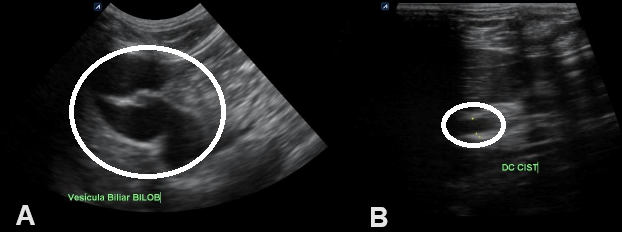
A. Gallbladder with a usual topography of bilobed appearance (circle) with adequate distension, thin and regular walls, filled with anechoic liquid content (bile). B. Cystic duct dilated by liquid content measuring approximately 0.31 cm of lumen indicating cholangitis/cholangiohepatitis (circle). Figure 1: Abdominal ultrasound.
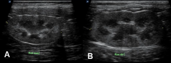
A. Left kidney with normal topography and normal size with high cortical echogenicity, suggestive of nephropathy. B. Right kidney with normal topography and normal size with high cortical echogenicity, suggestive of nephropathy Figure 2: Abdominal ultrasound.
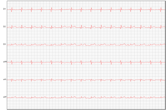
To confirm the suspicion of renal dysfunction, urea (70 mg/dL, normal value for the specie: 10 to 56 mg/dL), and creatinine (3,42 mg/dL, normal value for the specie: 0,80 to 1,80 mg/dL) was dosed, presenting increase in serum levels, and the quantitative immunofluorescence assay- VCHECK SDMA, a value was the value was with the normal range (12,60 ug/dL, normal value: up to 14 ug/dL). Urine biochemical analysis confirmed the presence of proteins in the urine type I examination. Due to the presence of a heart murmur during auscultation, an electrocardiogram and Doppler echocardiography were performed. The electrocardiogram indicated the presence of sinus rhythm with increased duration of QRS complex, suggesting left ventricular overload (Figure 3).
The doppler echocardiography revealed the presence of mild mitral insufficiency and thickening of the left ventricular wall, compatible with concentric hypertrophic cardiomyopathy (Figure 4). Not was observed stenosis of aortic valve.
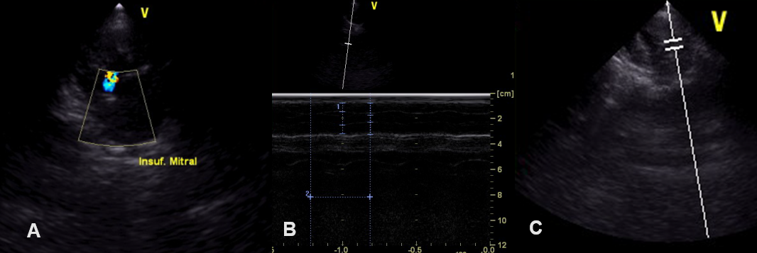
A. Presence of turbulent systolic flow within the left atrium, with mild mitral valve insufficiency. B. Thickening of the interventricular septum and left ventricular free wall, and decreased left ventricular systolic diameter (0,54 cm) compatible with concentric hypertrophy of the left ventricular wall. C. Normal aortic valve in appearance and movement of its cusps. Figure 4: Doppler echocardiography.
To exclude the possibility of hyperthyroidism, total T4 was measured (total T4: 2,5 μ/dL; normal values: 1,10-3,90 μ/dL), and free T4 (T4LD) by equilibrium dialysis (T4LD: 3,1 ng/dL; normal values: 0,76-3,94 ng/dL), with the values obtained being within normal parameters for the species.
Discussion
The hypertrophic cardiomyopathy is the most common heart disease in felines, being the main cause of congestive heart failure in this species [2]. The hypertrophic cardiomyopathy is reported most frequently in males and in Shorthair domestic cats, followed by Longhair domestic cats. At the time of diagnosis, most cats with hypertrophic cardiomyopathy are on average 5 to 7 years old, although it has been described in other age groups [5]. In our case report, the cat has short hair and was 4 years old at the time of diagnosis.
Most cats with hypertrophic cardiomyopathy do not present clinical signs at the time of diagnosis, and are generally referred for cardiac evaluation due to abnormal cardiac auscultation detected during routine examination, cardiovascular evaluation before anesthetic procedures or accidentally during routine clinical examination [6, 7]. In our case report, the cat was treated due to emetic symptoms, associated with an inflammatory process of bile ducts (cholangitis) and azotemia.
Although the literature reports symptoms associated with cardiac hypertrophy, such as open-mouth breathing, dyspnea, weakness, syncope and, in severe cases, acute bilateral and painful paresis of the pelvic limbs [8, 9], the cat described in our study was asymptomatic about the symptoms of heart disease, being the heart murmur detected during the routine examination.
The cardiac auscultation is abnormal in most cats with hypertrophic cardiomyopathy [10], the most common changes being the presence of a systolic murmur resulting from mitral regurgitation [1]. These murmurs are of variable degree, increasing with heart rate, and can be found in asymptomatic cats [1, 6], as described in our case report.
The use of the electrocardiogram is indicated in suspected feline hypertrophic cardiomyopathy, however the electrocardiographic findings have low specificity, although they are associated with a greater risk of developing sudden death or shorter survival time [9]. In our study, the electrocardiogram revealed the presence of sinus rhythm with increased duration of the QRS complex, suggesting left ventricular overload.
As a result, it was decided to perform Doppler
echocardiography to determine the cause of the heart murmur, because this examination allows the acquisition images of heart chambers, valves and large vessels [11], being considered the gold standard test for diagnosing cardiomyopathies in cats due to its high accuracy [12].
The consequences associated with hypertrophic cardiomyopathy include the presence of ventricular arrhythmias and myocardial dysfunction. The diastolic dysfunction occurs early in the course of hypertrophic cardiomyopathy, even before left ventricular wall remodeling is detected [1, 9].
The hypertrophic cardiomyopathy causes a progressive increase in pressure in the left atrium, with secondary atrial dilation and the development of congestive heart failure [13, 14, 15]. In our study case, the doppler echocardiography revealed the presence of mitral insufficiency and thickening of the left ventricular wall, compatible with concentric hypertrophic cardiomyopathy, such as described by literature [2].
Conclusion
In this case report we described the incidental diagnosis of hypertrophic cardiomyopathy in a cat with cholangitis and renal dysfunction. The early diagnosis or cardiac disease is fundamental to the continuous monitoring and the need of apropriated therapeutic instalation to the control of complications associated with hypertrophic cardiomyopathy in cats.
References
-
Kittleson MD, Côté E (2021) The feline cardiomyopathies: 2. Hypertrofic cardiomyopathy. J Feline Med Surg 23(11): 1028-1051.
-
Luís Fuentes V, Abbott J, Chetboul V, Côté E, Fox PR, et al. (2020) ACVIM consensus statement guidelines for the classification, diagnosis, and management of cardiomyopathies in cats. J Vet Intern Med 34(3): 1062- 1077.
-
Gil-Ortuño C, Sebastián-Marcos P, Sabater-Molina M, Nicolas-Rocamora E, Gimeno-Blanes JR, et al. (2020) Genetics of feline hypertrophic cardiomyopathy 98(3): 203-214.
-
Stern JÁ, Rivas VN, Kaplan JL, Ueda Y, Oldach MS, et al. (2023) Hypertrophic cardiomyopathy in purpose-bred cats with the A31P mutation in cardiac myosin binding protein-C. Sci Rep 13(1): 10319.
-
Kittleson MD, Côté E (2021) The feline cardiomyopathies: 1. General concepts. 23(11): 1009-1027.
-
Luís Fuentes V, Wilkie LJ (2017) Asymtomatic hypertrophic cardiomyopathy: diagnosis and therapy. 47(5): 1041-1054.
-
Fox PR, Keene BW, Lamb K, Schober KE, Chetboul V, et al. (2019) Long-term incidence and risk of noncardiovascular and all-cause mortality in apparently healthy cats and cats with preclinical hypertrophic cardiomyopathy. J Vet Intern Med 33(6): 2572-2586.
-
Rho J, Shin SM, Jhang K, Lee G, Song KH, et al. (2023) Deep learning-based diagnosis of feline hypertrophic cardiomyopathy. PLoS One 18(2): e0280438.
-
Kittleson MD, Côté E (2021) The feline cardiomyopathies: 3. Cardiomyopathies other than HCM. J Feline Med Surg 23(11): 1053-1067.
-
Saponaro V, Mey C, Vonfeld I, Chamagne A, Alvarado MP, et al. (2023) Systolic third sound associated with systolic anterior motion of the mitral valve in cats with obstructive hypertrophic cardiomyopathy. J Vet Intern Med 37(5): 16979-1684.
-
Novo Matos J, Payne JR (2023) Predicting development of hypertrophic cardiomyopathy and disease outcomes in cats. 53(6): 1277-1292.
-
Rohrbaugh MN, Schober KE, Rhinehart JD, Bonagura JD, Habing A, et al. (2020) Detection of congestive heart failure by Doppler echocardiography in cats with hypertrophic cardiomyopathy. J Vet Intern Med 34(3): 1091-1101.
-
Kiatsilapanan A, Surachetpong SD (2020) Assessment of left atrial function in feline hypertrophic cardiomyopathy by using two-dimensional speckle tracking echocardiography. BMC Vet Res 16(1): 344.
-
Matsuura K, Bach MBT, Takahashi K, Willesen JL, Koch J, et al. (2022) Non-invasive assessment of left ventricular relaxation property using color M-mode-derived intraventricular pressure gradientes in cats. J Vet Cardiol 41: 236-248.
-
Cheng WC, Lawson C, Liu HH, Wilkie L, Dobromyskyi M, et al. (2023) Exploration of mediators associated with myocardial remodeling in feline hypertrophic cardiomyopathy. Animals (Basel) 13(13): 2112.
- Mitochondrial Bio-Logistics: Steering Co-Enzyme Q10 and Lycopene Synergies within the Science 4.0 Bio-OS Framework
- Hymenoptera Specimens from the Caño Negro Wetland, of the National Museum Collection, Costa Rica
- Science 4.0: Comprehensive Architecture of the Biological Operating System (Bio-OS) A Framework for Systemic Resilience and Industrialized Bio-Governance
- Rabbit on, or Hare Back? Understanding Climate Change
- Clinical Validation of Science 4.0: Flow Steering and Epigenetic Drift Inversion on a 76-Year-Old Hybrid System
- Seeds Planted by another Mind