Elective Ovariohysterectomy in Paraguayan Hairy Dwarf Porcupine (Coendou spinosus) Using the Transversus Abdominis Plane Block-Case Report
Wild mammal orphans are challenging cases in rehabilitation centers since, usually, as a result of artificial care an emotional bond is created due to human-animal interaction. In these cases, in which reintroduction to the wild is not possible, the animals need to be sent to zoos, breeding sites or fauna holding facilities. This report describes an elective ovariohysterectomy (OVH) procedure in an adult female hairy dwarf porcupine (Coendou spinosus), kept in a zoo in southern Brazil. The aim of the procedure, besides reproductive control, was to reduce hormonal changes that were leading to bad and confrontational behavior of the female animal with others of the same species, therefore seeking to improve the well-being of the animals. For the anesthetic protocol, the pre-anesthetic medication consisted of midazolam (0.5 mg/kg, IM), ketamine (10 mg/kg, IM) and tramadol (2 mg/kg, IM). As maintenance of the anesthesia, isoflurane was utilized. As local anesthesia, a transversus abdominis plane block (TAP Block) was performed to improve analgesia. No complications were seen during or after the procedure. Elective OVH proved to be feasible in C. spinosus, as did the TAP Block technique, emphasizing the importance of having a technically qualified team for better outcome.
Introduction
The hairy dwarf porcupine (Coendou spinosus) is a small mammal that belongs to the Erethizontidae family and Sphiggurus subgenus. It can be found in southeast and south Brazil, Argentina, Uruguay and Paraguay [1]. At birth, these animals weigh approximately 40 g and can reach a weight of 1.5 to 2 kg in adulthood [2]. It has a coat that has yellowish-gray tones on its back and a range of tones that go from yellow to grayish-brown on its belly. Its coat is made up of rigid spines and longer, thinner hairs, which have the function to hide the spines [1]. It has arboreal and nocturnal habits, using its prehensile tail to help moving through the trees. They are herbivorous animals that feed on leaves, flowers, seeds and fruits [3].
Individuals belonging to the Erinaceidae family reach sexual maturity at 19 months and can produce litters of one to two offspring, after a gestation period lasting approximately 200 days. The reproduction rate is one litter per year and their life expectancy in captivity can reach up to 12 years [2]. They are classified on the International Union for Conservation of Nature (IUCN) Red list as least concern (LC) [4]. In general, the longer time in captivity, the chances of a wild animal returning to its natural habitat decrease due to the bond and dependence developed with human caregivers [5].
Considering the current handling practices, ongoing research about animal nutrition, health and well-being, as well as providing a suitable environment with protection against predators, it is possible to perceive an increase of life expectancy as well as a growing birth rate of captive animals.
One of the consequences of this situation is that zoos can no longer accommodate the animal surplus of some species without compromising their well-being [6]. Therefore, species that are classified as LC and that do not belong to special programs for captive reproduction are subjected to procedures of birth control via surgeries, such as elective ovariohysterectomy for females and orchiectomy for males.
The transversus abdominis plane block (TAP Block) technique consists in applying local anesthetic between the transversus abdominis and internal oblique muscles, providing analgesia for the abdominal wall [7]. It was first described in wild mammals in a Canadian lynx (Lynx canadensis) in a laparotomy [8]. The technique was also described in cadaveric studies in rabbits [9] and rats [10].
The elective orchiectomy technique was already described for the species [11] and the aim of this report is to describe the elective ovariohysterectomy in a C. spinosus kept in a zoo as a method of contraception and well-being.
Case Report
A female hairy dwarf porcupine (Coendou spinosus), approximately two years old and weighing 1.6 kg, was received by a zoo in southern Brazil in August 2021. The animal was an orphan (the mother was not around when it was found), so it was raised outside its natural environment using artificial breeding techniques. Thus the animal formed an emotional bond with its caregivers, making it impossible to reintroduce it to nature. The animal lived in an enclosure with three other castrated males of the same species, however, it began to exhibit aggressive behavior, probably attributed to hormonal territorial behavior.
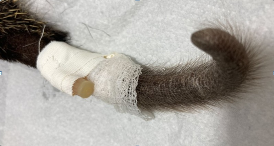
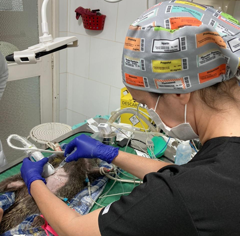
Due to the fact that the zoo was not interested in their reproduction, since it is not an endangered species, and with the aim to reduce hostility between the animals, an elective ovariohysterectomy (OVH) was performed in October, 2023. The anesthetic protocol established was pre-anesthetic medication using midazolam (0.5 mg/kg, Intramuscular (IM)), ketamine (10 mg/kg, IM) and tramadol (2 mg/kg, IM). Induction was performed using a face mask with 3% isoflurane diluted in 100% oxygen. After the loss of reflexes, the animal was positioned in the supine position and maintained with isoflurane in 100% oxygen saturation. Venous access was performed through the lateral vein with a 24G catheter (Figure 1), through which continuous fluid therapy with Ringer’s lactate (2 mg/kg/h) was carried out during the procedure. A wide trichotomy of the abdominal region was performed. Furthermore, ultrasound-guided transversus abdominal plane block (TAP Block) was performed with lidocaine (6 mg/kg) (Figures 2 & 3).
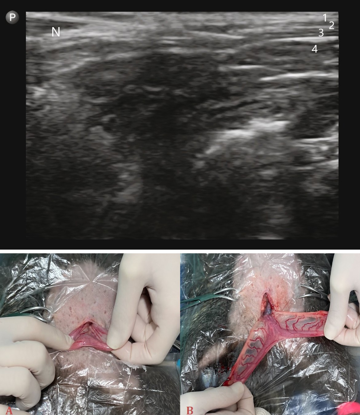
- External oblique muscle.
- Internal oblique.
- Transversus abdominis.
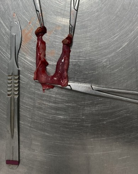
- Peritoneal cavity. N. Needle. Figure 3: Ultrasound image of the TAP Block Antisepsis of the operative field was done with 70% ethanol alcohol and 0.2 % chlorhexidine digluconate solution. Ovariohysterectomy was performed through a retro- umbilical celiotomy, with an incision in the ventral midline of the abdomen of about 2 cm. The uterine cervix was palpated dorsal to the bladder and then exteriorized (Figure 4). The mesovarium is very short and exteriorization of ovaries was difficult, so the suspensory ligament was torn using digital pressure. Hemostasis was done with 3.0 polyglycolic acid ligatures around the vessels cranial to the ovaries bilaterally. The mesometrium was torn without damaging the local vessels. Finally the uterus was removed (Figure
- by a transfixing ligature just cranial to the cervix with the same suture material.
The musculature and subcutaneous tissue were closed in a continuous simple pattern with polyglycolic acid 3.
The surgical procedure was successful, with no complications during or after. At the end of the surgery, the animal recovered well, and within a few minutes was already conscious it was placed in a transport box until full recovery and then kept in an adapted enclosure to ensure better treatment during the postoperative period.
While in the post_operative period the animal received the following medication: oxytetracycline (5 mg/kg IM, single dose), metamizole (25 mg/kg IM, twice a day for 5 days), tramadol (2 mg/kg IM, twice a day for 3 days) and meloxicam (0,1 mg/kg IM, once a day for 3 days).
A: Exteriorization of the uterus after laparotomy. B: Exteriorization of the uterus after section of the ovarian pedicles, with uterine vessels easily visualized in the myometrium. Figures 4A & 4B: Transsurgical images during elective ovariohysterectomy in a C. spinosus.
Discussion
Ovaryohisterectomy (OVH) refers to the surgical removal of the ovaries and uterus [12]. The female reproductive system of rodents, as in most mammals, consists in ovaries, uterine tube, uterus, vagina, vulva and accessory glands and the clitoris [13]. The surgical technique used for OVH through the retroumbilical ventral midline in the patient was the same as already described in other rodent species, such as guinea pigs (Cavia porcellus) [14]. It is a well- known technique in domestic animals, However, it is not as commonly done in zoological animals due to complications that may occur, including stitch dehiscence, inflammation, infection of the surgical site and the impossibility to use protective gear due to stress or anatomical incompatibility, for example.
As described in the literature [15], along with the reduction of the chances of self-induced trauma by the animal due to the removal of stitches, some other advantages that can be cited in relation to intradermal suturing such as lesser tension and better skin opposition, reduction of the risks of inflammation and infection, and no need for stitches removal. Therefore, the use of this kind of suture is appropriate for wild animals, where daily handling for stitches cleaning could be an extra stress-inducing factor. For this case reported, it was decided to perform the cutaneous synthesis with intradermal suture, where, besides all the previous advantages mentioned, it would not be necessary to use dressing, and the use of after surgery clothing or an Elizabethan collar, items normally used post-surgery in dogs and cats, would not be viable due to the presence of spines.
Although the case reported involves elective ovariohysterectomy, it is important to note that this procedure prevents uterine illnesses that are common occurrences in hedgehogs (a close species). These conditions encompass a rage of problems such as endometrial hyperplasia, polyp formation and tumors, including endometrial stromal sarcoma, adenocarcinoma, mammary gland carcinoma and mammary papilla adenoma [16]. Furthermore, hormonal territorial behavior can also be a condition prevented with this procedure [17], which was one of the primary objectives of the reported case.
The TAP Block technique had not already been described for C. spinosus. Despite the fact that mammals can differ the anatomy of the abdominal wall [18], C. spinosus seems to have the same muscular layers as the other domestic animals, making it possible to perform the TAP Block.
Conclusion
Ovariohysterectomy is a well-known technique in domestic animals, however, it is not as commonly done in zoological animals due to complications that may occur, including stitch dehiscence, inflammation, infection of the surgical site and the impossibility to use protective gear due to stress or anatomical incompatibility, for example. Despite these challenges, this case proved that the porcupine. (Coendou spinosus), as well as the TAP Block technique can be successfully performed in a hairy dwarf porcupine. Anatomy, physiology and behavior are important topics to study before performing surgery in any animal, especially if they are not well-described in literature. The TAP Block technique was successful as well. It is important to have a technically qualified team for a better outcome of procedures.
References
-
Reis NR, Peracchi AL, Pedro WA, Lima IP (2011) Mamíferos do Brasil. In: 2nd (Edn.), Londrina, Brazil, pp: 439.
-
Lange RR, Schmidt EMS (2014) Rodentia-Roedores Selvagens (Capivara, Cutia, Paca e Ouriço). In: Cubas ZS, Silva JCR, et al. (Eds.), 2nd (Edn.), Tratado de animais selvagens: Medicina Veterinária. São Paulo: Roca/GEN 1: 1137-1168.
-
Silveira FF (2018) Fauna Digital do Rio Grande do Sul: Ouriço-cacheiro (_Coendou spinosus_). Bird and Mammal Evolution, Systematics and Ecology Lab-UFRGS.
-
Roach N, Naylor L (2016) _Coendou spinosus_. The IUCN Red List of Threatened Species.
-
Gomes CWC (2020) Neonatologia de Animais Silvestres. Boletim técnico - Associação Brasileira de Veterinários de Animais Selvagens (ABRAVAS), n.51, Ano V, nº51.
-
Wallace PY, Asa CS, Agnew M, Cheyne S (2016) A review of population control methods in captive-housed primates. Animal Welfare 25(1): 7-20.
-
Tsai HC, Yoshida T, Chuang TY, Yang SF, Chang CC, et al. (2017) Transversus abdominis plane block: an updated review of anatomy and techniques. Biomed Res Int.
-
Schroeder CA, Schroeder KM, Johnson RA (2010) Transversus Abdominis Plane Block for Exploratory Laparotomy in a Canadian Lynx (_Lynx canadensis_). Journal of Zoo and Wildlife Medicine 41(2): 338-341.
-
Di Bella C, Pennasilico L, Staffieri F, Serino F, Palumbo Piccionello A (2021) Ultrasound-Guided Lateral Transversus Abdominis Plane (TAP) Block in Rabbits: A Cadaveric Study. Animals (Basel) 11(7).
-
Burrows CS, Duncan JC, Martinez-Taboada F (2023) Transversus abdominis plane block in rats: Preliminary cadaveric studies. Laboratory Animals 57(1): 50-58.
-
Silva IRMN, Da Silva HO, Ohyama BM, Bernardi A, Morel AP, et al. (2022) Orchiectomy in Paraguayan hairy dwarf porcupine (_Coendou spinosus_)-case report. Brazilian Journal of Development 8(8): 56503-56510.
-
MacPhail CM (2014) Cirurgia do sistema reprodutivo e genital. _In_: Fossum, TW. In: 4th (Edn.), Cirurgia de Pequenos Animais. Rio de Janeiro: Elsevier, pp: 797-817.
-
Rugh R (1968) Minneapolis: Burgess Publishing Company. pp: 430.The mouse-reproduction and development.
-
Quesenberry KE, Orcutt CJ, Mans C, Carpenter JW (2020) Ferrets, Rabbits, and Rodents: Clinical Medicine and Surgery. 4th (edn), St. Louis: Elselvier, pp: 650.
-
Papazoglou LG, Tsioli V, Papaioannou N, Georgiadis M, Savvas I, et al. (2010) Comparison of absorbable and nonabsorbable sutures for intradermal skin closure in cats. Can Vet J 51(7): 770-772.
-
Duffy DJ, Bennett RA (2022) Soft Tissue Surgery in Hedgehogs. In: Bennett RA, Pye GW (2022). Surgery of Exotic Animals. 1st (Edn.), New Jersey: Wiley Blackwell, pp: 322-331.
-
Richardson C, Flecknell P (2006) Routine neutering of rabbits and rodents. In Pract. 28(2): 70-79.
-
Cevik J, Hunter-Smith DJ, Rozen WM (2022) Anatomical differences in the abdominal wall between animal species with implications for the transversus abdominis plane block: a systematic review. Surg Radiol Anat 44(8): 1171-1180.
- Mitochondrial Bio-Logistics: Steering Co-Enzyme Q10 and Lycopene Synergies within the Science 4.0 Bio-OS Framework
- Hymenoptera Specimens from the Caño Negro Wetland, of the National Museum Collection, Costa Rica
- Science 4.0: Comprehensive Architecture of the Biological Operating System (Bio-OS) A Framework for Systemic Resilience and Industrialized Bio-Governance
- Rabbit on, or Hare Back? Understanding Climate Change
- Clinical Validation of Science 4.0: Flow Steering and Epigenetic Drift Inversion on a 76-Year-Old Hybrid System
- Seeds Planted by another Mind