New Annotated Records of Helminth Parasites with Light Microscopy III Acanthocephala (Archiacanthocephala and Eoacanthocephala)
We give an account of 8 species of acanthocephalans in two classes and 4 families collected from hosts in different geographical locations in Iran, India, Canada, Russia, and the USA. Each species account is annotated and morphologically presented using light microscopy showing characteristic diagnostic features. None of these accounts has been previously published. All geographical records and many host associations are new. In the Class Archiacanthocephala, we include Moniliformis kalahariensis Meyer, 1931 cystacanths (Moniliformidae) from Blattella (Phyllodromia) germanica Linn. in Bombay, India. In the Class Eoacanthocephala, we include Neoechinorhynchus (Hebesoma) carinatus Buckner and Buckner, 1993 from Erimyzon tenuis (Agassiz) in Louisiana, Neoechinorhynchus (Hebesoma) pungitius Dechtiar, 1971 in Lake Huron, Neoechinorhynchus (Neoechinorhynchus) rutili (Müller, 1780) Hamann in Stiles and Hassall, 1905 from Leuciscus idus (Linn.) in Lake Baikal, Russia Neoechinorhynchus (Neoechinorhynchus) tumidus Van Cleave and Bangham, 1949 from Coregonus sp. in Baunt Lake, Russia, and Tanaorhampus longirostris Van Cleave, 1913 cystacanth (Neoechinorhynchidae) in Ohio. In the eoacanthocephalan family Quadrigyridae, we also describe specimens of Acanthocephalorhynchoides cholodkowskyi (Kostylew, 1928), from Capoeta damascina (Valenciennes) in Iran and provide morphometric comparisons with other collections from elsewhere.
Omar M Amin* and Nataliya Y Rubtsova
Introduction
We have collected thousands of parasitic organisms from vertebrates over the years as part of an overall research program from North America and elsewhere in the world. Other collections were gifted to us for diagnosis or for joint research efforts. Significant collections were published. A few others were just saved and put aside after having been processed as whole mounts as reference material for future examination. We have started this series of investigations by exploring a group of digeneans, cestodes, and nematodes that have not been previously studied or published [1]. The polymorphid acanthocephalans are the subject matter of the second number of this series. In this volume, we describe and illustrate acanthocephalans of two classes, Archiacanthocephala and Eoacanthocephala, and note their host and geographical distribution [2]. A species of moniliformid acanthocephalans from India is described and its host and geographical associations reported. Four species of the genus Neoechinorhyncus Stiles and Hassall, 1905 (Neoechinorhynchidae) from North America and East Asia are similarly described and unusual diagnostic features and host and geographical distributions reported. Tanaorhampus longirstris Van Cleave, 1913 (Neoechinorhynchidae) cystacanths in the copepod crustacean intermediate host Diaptomus pallidus Herrick were reported in Ohio [3] but our material from the same system offers new perspectives. Another eoacanthocephalan of the family Quadrigyridae discussed in this presentation is Acanthocephalorhynchoides cholodkowskyi (Kostylew, 1928) from Iran. Many new morphological features are noted for that species too.
Materials and Methods
The specimens reported in this presentation were collected using routine methods for the examination of relevant hosts.
Processing for Microscopy
Specimens were placed in water overnight or until fully extended then fixed in 70% ethanol. Acanthocephalans were punctured with a fine needle and often subsequently stained in Mayer’s acid carmine, de-stained in 4% hydrochloric acid in 70% ethanol, dehydrated in ascending concentrations of ethanol (24 hours each), and cleared in 100% xylene then in 50% Canada balsam and 50% xylene (24 hours each). Whole worms were then mounted in Canada balsam.
Optical Microscopy
Images were acquired using a Zeiss Axioskop Transmitted Nomarski DIC Phase Contrast Microscope Trinocular (Munich, Germany) and a Canon T3i EOS 600D DSLR Camera (Melville, New York). Measurements are in micrometers unless otherwise noted; the range is followed by the mean values between parentheses when appropriate. Width measurements represent maximum width.
Results and Discussion
Seven species of acanthocephalans reported from vertebrate hosts in different geographical regions of the world are listed in Table 1 along with their hosts and localities from where they were collected. Morphological notes along with light microscopy images of major diagnostic features are included. Collectors’ information when known and dates are also included.
Class Archiacanthocephala
Family Moniliformidae Moniliformis kalahariensis Meyer, 1931 cystacanth (Fig. 1) We have studied 9 cystacanths of Moniliformis kalahariensis from Blattella (Phyllodromia) germanica originally collected in Bombay, India (Table 1) from the Meyer [4] collection provided courtesy of Dr. Birger Neuhaus, curator of the Museum of Natural Science, Berlin. We were permitted to keep 3 specimens; 2 for SEM and one that we stained and whole mounted to study the internal anatomy which is reported here for the first time. In the first description of M. kalahariensis from South Africa since its original description from Botswana by Meyer [4], Amin et al. [6] included 5 SEM images of cystacanths showing a whole worm, a proboscis, apical end of a proboscis showing 2 eccentric apical sensory pores, immature hooks without lateral slits, unlike adult hooks, and a posterior gonopore (Figs. 8-12). The one whole mount that we studied agrees with Meyer’s [4] description and line drawing (Fig. 35), provides measurements of hook roots and delineates their shape for the first time. None of the hooks described by Meyer (Fig. 32) [4] or by Amin et al. [5] (Figs. 3,4) showed roots or made any reference to them. Measurements of our cystacanth follow. Trunk 1.00 by 0.64 mm. Neck 375 by 200 posteriorly. Proboscis 416 by 146 anteriorly, with 10 rooted hooks per row. Roots simple with slightly longer and posteriorly directed roots. Apical hooks and roots 25 & 25 long. Basal hooks and roots 15-22 & 13-17 long with small anterior manubria. All other hooks 25 & 30- 35 long. Lemnisci 650 by 114-125 (Fig. 1). Specimens: Deposited at the Harold W. Manter Laboratory (HWML) Parasitology Collection no. 217494.
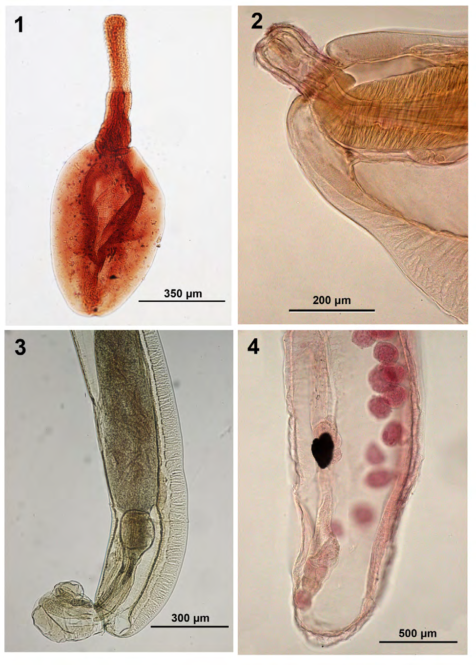
Figure 1-4: Morphology of acanthocephalans from wildlife.
Figure 1-4: Morphology of acanthocephalans from wildlife. 1. Cystacanth of Moniliformis kalahariensis from Blatella germanica in Bombay, India. Note the darker stained lemnisci. 2-4. Adult Neoechinorhynchus (Hebesoma) carinatus from Erimyzon tenuis in Louisiana. 2. Anterior end. 3. Male reproductive system with everted bursa and large cement gland. 4. Sub-ventral female reproductive system with uterine bell longer than uterus. Black blot is an artifact.
Class Eoacanthocephala
Family Neoechinorhynchidae Neoechinorhynchus (Hebesoma) carinatus Buckner and Buckner, 1993 (Figs. 2-4) Dr. Richard Buckner kindly provided 7 mature adults (5 females, 2 males) collected from the sharpfin chubsucker Erimyzon tenuis (Agassiz) (type host) in Finney Creek, Washington Parish, Louisiana (type locality) on July 20, 1974, almost 20 years before the original description in 1993. Amin [6] relegated this species to the subgenus Hebesoma Van Cleave, 1928 because of the polar prolongation of the egg fertilization membrane. Buckner and Buckner [7] found that 36 male and 36 female specimens from the type locality in Louisiana were markedly smaller in all structures than the 6 male and 6 female specimens measured from the same host species in Mississippi. We attribute this morphological disparity to geographical variation among two populations separated by about 350 km. The Louisiana specimens were collected from two rivers in the Portchartrain drainage basin and the Mississippi specimens were obtained from the Tchoutacabouffa River which is confluent with Biloxi Bay [7]. Geography-based morphological variations in the genus Neoechinorhynchus were previously noted in N. crassus Van Cleave, 1919 by Lynch [8], in N. cristatus Lynch, 1936 by Bullock [9], and in N. notemigoni Dechtiar, 1967 by Buckner [10]. Comparable geographically based morphological variations are known in other genera of acanthocephalans.
Our specimens appear to be a subset of the original population described from E. tenuis in Louisiana and their measurements were similar to those in Tables 1 & 2 in Buckner and Buckner [7]. However, we noted a few qualitative variations. The distance between middle and posterior hooks in our specimens was also much wider than shown in the original description (Fig. 2) [7]. The receptacle wall was much thicker in our specimens (Fig. 2). Male specimens were described with posterior testis usually in contact with cement gland (Buckner and Buckner, p. 32 & Fig. 1) [7] but that distance was much wider in our specimens. Otherwise, the male reproductive system (Fig. 3) was comparable. The female reproductive system (Fig. 4) was comparable to that shown by Buckner and Buckner (Fig. 3) [7] but additional information on the genital vestibule was provided [11]. Other features were, otherwise, comparable. Specimens: Deposited at the Harold W. Manter Laboratory (HWML) Parasitology Collection nos. 217495, 217496.
| Host | Distribution | |
|---|---|---|
| Class Archiacanthocephala | ||
| Family Moniliformidae | ||
| Moniliformis kalahariensis Meyer, 1931 cystacanth | Blattella (Phyllodromia) germanica Linn. | Bombay, India |
| Class Eoacanthocephala | ||
| Family Neoechinorhynchidae | ||
| Neoechinorhynchus (Hebesoma) carinatus Buckner and Buckner, 1993 | Erimyzon tenuis (Agassiz) | Louisiana |
| Neoechinorhynchus (Hebesoma) pungitius Dechtiar, 1971 | Not identified | Lake Huron, Canada |
| Neoechinorhynchus (Neoechinorhynchus) rutili (Müller, 1780) Hamann in Stiles and Hassall, 1905 | Leuciscus idus (Linn.) | Lake Baikal, Russia |
| Neoechinorhynchus (Neoechinorhynchus) tumidus Van Cleave and Bangham, 1949 | Coregonus sp. | Baunt Lake, Russia |
| Tanaorhampus longirostris Van Cleave, 1913 cystacanth | Diaptomus pallidus Herrick | Caesar Creek Lake, Ohio |
| Family Quadrigyridae | ||
| Acanthocephohalorhynchoides cholodkowskyi (Kostylew, 1928) Williams, Gibson, Sadighian, 1980 | Capoeta damascina (Valenciennes) | Mand River, Iran |
Table 1: Acanthocephala (Archiacanthocephala and Eoacanthocephala) reported from wildlife in this paper and their hosts and geogr
• Neoechinorhynchus (Hebesoma) pungitius Dechtiar, 1971 (Figs. 5-8) Alex Dechtiar kindly provided 4 specimens (2 males, 2 females) of N. (H.) pungitius probably from a nine-spine stickleback, Pungitius pungitius (Linn.) in Lake Huron, collected in 1969. Both are type host and type locality. Our specimens were similar to those described by Dechtiar [12] and the measurements of our specimens fit within those reported in the original description. The line drawings (Figs. 1-5 in Dechtiar) [12] were schematic and occasionally inaccurate. We have provided light microscope images of certain structures complementing the original description and correcting some of the line drawings of Dechtiar [12]. We are adding light microscope images of a whole male and a whole female showing their overall shape and a male reproductive system and lemnisci. The line drawings of males in Dechtiar (Fig. 2) [12] and Arai (Fig. 27C) [13] show testes in anterior trunk and inflated cement gland. Our specimens show detail of the convoluted lemnisci (Fig. 5) detail of the deltoid vagina and sphincters (Fig. 6), testes in the posterior field of trunk with less inflated cement gland (Fig. 7), and more defined posterior reproductive structures (Fig. 8). Specimens: Deposited at the Harold W. Manter Laboratory (HWML) Parasitology Collection no. 217497.
| Geography | Lake Sevan (Armenia) Lake Issyk-Kul (Kirgis) | River Zayande at Esfahan, Central Iran | Mand River, Fars, Southwest Iran | |||
|---|---|---|---|---|---|---|
| Authority | Petrochenko (1956) (after Kostylew, 1928) | Williams, Gibson, Sadighian (1980) | This paper | |||
| Hosts | Varicorhinus capoeta sevangi (Fillippi), Schizothorax pseudoaksaiensis issykkuli Berg & Diptychus dybowskii Kessler | Capoeta capoeta (Güldenstädt) Capoeta bushei Kessler & Other cyprinid hosts in other localities | Capoeta damascina Valenciennes | |||
| Sex | Males | Females | Males | Females | Males | Females |
| Sample size | Unknown | Unknown | 5 | 5 | 6 | 3 |
| Trunk L X W (mm) | 11-14 X 1.2-1.4 | 18-22 X 2.0* | 7.5-8.4 X 0.69- 0.8 | 10.5-12.8 X 0.61-0.8 | 6.50-10-00 X 0.42- 0.87 | 7.00-11.10 X 0.70-1.05 |
| Subcutaneous giant nuclei | 8 dorsal 2 ventral | 8 dorsal 2 ventral | --- | --- | 7-9 dorsal 1-2 ventral | 8-9 dorsal 1 ventral |
| Ant. trunk spines & spines / circle | 4-5 circles & 14-16 | 4-5 circles & 15-16 | 6-8 circles & 16 | 6-8 circles & 16 | Absent | Absent |
| Post. trunk spines & spines / row | Only ventral rows & 5-6 | Only ventral rows & 6-7 | 18 ventral rows | 18 ventral rows | Absent | Absent |
| Proboscis L X W | 135-160 X 132-145 | --- X --- | 150-160 X --- | 150-200 X --- | 104-125 X104-120 | 125-135 X114- 125 |
| Ant. Rooted hook L | 51-61 | Up to 77 | 63-70 | 65-74 | 37-43 | 43-52 |
| Middle hook L | 32-42 | --- | 52-58 | 57-63 | 33-36 | 38-42 |
| Basal hook L | 32 | --- | 34-38 | 32-42 | 31-36 | 32-40 |
| Receptacle L X W | 600-700 X --- | --- | --- | --- | 520-666 X135-177 | 489-676 X146- 177 |
| Long Lemniscus L X W (mm) | 3.0-5.0 X --- | 7.0 X --- | 2.8-3.1 X --- | 3.0-4.3 X --- | 2.55-3.65 X 0.15-0.28 | 2.37-3.60 X 0.14-0.24 |
| Short lemniscus | --- | --- | --- | --- | 2.45-3.52 X | 2.05-3.22 |
| L X W (mm) | 0.12-0.22 | 0.14-0.24 | ||||
| Nuclei in lemnisci | 3 | 3 | 3 | 3 | 3-4 & 2-3 | 3-4 & 3 |
| Ant. Testis L X W (mm) | --- | XXX | 0.75-1.00 X --- | XXX | 0.62-1.17 X 0.25-0.45 | XXX |
| Post. Testis L X W (mm) | --- | XXX | 0.75-1.00 X --- | XXX | 0.42 X 0.97 X 0.35- 0.40 | XXX |
| Cement gl. L X W & giant nuclei | Syncytial | XXX | Syncytial 6 giant nuclei | XXX | 525-1,050 X 250- 4506-7 giant nuclei | XXX |
| Cement reservoir L X W | --- | XXX | --- | XXX | 200-375 X 125-200 | XXX |
| Saefftigen’s pouch L X W | --- | XXX | --- | XXX | 625-1,000 X 100-175 | XXX |
| Sperm vesicle L X W | --- | XXX | --- | XXX | 625-1,100 X 175-250 | XXX |
| Bursa L X W | --- | XXX | --- | XXX | 500-825 X 375-500 | XXX |
| Female reproductive syst. L (mm) | XXX | --- | XXX | Vagina 0.29- 0.37 Uterus 0.30 long | XXX | 0.80-1.35 |
| Female gonopore | XXX | --- | XXX | Subterminal | XXX | Subterminal |
| Egg L X W | XXX | 33-37 X 19-23 | XXX | 25-31 X 13-14 | XXX | 22-28 X 11-13 |
Table 2: Comparative measurements of specimens of Acanthocephalorhynchoides cholodkowskyi from Armenia, Kirgiz, and Iran.
*Extreme measurements or states are bolded. Table 2: Comparative measurements of specimens of Acanthocephalorhynchoides cholodkowskyi from Armenia, Kirgiz, and Iran.
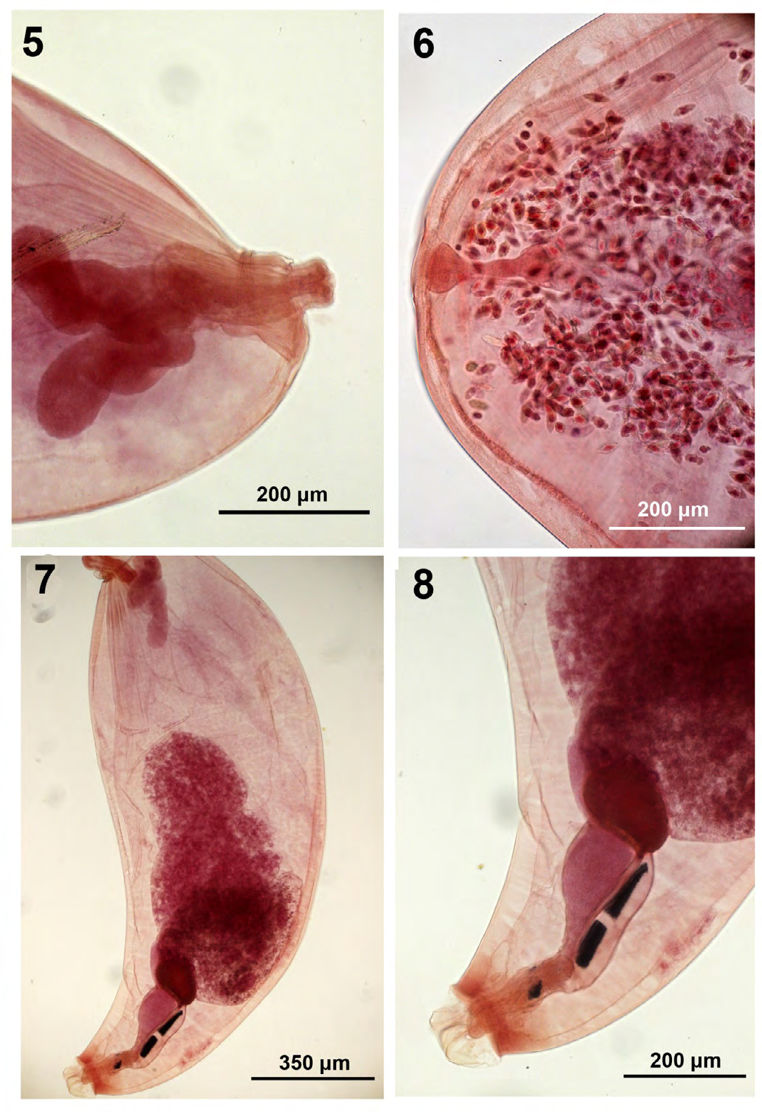
Figure 5-8: Morphology of acanthocephalans from wildlife. 5-8. Adults of Neoechinorhynchus (Hebesoma) pungitius from an unidentified host in Lake Huron, Canada. 5. Anterior end showing convoluted lemnisci. 6. Posterior end of a female showing the reproductive system with its deltoid vagina. 7. A male specimen showing the posteriorly placed testes compared to the anterior testes in the original description. 8. Detail of the male reproductive system past the testes. Saefftigen’s pouch is dorsal with black stain.
• Neoechinorhynchus (Neoechinorhynchus) rutili (Müller, 1780) Hamann in Stiles and Hassall, 1905 (Figs. 9-11) Two adult females in the ovarian ball stage collected from Leuciscus idus (Linn.) in northern Lake Baikal in 2015 were kindly provided by Dr. Darima R. Baldanova for study. Baldanova, et al. [14] described only one species in the genus Neoechinorhynchus Hamann in Stiles and Hassall, 1905 from Lake Baikal, N. rutili from L. idus and 3 other species of cyprinids from Lake Baikal and its basin including the littoral mouth of the River Selenga and reported it in other Palaearctic waters in Arctic Ocean, Baltic, Black, Aral, and Caspian seas, Turkmenistan, Western Mongolia province, and the Amur transitional region as well as from the Nearctic in Lavrentine Province. Van Cleave and Lynch [15] described N. rutili from North American populations and provided evidence of its continuous geographical circumpolar distribution in North America and Europe. Van Cleave and Lynch [15] listed 17 species in 8 families of fish hosts from Wisconsin, Washington, Alaska, and the Arctic circle of Canada in North America as well as from Northern Europe (Sweden, Finland, and Russia). Lisitsyna [16] described it from Ukraine and Petrochenko [17] listed 61 fish hosts from northern waters of former USSR basins as well as from Hungary, Switzerland, Germany, and North America. In the Nearctic, Amin [18] also reported N. rutili from the brook stickleback Culaea inconstans (Kirtland) in Tichigan Lake Canal, Wisconsin in June 1981 and Arai [13] provided a description based on Van Cleave and Lynch [15] and reported it from 38 species of fish from Canada in various localities in British Columbia, Ontario, the Pacific and Atlantic, Newfoundland, New Brunswick, North-West Territories, and Manitoba. In the Palearctic, Amin and Gunset [19] studied the pattern of giant nuclei in 300 specimens of N. rutili collected by Dr. F. Moravec between 1966 and 1987 from Cyprinus carpio Linn. in Mácha Lake, northern Bohemia, Czech Republic. We shall not list any further records of N. rutili from various hosts in the Palearctic and the Nearctic confirming the pattern of circumpolar distribution of N. rutili as originally proposed by Van Cleave and Lynch.
Our two female specimens from Lake Baikal had prominent giant subcutaneous nuclei (5 dorsal and 1 ventral) (Fig. 9) and measured 5.25-8.50 mm long by 0.65- 1.02 mm wide. Proboscis with prominent apical organ, 146 by 135. Rooted anterior hook (Fig. 10), rootless middle, and basal hooks 82-87, 42-45, 40-47 long, respectively; middle and basal hooks equal. Receptacle 364-395 by 112-114 with large triangulate cephalic ganglion at base. Lemnisci (Fig. 9) subequal, 3.25-3.50 mm long by 0.15 mm wide consistent in both unequal size females. Gonopore terminal (Fig. 11). These two females fit in the maximum range in females measured by Baldanova from Lake Baikal (hooks: 60-80, 30-40, 20-40 & longer lemniscus 0.74-5.24 mm long). The female specimens measured by Van Cleave, however, had slightly smaller proboscis (90-132 X 93-132) and smaller hooks (64-84, 31-46, 22-34) with basal hooks being smaller than middle hooks. Specimens: Deposited at the Harold W. Manter Laboratory (HWML) Parasitology Collection no. 217498.
• Neoechinorhynchus (Neoechinorhynchus) tumidus Van Cleave and Bangham, 1949 (Figs. 12-14) Two gravid females collected from one specimen of Coregonus sp. in Baunt Lake in 2015 were kindly provided by Dr. Darima R. Baldanova for study. Baldanova and Pronin (2002) did not include this species among the acanthocephalans reported in Lake Baikal. Baunt Lake (55°12’20”N,112°59'11"E) is a large 33 m deep lake in the northern Transbaikal Region. Neoechinorhynchus tumidus was originally described in North America from Coregonus artedi (LeSueur) in Trout Lake, Wisconsin and in Waskesin Lake, Saskatchewan, Canada as well as from Coregonus clupeaformis (Mitchill) of Waskesin Lake by Van Cleave and Bangham [20]. It was subsequently reported from additional Canadian locations by Arai [13] as well as from Coregonus spp. in the Pechora and Ob` basins of the USSR [17] and in unspecified Russian locations [21]. Our record from Baunt Lake, thus represents a new geographical record. The finding of N. tumidus, like the finding of N. rutili, (above), represents another case of circumpolar distribution of another species of Neoechinorhynchus between North America and Europe which has never been previously reported as such.
Our two female specimens from Baunt Lake were enlarged anteriorly (Fig. 12) with 5 dorsal and 1 ventral giant subcutaneous nuclei and measured 9.50-11.30 mm long by 1.77-2.02 mm wide anteriorly. Proboscis with prominent apical organ (Fig. 13) measuring 146-166 by 177- 208. Rooted anterior and middle hooks almost equal, 70 & 68 long, and rootless basal hooks 45-52 long. Receptacle 520- 530 by 198-218 with large triangulate cephalic ganglion at base. Lemnisci equal-subequal, 2.92 mm long by 0.19 mm wide. Gonopore distinctly subterminal (Fig. 14). Eggs 30-32 by 15-18. These two females had comparable measurements to those measured by Van Cleave and Bangham [20], Petrochenko [17], and Arai [13] but nearer to the upper part of the range. Unlike females drawn by Van Cleave and Bangham [20] and Petrochenko [17], Fig. 24 in Arai [13] showed an uninflated female trunk anteriorly and a terminal gonopore even though he described it as subterminal (p. 47). Specimens: Deposited at the Harold W. Manter Laboratory (HWML) Parasitology Collection no. 417499.
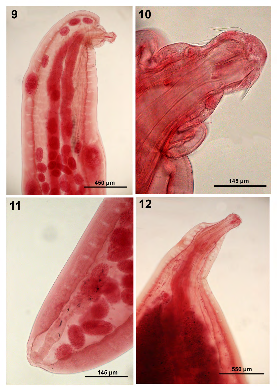
Figure 9-12: Morphology of acanthocephalans from wildlife. 9-11. Neoechinorhynchus (Neoechinorhynchus) rutili from Leuciscus idus in Lake Baikal, Russia. 9. Anterior portion of a female specimen showing the long lemnisci and large subcutaneous giant nuclei. 10. Proboscis with long apical hooks. 11. Detail of female reproductive system. 12. Anterior part of a female specimens of Neoechinorhynchus (Neoechinorhynchus) tumidus from Coregonus sp. in Baunt Lake, Russia. Note the triangulate cephalic ganglion at base of receptacle.
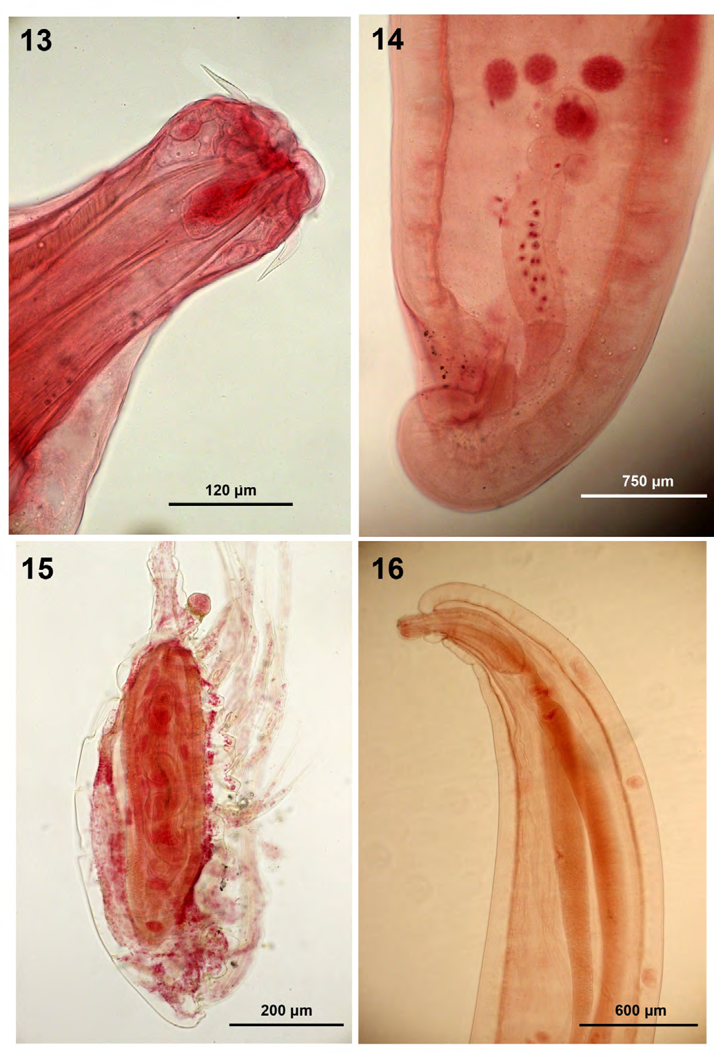
Figures 13-16: Morphology of acanthocephalans from wildlife. 13,14. female specimens of Neoechinorhynchus (Neoechinorhynchus) tumidus from Coregonus sp. in Baunt Lake, Russia. 13. Proboscis with long anterior hooks and prominent apical organ. 14. Detail of the female reproductive system showing the characteristic subterminal gonopore. 15. A cystacanth of Tanaorhampus longirostris in the body cavity of Diaptomus pallidus in Caesar Creek Lake, Ohio. 16. Adults of Acanthocepohalorhynchoides cholodkowskyi from Capoeta damascina in Mand River, Iran. A specimen showing the relative size of the proboscis to the anterior part of the trunk housing the long lemnisci and some of the dorsal giant subcutaneous nuclei.
• Tanaorhampus longirostris Van Cleave, 1913 cystacanth (Fig. 15) One cystacanth of T. longirostris in the body cavity of one crustacean specimen of Diaptomus pallidus Herrick collected from Caesar Creek Lake in Ohio by Dr. Jerry H. Hubschman on July 18, 1981 was graciously gifted to us for further study. A description of this find was published in Hubschman [3] along with 3 figures, shortly thereafter. Specimens of T. longirostris were collected from a number of fish species but mature adults were found only in Gizzard shad, Dorosoma cepedianum (Lesueur) in July. Our specimen was fixed in formalin, stained in Semichons, and whole-mounted in Permount and our observations expand on those reported by Hubschman [3]. The relationship between T. longirostris cystacanth and the crustacean intermediate host D. pallidus is best seen in Figure 15.
Specimen: Deposited at the Harold W. Manter Laboratory (HWML) Parasitology Collection no. 217500.
Class Eoacanthocephala
Family Quadrigyridae • Acanthocephalorhynchoides cholodkowskyi (Kostylew, 1928) Williams, Gibson, Sadighian, 1980 (Figs. 16-20) This acanthocephalan was originally described as Quadrigyrus cholodkowskyi by Kostylew from the intestine of Varicorhinus capoeta sevangi (De Filippi) from Lake Sevan in Armenia. This assignment to Quadrigyrus Van Cleave, 1920 was accepted by Meyer [22]. The uncertain organization of the 18 hooks on the proboscis also noted by Williams et al. [23] apparantly prompted Kostylew (Fig. 2) [24] to include a fourth posterior hook to the proboscis armature. It was later reassigned to Acanthogyrus (Acanthosentis) Verma and Datta [25] and to Pallisentis Van Cleave, 1928 by Amin [26] who later on [27] reassigned it to Acanthocephalorhynchoides. It was erroneously relegated to the genus Neoechinorhynchus by Dinnik [28] and Mikailov [29]. The organization of trunk spines in one anterior trunk zone with anterior rings and extended posterior ventral extension precludes its relegation to Acanthogyrus which has only a few anterior spine circles or to Pallisentis with two separate sets of circular trunk spines. Williams, et al. [23] and Schmidt and Huggins [30], made a good case of assigning this species to Acanthocephalorhynchoides Kostylew. Williams, et al. [23] noted records of A. cholodkowskyi outside of Armenia in Azerbaijan and Georgia and east to Kazakhstan, Kyrgyzstan and Uzbekistan.
Our adult specimens (6 males, 3 females) were collected from Capoeta damascina in the Mand River, Fars, south- western Iran draining in the Persian Gulf, in 1997 by Coed representing a new host and locality records. The Williams, et al. [23] specimens were also collected in Iran between 1972 and 1975 from a distant location in central Iran, the River Zayande, which is a part of a land-locked system draining in a seasonal salt marsh in Esfahan.
Our specimens are distinguished by the absence of trunk spines and the presence of 7-9 dorsal and 1-2 ventral giant subcutaneous nuclei (Figs. 16,17), 18 small and atypically arranged proboscis hooks (Fig. 18), thick-walled receptacle with large cephalic ganglion at its base, long and thick unequal lemnisci (Fig. 16) not reaching testes with 3-4 and 2-3 giant nuclei (Fig. 17) in the longer and shorter lemniscus, respectively, and most nuclei at the anterior end of each lemniscus (Fig. 17), two contiguous post-equatorial testes with the anterior being larger than the posterior, large cement gland (Fig. 19) with 6-7 large oval giant nuclei, long Saefftigen’s pouch occasionally extending into the bursa, Females with uterine wall thinner than that of vagina and with clearly subterminal gonopore (Fig. 20), and eggs small, ovoid, with concentric shells. See Table 2 for details.
Specimens: Deposited at the Harold W. Manter Laboratory (HWML) Parasitology Collection no. 217501.
Remarks
Our specimens are compared with those of Williams, et al. [23] also from Iran and those of Kostylew [24] as supplemented by Petrochenko [17]. Differences are detailed in Table 2, with many new characters that we introduce for the first time from our research of our Mand River specimens. Specimens in the original description from Armenia were unusually large with females reaching 22.00 mm long by 2.00 mm wide having lemnisci as long as 7.0 mm long and eggs as large as 33-37 by 19-23 (Table 2). New features in our Mand River specimens in Iran include the absence of trunk spines. Williams, et al. (p. 690) [23] remarked that trunk spines “are apparently lost in some specimens.” We observed specimens with 3-4 and 2-3 giant nuclei in the longer vs. the shorter lemnisci. Kostylew [24] and Williams, et al. [23] noted 3 and 3 nuclei but Petrochenko’s (p. 244) [17] original specimens included 4 and 3 nuclei (Fig. 88e) like some of ours. Kostylew [24] noted 8 dorsal and 2 ventral giant subcutaneous nuclei but we noted a wider diversity of 7-9 (usually 8) dorsal and 1-2 (usually 1) ventral nuclei. Williams, et al. [23] counted 6 giant nuclei in the cement gland but we have seen 7 in some specimens.
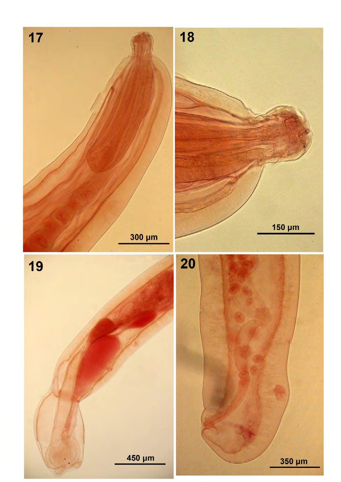
Figure 17-20: Morphology of acanthocephalans from wildlife. 17-20. Adults of Acanthocepohalorhynchoides cholodkowskyi from Capoeta damascina in Mand River, Iran. 17. Proboscis, receptacle with posterior cephalic ganglion, and anterior part of lemniscus with 4 giant nuclei in one specimen. 18. A proboscis showing the relative size of hooks of various circles. 19. Detail of a male reproductive system with everted bursa. 20. Detail of a female reproductive system showing the subterminal gonopore and the comparative length of the uterine bell vs. the uterus.
Discussion
We have collected thousands of parasitic organisms from vertebrates over the years as part of an overall research program from North America and elsewhere in the world. We have recently described and illustrated acanthocephalans of two classes, Archiacanthocephala and Eoacanthocephala, and noted their host and geographical distribution [2]. In this presentation, the relationship between cystacanths of two acanthocephalan species and their hosts; Moniliformis kalahariensis (Moniliformidae) in Blattella germanica from India and Tanaorhampus longirostris (Neoechinorhynchidae) in the copepod Diaptomus pallidus from Ohio were best illustrated by light microscopy providing additional perspectives than have been previously described by Amin, et al. [5] and Hubschman [3], respectively. Specimens of 4 species of the genus Neoechinorhyncus from North America and Russia are described. Neoechinorhynchus carinatus from Louisiana, USA provided an opportunity to document geography-based morphological variations and distinct morphological dissimilarities were noted between our specimens and those in the original description from the same locality. The line drawings of N. pungitius males in Dechtiar [12] and Arai [13] show anterior testes and inflated cement gland but our specimens from the same host and locality show posterior testes, less inflated cement glands and more defined posterior reproductive structures. The circumpolar distribution of N. rutili originally outlined by Van Cleave and Lynch [15] in North America and Europe has been reconfirmed by our reporting it from Lake Baikal, Russia. We have also found significant morphometrical variability between our specimens and those described by Van Cleave and Lynch [15] and by Baldanova and Pronin [14]. We have also found that N. tumidus that we examined from Baunt Lake, Russia, and that was described from the USA [20] does exhibit circumpolar distribution like N. rutili. It has similarly unusual diagnostic features and new host and geographical distributions are now reported for the first time.
Another eoacanthocephalan of the family Quadrigyridae discussed in this presentation is A. cholodkowskyi from Iran and compared with another Iranian population [23] and that from other populations from Armenia [24] and Russia [17]. Our comparative morphometrical data suggest that A. cholodkowskyi is quite a variable species with some of the measurements of our Mand River specimens exceeding the range of those reported by other observers. Our new geographical and host record from C. damascina give additional perspectives of the distribution of this species.
Acknowledgments
This project was partially supported by an ongoing institutional grant from the Parasitology Center, Inc., Scottsdale, Arizona, USA. We are immensely grateful to the many collaborators who contributed the study materials that made the execution of this work possible. Collaborators simply shared research material as part of joint research understandings. We also gratefully recognize the help of Dr. Gabor Racz., Collection Manager, Harold W. Manter Laboratory of Parasitology, University of Nebraska-Lincoln for kindly accessing and cataloging our specimens.
References
-
Amin OM, Rubtsova NY (2023a) New Annotated Records of Helminth Parasites, Mostly from North America, with Light Microscopy. I. Trematoda (Digenea), Cestoda, Nematoda. Int J Zoo Animal Biol 6(5): 000507.
-
Amin OM, Rubtsova NY (2023b) New annotated records of helminth parasites with light microscopy II. Acanthocephala (Polymorphida). Int J Zoo Animal Biol 6(6): 000530.
-
Hubschman J H (1983) _Diaptomus pallidus_ Herrick, 1879 (Crustacea: Copepoda) as an intermediate host for _Tanaorhamphus longirostris_ (Van Cleave, 1913) (Acanthocephala: Neoechinorhynchidae). J Parasitol 69 (5): 930-932.
-
Meyer A (1931) Neue Acanthocephalen aus dem Berliner Museum. Burgründung eines neue Acanthocephalen systems auf Grund einer Untersuchung der Berliner Sammlung. Zool Jahrbücher Abteilung Syst, Ökologie Geograph Tiere 62: 53-108.
-
Amin OM, Heckmann RA, Halajian A, El-Naggar A, Tavakol S (2014) Description of _Moniliformis kalahariensis_ (Acanthocephala: Moniliformidae) from the South African Hedgehog, _Atelerix frontalis_ (Erinaceidae) in South Africa. Comp Parasitol 81 (1): 33-43.
-
Amin OM (2002) Revision of _Neoechinorhynchus_ Stiles and Hassall, 1905 (Acanthocephala: Neoechinorhynchidae) with keys to 88 species in two subgenera. System Parasitol 53: 1-18.
-
Buckner RL, Buckner SC (1993) Description of _Neoechinorhynchus_ _carinatus_ n. sp. (Acanthocephala: Neoechinorhynchidae) from the sharpfin chubsucker, _Erimyzon tenuis_, of Louisiana and Mississippi. J Parasitol 79 (1): 32-36.
-
Lynch LE (1936) New species of _Neoechinorhynchus_ from the western sucker, _Catostomus_ _macrocheilus_ Girard. Trans Amer Microsc Soc 55: 21-43.
-
Bullock WL (1955) The occurrence of _Neoechinorhynchus_ _cristatus_ Lynch, 1936 (Acanthocephala) in eastern North America. J Parasitol 41: 323-324.
-
Buckner RL (1983) Occurrence of two species of _Neoechinorhynchus_ (Acanthocephala) in golden shiners of Alabama and Mississippi. Proc Helminthol Soc Wash 50: 176-178.
-
Oetinger DF, Buckner RL (1993) Morphology of the genital vestibule of _Neoechinorhynchus_ _carinatus_ (Acanthocephala: Neoechinorhynchidae). J parasitol 79(6): 930-934.
-
Dechtiar AO (1971) _Neoechinorhynchus pungitius_ n. sp. (Acanthocephala: Neoechinorhynchidae) from ninespine stickleback of Lake Huron. Canad J Zool 49: 483-486.
-
Arai HP (1989) Acanthocephala, p 1-90. _In_ L. Margolis and Z. Kabata eds.). Guide to the Parasites of fishes of Canada. Part III. Can Spec Publ Fish Aquat Sci 107: 95.
-
Baldanova DR, Pronin NM (2001) Acanthocephalans (Phylum Acanthocephala) of Baikal. Morphology and ecology. NAUKA, Novosibirsk, Russia, pp: 157.
-
Van Cleave HJ, Lynch JE (1950) The circumpolar distribution of _Neoechinorhynchus_ _rutili_, an acanthocephalan parasite of fresh-water fishes. Trans Amer Micros Soc 69: 156-171.
-
Lisitsyna OI (2019) Fauna of Ukraine. Acanthocephala. NVP “Naukova Dumka Ukrainy” 31: 223.
-
Petrochenko VI (1956) Acanthocephala of Domestic and Wild Animals, I, Moscow, Acad Sci, pp., 465.
-
Amin OM (1986) Acanthocephala from lake fishes in Wisconsin: Host and seasonal distribution of species of the genus _Neoechinorhynchus_ Hamann, 1892. J Parasitol 72(1): 111-118.
-
Amin OM, Gunset M (1992) The pattern of giant nuclei in _Neoechinorhynchus rutili_ (Acanthocephala: Neoechinorhynchidae). Trans Amer Micros Soc 111 (1): 65-69.
-
Van Cleave HJ, Bangham RV (1949) Four new species of the acanthocephalan family Neoechinorhynchidae from fresh-water fishes of North America, one representing a new genus. J Wash Acad Sci 39 (12): 398-409.
-
Yamaguti S (1963) Acanthocephala. In Systema Helminthum. Wiley Interscience, New York, USA, 5: 1-423.
-
Meyer A (1932-1933) Acanthocephala. Dr. H.G. Bronns, Klassen und Ordnungen Tier-Reichs, Leipzig, Bd. 1-332, 333-582.
-
Williams JS, Gibson DI, Sadighian A (1980) Some helminth-parasites of Iranian freshwater fishes. J Natur Hist 14 (5): 685-699.
-
Kostylew NN (1928) Acanthocephalen der Fische der Goktscha-Sees. Centr Bakteriol, Parasit Infekt. Abteil I Orig 108: 146-150.
-
Golvan YJ (1959) Le phylum des Acanthocephala. Deuxième note. La Classe des Eoacanthocephala (Van Cleave, 1936), Ann Parasitol Hum Comp. Paris, USA, 34: 5-52.
-
Amin OM (1985) Classification. In Biology of the Acanthocephala. In: Crompton DWT, Nickol BB (Eds.), Cambridge University Press, London, UK, pp: 27-71.
-
Amin OM, Heckmann RA, Ha NV, Luc PV, Doanh PN (2000) Revision of the genus _Pallisentis_ (Acanthocephala: Quadrigyridae) with the erection of three new subgenera, the description of _Pallisenti_s (_Brevitritospinus_) _vietnamensis_ subgen. et sp. n., a key to species of _Pallisentis_, and the description of a new quadrigyrid genus, _Pararaosentis_. Comp Parasitol 67(1): 40-50.
-
Dinnik JA (1933) The parasitic worms of fish from Lake Sevan, Trudy Sevanskoi Ozernoi Stantsii 4: 105-128.
-
Mikailov TK (1975) Parasites of fish in the reservoirs of Azerbaydzhan (systematics, dynamics and origin) (Baku: Elm), pp: 297.
-
Schmidt GD, Hugghins EJ (1973) Acanthocephala of South American fishes, Part I, Eoacanthocephala, J Parasitol 59: 829-835.
- Mitochondrial Bio-Logistics: Steering Co-Enzyme Q10 and Lycopene Synergies within the Science 4.0 Bio-OS Framework
- Hymenoptera Specimens from the Caño Negro Wetland, of the National Museum Collection, Costa Rica
- Science 4.0: Comprehensive Architecture of the Biological Operating System (Bio-OS) A Framework for Systemic Resilience and Industrialized Bio-Governance
- Rabbit on, or Hare Back? Understanding Climate Change
- Clinical Validation of Science 4.0: Flow Steering and Epigenetic Drift Inversion on a 76-Year-Old Hybrid System
- Seeds Planted by another Mind