A Case Report of Association between Canine Hypothyroidism and Diabetes
Hypothyroidism is the most common hormonal disorder in dogs because of the low production of the hormones thyroxine (T4) and triiodothyronine (T3) by the thyroid gland. The main cause of hypothyroidism in adult dogs is primary hypothyroidism associated with destruction of the thyroid gland in lymphocytic or autoimmune thyroiditis, followed by idiopathic thyroid degeneration or idiopathic follicular atrophy. Dogs affected by hypothyroidism may develop diabetes mellitus due to the propensity for the development of other endocrine diseases associated with the immune-mediated attack against cellular antigens associated with endocrine system. In this case report, an eleven-year-old male Lhasa Apso was presented to the clinical facility with signs of apathy, anorexia and non-pruritic bilateral symmetric alopecia that extended to the tip of the tail but sparing the head and limbs. Other clinical manifestations included polyuria, polydipsia, and bilateral cataracts. The animal was submitted to the blood count, T4, T3, TSH dosage, biochemical tests, and imaging tests (cervical and abdominal ultrasonography). Exam results demonstrate the presence of hypothyroidism and diabetes mellitus. Since hypothyroidism corresponds to the most common endocrine disorder in dogs, and they are prone to the development of diabetes mellitus, the veterinarian must be aware of the clinical manifestations of these diseases.
Introduction
The canine hypothyroidism occurs due to a decrease in the production of thyroxine (T4) and triiodothyronine (T3) by the thyroid gland, leading to a metabolic multisystem disorder. Hypothyroidism can be classified as primary, secondary, tertiary [1]. Primary hypothyroidism occurs in 95% of clinical cases due to the destruction of thyroid glandular tissue by lymphocytic thyroiditis, idiopathic follicular atrophy, thyroidectomy, and thyroid tumors [2].
The secondary hypothyroidism accounts for 5% of cases. by the decrease in TSH secretion by the pituitary gland, causing secondary follicular atrophy, due to the presence of pituitary tumors, pituitary malformations and isolated TSH deficiency [1]. The tertiary hypothyroidism is extremely rare, being caused by a deficiency in the release of TRH produced by the hypothalamus, and may be associated with malformation of the hypothalamus, neoplasms, abscesses, or inflammation [2].
Canine hypothyroidism is most frequently reported in middle-aged animals, between 4 and 10 years old, castrated and of both sexes. The dog breeds with the greatest predisposition are Golden Retriever, Doberman Pinscher, Dachshund, Irish Setter, Miniature Schnauzer, Poodle, Boxer, Labrador, Cocker Spaniel, Poodle and Shetland Shepherd [1, 3].
Dogs with hypothyroidism may be prone to polyendocrine syndromes, such as those associated with diabetes mellitus, due to the immune-mediated attack against endocrine system antigens. Apparently this attack is related to the dog’s immunogenetic status in association with environmental factors [1, 4].
Case Report
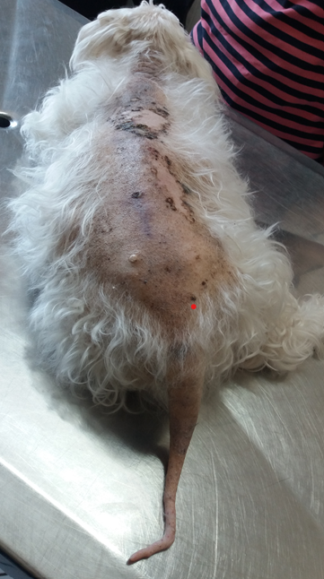
In November 2023, an animal of the canine species, Lhasa Apso breed, eleven-year-old, male, neutered and vaccinated, was attended. The owner reported that the animal presented apathetic behavior, ate a lot and spent most of the day sleeping, being reluctant to exercise and play. It also reported that the dog had polydipsia, polyuria and progressive blindness. On the day of consult, he had no reaction to sound stimuli, was unable to move and had shallow breathing. During inspection, the dog presented an alopecic area that covered the dorsal region of the trunk up to the insertion of the tail, and which extended to distal end (Figure 1).
A complete blood count, measurement of alanine aminotransferase (ALT), alkaline phosphatase (AF), glucose and cholesterol, thyroxine (free T4), TSH, type I urine, abdominal ultrasound and doppler echocardiography were performed.
In the blood count, a smaller number of erythrocytes (5.0 million/mm3; normal values=5.7-7.4 million/mm3) and leukocytes (5.7 thousand/mm3; normal values=6.0-16, 0 mil/mm3) and hematocrit (35%; normal=38.0-47.0%) was observed.
The dosages of ALT (158 U/L; normal values=9-92 U/L), FA (168; normal values=10-155 U/L), glucose (392 mg/ dL; normal=60-118 mg/ dL) and cholesterol (382 mg/dL; normal=135-270 mg/dL) and TSH by chemiluminescence (0.93 ng/mL; normal=0.05-0.40 ng/mL) were elevated. On the other hand, serum levels of free T4 by dialysis (0.26 ng/ dL; normal=0.80-3.50 ng/dL) were below normal values for canine species.
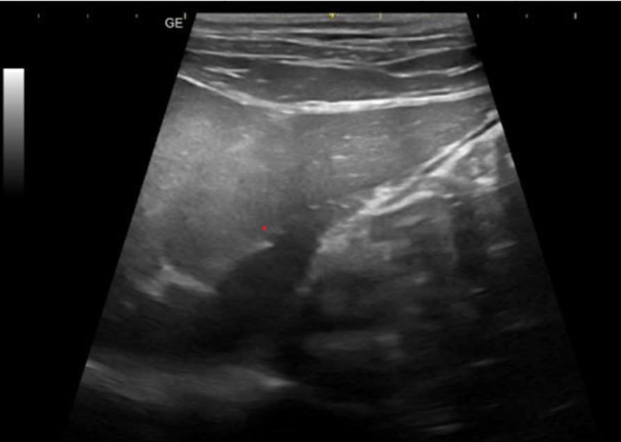
Type I urinalysis was performed by collecting urine through a urethral probe, and the biochemical examination showed the presence of glucose (++++ or 100 mg/dL), ketone bodies (+++ or 150 mg/dL) and the presence of occult blood (+++ or 50 erythrocytes/μL). In sedimentoscopy, erythrocytes (50; normal=1-2/field), leukocytes (5-8; normal=1-2/field) and bacteria (+; normal=absent) were observed. Abdominal ultrasound revealed hepatosplenomegaly compatible with inflammatory disease and liver changes suggestive of hepatic steatosis (Figure 2).
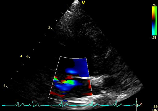
During auscultation, the dog had a heart rate of 55 beats per minute (normal values = 60-160 beats/minute). The Doppler echocardiogram and color flow mapping showed the presence of mild mitral insufficiency (Figure 3).
Discussion
In adult dogs, 60% of cases of hypothyroidism are associated with autoimmune disease. Large dogs, as well as neutered males and females, are at greater risk for developing hypothyroidism [3, 5]. In this case report, the patient was a small and neutered dog. Dogs with hypothyroidism show clinical signs of lethargy, intolerance and reluctance to exercise, and a tendency to gain weight without an increase in appetite. Disease progression leads to obesity, lethargy and dermatological changes [1, 2]. The dog in this case report was apathetic, ate a lot and spent most of the day sleeping, being reluctant to exercise and play.
Sixty to 80% of dogs with hypothyroidism present with cutaneous manifestations; The most commonly observed dermatological signs are weakened hair, generally bilateral and symmetrical alopecia affecting the body, ventral thorax and tail. It may also present peeling of the skin, dry or old seborrhea, superficial pyoderma, hyperkeratosis, hyperpigmentation, comedo formations, ceruminous otitis and myxedema [1]. The dog reported in this case had an alopecic area from the dorsal region of the body to the insertion of the tail, which extended to its tip, giving a “rat tail” appearance. Tail alopecia, called “rat tail” is a common dermatological sign of this disease [6].
Other clinical signs include generalized neuropathies or myopathy, megaesophagus, central nervous system abnormalities, dwarfism, reproductive abnormalities, diabetes mellitus, ocular abnormalities, cardiovascular abnormalities, stupor with myxedema, and coma [7, 8].
Dogs with hypothyroidism may be prone to polyendocrine syndromes, with this disease being associated with other endocrinopathies, such as diabetes mellitus [9, 10, 11].
Through blood glucose and urine chemistry, it was possible to confirm that the dog also had diabetes mellitus, which justified the polyphagia, polyuria and polydipsia signals. The definitive test for diagnosing hypothyroidism is serum TSH measurement, as increased levels of this hormone are confirmatory, regardless of T3 and T4 values [12].
When measuring hormones produced by the thyroid, the free T4 is more reliable; because it is free in plasma, it can maintain its concentrations even if there are variations in the release and metabolism of total T3 and T4 or in the concentration of plasma proteins [2, 12]. In the dog under study, TSH values by chemiluminescence were elevated, and serum levels of free T4 by dialysis were below normal values for the canine species.
As thyroid hormones influence the catabolism of adipose tissue and regulate the synthesis and degradation of cholesterol, dogs with hypothyroidism present obesity and hypercholesterolemia, such as observed in this case report. Obesity can occur in 41% of dogs with hypothyroidism, and fasting hypercholesterolemia occurs in up to 75% of cases [1, 13]. However, other diseases such as diabetes mellitus and hyperadrenocorticism can also cause an increase in serum cholesterol levels [14].
The literature reports that 30% of dogs with hypothyroidism have moderate non-regenerative anemia, as seen in this case report. It is common for dogs with hypothyroidism to present anemia related to physiological adaptation to decreased oxygen demand or associated with erythroid differentiation and interference with erythropoietin synthesis [1, 6].
Less common changes include discrete increases in serum FA and ALT levels, related to lipid metabolism disorder in the liver and disease-induced myopathy Strey S, et al. [1] and Bolton TA, et al. [15], as described in the liver changes observed in this case report.
Bradycardia is a common finding in dogs with hypothyroidism, since thyroid hormones exert inotropic and chronotropic effects on the heart Debmalya S, et al. [16] which is in accordance with what was observed in the animal in this study. Abnormalities such as sinus bradycardia, weak apical beat, low QRS voltage and inverted T waves can also be observed in this disease [17]. However, in the dog under study, a Doppler echocardiogram was performed, with the observation of mild mitral insufficiency.
Conclusion
The hypothyroidism corresponds to the most common endocrine disorder in dogs, which are prone to polyendocrine syndromes, with the possibility of developing diabetes mellitus. Therefore, the veterinary clinician must be aware of the clinical manifestations of this disease, and it is necessary to carry out additional tests to determine the correct diagnosis and the presence of other endocrinopathies, so that the correct treatment can be instituted.
References
-
Strey S, Mischke R, Rieder J (2021) Hypothyroidism in dogs: an overview. Prax Ausg K Kleintiere Heimtiere 49(3): 195-205.
-
Bugbee A, Rucinsky R, Cazabon S, Kvitko-White H, Lathan P, et al. (2023) 2023 AAHA selected endocrinopathies of dogs and cat’s guidelines. J Am Anim Hosp Assoc 59(3): 113-135.
-
O`Neill DG, Khoo JSP, Brodbelt DC, Church DB, Pegram C, et al. (2022) Frequency, breed predispositions and other demographic risk factors for diagnosis of hypothyroidism in dogs under primary veterinary care in the UK. Canine Med Genet 9(1): 11.
-
Ringstad NK, Lingaas F, Thoresen SI (2022) Breed distributions for diabetes mellitus and hypothyroidism in Norwegian dogs. Canine Med Genet 9(1): 9.
-
Bucalo O, Satué K, Medica P, Cravana C, Fazio E (2023) Thyroid evaluation in suspicious hypothyroid adult dogs before and after treatment. Pol J Vet Sci 26(1): 99-108.
-
Scott-Moncrieff JC (2007) Clinical signs and concurrent diseases of hypothyroidism in dogs and cats. Vet Clin North Am Small Anim Pract 37(4): 709-722.
-
Violette NP, Ledbetter EC (2017) Intracorneal stromal hemorrhage in dogs and its associations with ocular and systemic disease: 39 cases. Vet Ophtalmol 20(1): 27-33.
-
Bertalan A, Kent M, Glass E (2013) Neurologic manifestations of hypothyroidism in dogs. Compend Contin Educ Vet 35(3): E2.
-
Hwang SY, Na JH, Lee JH, Park SM, Kyu Chae H, et al. (2021) Autoimmune polyendocrine syndrome with hypoadrenocorticism and diabetes mellitus in a dog: A rare case. Vet Med Sci 7(6): 2120-2123.
-
Radosta L (2024) Behavior changes associated with metabolic disease of dogs and cats. Vet Clin North Am Small Anim Pract 54(1): 17-28.
-
Strey S, Mischke R, Rieder J (2023) Polyendocrine syndrome in dogs. Tierarztl Prax Ausg K Kleintiere Heimtiere 51(5): 313-325.
-
Lathan P (2023) Laboratory diagnosis of thyroid and adrenal disease. Vet Clin North Am Small Anim Pract 53(1): 207-224.
-
Sieber-Ruckstuhl NS, Tham WK, Baumgartner F, Selva JJ, Wenk MR, et al. (2022) Serum lipidome signatures of dogs with different endocrinopathies associated with hyperlipidemia. Metabolites 12(4): 306.
-
Miceli DD, Pignataro OP, Castillo VA (2017) Concurrent hyperadrenocorticism and diabetes mellitus in dogs. Res Vet Sci 115: 425-431.
-
Bolton TA (2021) Acute hepatopathy in a dog secondary to hypothyroidism-induced atherosclerosis infarction and necrosis. J Am Anim Hosp Assoc 57(1): 47-50.
-
Debmalya S, Saumitra R, Singh MH (2022) Interplay between cardiovascular and thyroid dysfunctions: a review of clinical implications and management strategies. Endocr Regul 56(4): 311-328.
-
Guglielmini C, Berlanda M, Fracassi F, Poser H, Koren S, et al. (2019) Electrocardiographic and echocardiographic evaluation in dogs with hypothyroidism before and after levothyroxine supplementation: a prospective controlled study. J Vet Intern Med 33(5): 1935-1942.
- Mitochondrial Bio-Logistics: Steering Co-Enzyme Q10 and Lycopene Synergies within the Science 4.0 Bio-OS Framework
- Hymenoptera Specimens from the Caño Negro Wetland, of the National Museum Collection, Costa Rica
- Science 4.0: Comprehensive Architecture of the Biological Operating System (Bio-OS) A Framework for Systemic Resilience and Industrialized Bio-Governance
- Rabbit on, or Hare Back? Understanding Climate Change
- Clinical Validation of Science 4.0: Flow Steering and Epigenetic Drift Inversion on a 76-Year-Old Hybrid System
- Seeds Planted by another Mind