A Case Report of Primary Bone Tumor with Metastasis in Lung and Liver
Primary bone tumors in dogs have a low frequency, however, among bone neoplasms, osteosarcoma accounts for more than 80% of malignant bone neoplasms, the occurrence of which is higher in dogs aged 5 to 9 years, mainly affecting large breed animals. Osteosarcoma has an aggressive biological behavior both at the site of growth and in the formation of metastases, being characterized by the proliferation of malignant primitive mesenchymal cells, with osteoblastic differentiation that produces bone matrix called osteoid or immature bone. Affected animals present pain, lameness, and increased volume of the affected area. The diagnosis is based on history, physical, laboratory and radiographic examination, the latter characterized by the presence of areas of bone destruction and new formation, periosteal reaction and increased volume of soft tissues. Diagnostic confirmation is carried out through histopathological examination. Since it mainly affects the appendicular skeleton, treatment includes surgery to amputate the affected limb, chemotherapy and radiotherapy. In this case report we describe an animal that suffered a tibial fracture after being run over and underwent surgery, with subsequent bone growth of a malignant nature and metastases to the lung and liver.
Introduction
The primary bone neoplasms represent 3 to 4% of malignant tumors in canine animals, with osteosarcoma, chondrosarcoma, hemangiosarcoma and fibrosarcoma being the most common. Low frequency primary bone tumors are represented by multiple myeloma, lymphosarcoma and liposarcoma [1]. Osteosarcoma is the most common malignant bone neoplasm, accounting for 85% of primary bone tumors in dogs [2]. This neoplasm occurs most frequently in dogs of large or giant breeds, with an average age of seven and a half years, mainly affecting the metaphyseal region of the bones [3]. The predilection sites for osteosarcoma are the regions of the long bones (humerus, femur, radius, tibia and ulna). About 25% of osteosarcomas arise in the axial skeleton, flat bones of the skull, ribs, vertebrae, sternum and pelvis [4, 5]. Due to its aggressive nature, the presence of metastases, mainly pulmonary, can be evident early in the course of the disease, being the most common cause of death in dogs. About 90% of metastatic lesions are found in the lungs, with the remaining 10% located in other organs such as liver [6, 7]. Amputation of affected limb is the most common surgical procedure recommended to remove neoplastic tissue and relieve pain [2]. In an attempt to increase life expectancy and in cases of metastases, adjuvant chemotherapy is performed [2, 8, 9]. The objective of this work was to report a case of canine primary bone neoplasia with the presence of lung and liver metastasis.
Case Report
In February 2024, a 12-year-old, female, medium-sized, mixed-breed canine animal was treated after being run over and presenting a complete fracture of middle third of right tibia, where surgery was performed to place a plate and screw. The owner reported that after surgery the animal was lame. It was decided to exchange for external fixators. After eight weeks, the region where the surgery was performed showed an increase in volume accompanied by exudation of purulent secretion in the places where the fixators were placed. An important alteration of bone trabeculae was observed, evidenced by osteolysis and lack of definition of the cortical bone of middle and distal third of right tibia and fibula, compatible with an aggressive bone lesion, suggesting neoplasia/osteomyelitis (Figure 1).
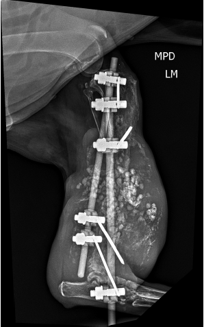
Figure 1: Lateral X-ray right tibia. Presence of two vertical fixators and five horizontal pins, 3 in the proximal portion and 2 in the distal portion of the right tibia. Presence of osteolysis and lack of definition of the cortical bone of middle and distal third of right tibia and fibula compatible with an aggressive bone lesion (bone neoplasia/osteomyelitis). Increased dimensions of adjacent soft tissues associated with several delimited radiopaque images measuring approximately 0.51 cm in diameter each adjacent to the topography of the right tibia and fibula, suggesting polymethyl methacrylate bone cement.
Due to these radiological findings, a complete blood count, serum biochemistry, chest X-ray, abdomen and abdominal ultrasound were requested. In the blood count, a lower number of erythrocytes (3.5 million/mm3; normal values=4.7-8.5 million/mm3), hemoglobin (9 mg/ DL; normal values=11.0-18.0 mg/DL), and hematocrit (28.0 %; normal=35.0-55.0%) was observed. A high number of leukocytes (22800 thousand/mm3; normal values=6.0-17,0 mil/mm3) was observed. The dosage of alanine aminotransferase (ALT) (286 U/L; normal values=88 U/L), aspartate aminotransferase (AST) (184 U/L; normal values=88 U/L), alkaline phosphatase (ALP) (1243,0 U/L; normal values=10,0-150 U/L), demonstrate high serum values.
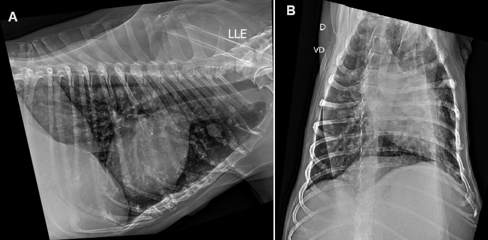
The lung X-ray demonstrated the presence of numerous disperses radiopaque images around pulmonary parenchyma, suggestive of pulmonary metastasis (Figure 2).
The abdominal x-ray indicated the presence of a large mass in cranial portion of abdominal cavity, suggestive of neoplasia (Figure 3).
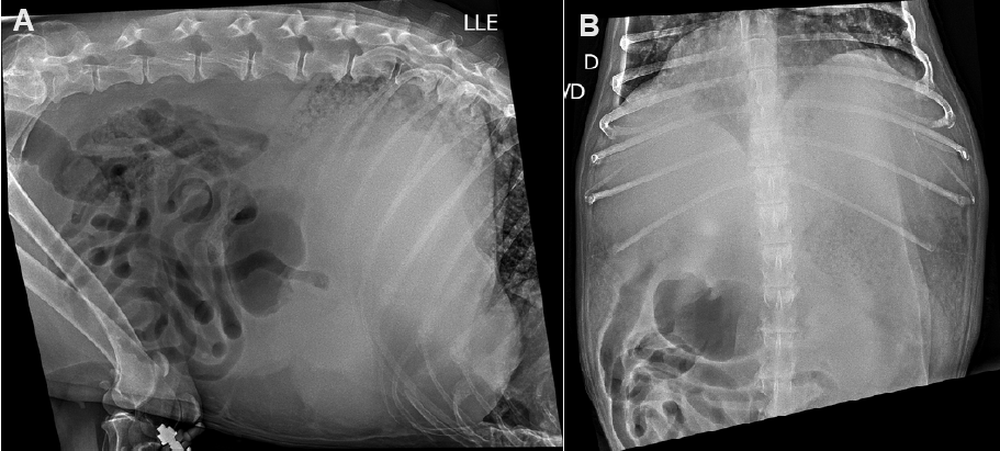
Important homogeneity in cranial portion of abdominal cavity, without delimitation, promoting a mass effect, displacing the gastric cavity dorsally and intestinal segments caudally.
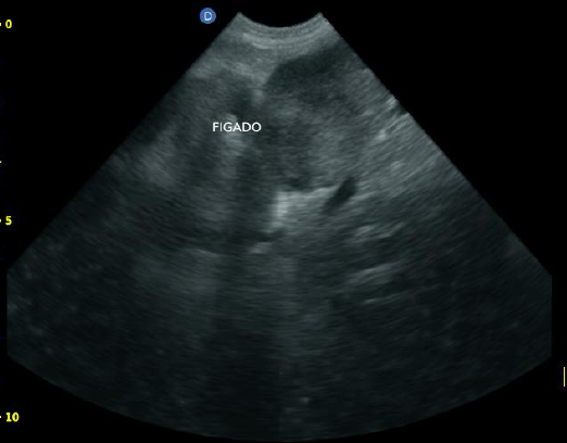
The abdominal ultrasound demonstrated the presence of a large mass that exceeded the limits of the costal margin, reaching the middle of abdomen (Figure 4).
Even with the suspicion of aggressive bone neoplasia with foci of metastasis in the lung and liver, it was decided by the veterinarians and authorized by the owner to amputate the right thoracic limb. Two days after surgery, the animal died. Due to what happened, the owner did not authorize a necropsy and histopathological analysis of neoplastic material.
Discussion
The primary malignant bone tumors that affect the axial skeleton of small animals are exemplified by osteosarcoma, chondrosarcoma, hemangiosarcoma and fibrosarcoma. The osteosarcoma is considered the most common malignant bone tumor in dogs, mainly affecting dogs aged 5 to 9 years old, with greater prevalence in large animals, but without sexual predisposition [2]. The animal in this report was 12 years old and medium size, initially treated after a tibia fracture caused by being run over, with no previous history of pain, local tibial edema or lameness. Factors predisposing to the development of osteosarcoma in dogs include the fixation of fractures with metallic instruments [10, 11, 12], fact described in this case report.
Most osteosarcomas develop from the appendicular skeleton in metaphyseal region of long bones. However, some develop from the axial [13, 14] or from extraskeletal tissues [15, 16, 17].
The clinical signs associated with osteosarcoma are nonspecific and depend on the primary site affected and involvement of underlying structures. The most common clinical sign is lameness resulting from pain. Due to its invasive nature, osteosarcoma causes obstruction of lymphatic drainage, with subsequent edema of the area adjacent to the tumor [2]. In our case report, the presence of edema and pain in the affected limb was described weeks after use of external fixation. The osteosarcomas originating from flat bones of head have a clinical course and longer survival time for affected animals [16], while those originating from the appendicular skeleton present an acute course, leading to the death of animal in a short period [3, 4, 5]. Most dogs die or are euthanized due to metastases, usually to lungs [18], as mentioned in this case report.
Animals with cancer may present with anemia, thrombocytosis and leukocytosis [19]. In biochemical analysis, increased serum levels of alkaline phosphatase can be observed in bone tumors [6, 20], and ALT and AST in primary or metastatic liver tumors [2, 21], facts also described in this case report.
In more than 75% of cases of osteosarcomas, metastases disseminated via the hematogenous route to the lungs can be observed (stage III), making the survival of animals 2 to 4 months after diagnosis [22], and 1 year in just 6.6% of cases [23].
The radiological characteristics of osteosarcomas are variable, with the presence of bone destruction, formation of reactive bone by endosteum and periosteum, and production of osteoid [24], as observed in the animal described in this report. When located in epiphyseal region of long bones, osteosarcoma generally does not invade the joint cavity, respecting the limits of joint capsule [25]. Osteolytic (radiolucent) tumors are hemorrhagic and soft, contain areas of light yellow necrosis, cause erosion of bone cortex and invade neighboring soft tissues. Osteoplastic (radiodense) tumors are characterized by excessive production of bone matrix, present various shades of gray, areas of bone and cartilage and incite a marked periosteal reaction, whereas mixed osteosarcoma is characterized by concomitant bone lysis and production [2, 24, 26].
According to histological characteristics, osteosarcoma is divided into osteoblastic, chondroblastic, fibroblastic, poorly differentiated teleangiectatic, and giant cell type. When there is no predominant pattern, the tumor is classified as combined or mixed osteosarcoma. Histological sections from various parts of tumor must be evaluated to diagnose type of osteosarcoma [24, 27].
Due to the death of animal two days after surgery to amputate the affected limb, the owner chose not to authorize necropsy or histopathological analysis, limiting the diagnostic conclusion.
Conclusion
Primary malignant bone tumors in dogs are uncommon, with osteosarcoma being considered the most common primary bone tumor. The initial symptoms are lameness, pain and increased volume in tumor focus. Diagnosis is based on history, physical and laboratory examination, radiographic findings and histopathological examination. Treatment includes amputation of affected limb, chemotherapy and radiotherapy, however, the prognosis of these animals is considered poor, especially in the presence of metastases.
References
-
Jongeward SJ (1985) Primary bone tumors. Vet Clin North Am Small Anim Pract 15(3): 609-641.
-
Bryan JN (2024) Updates in osteosarcoma. Vet Clin North Am Small Anim Pract 54(3): 523-539.
-
Williams K, Parker S, MacDonald-Dickinson V (2023) Risk factors for appendicular osteosarcoma occurrence in large and giant breed dogs in western Canada. Can Vet J 64(2): 167-173.
-
Amsellem PM, Selmic LE, Wypij JM, Bacon NJ, Culp WT, et al. (2014) Appendicular osteosarcoma in small-breed dogs: 51 cases (1986-2011). J Am Vet Med Assoc 245(2): 203-210.
-
Guim TN, Bianchi MV, De Lorenzo C, Gouvêa AS, Gerardi DG, et al. (2020) Relationship between clinicopathological features and prognosis in appendicular osteosarcoma in dogs. J Comp Pathol 180: 91-99.
-
Silver KI, Patkar S, Mazcko C, Berger EP, Beck JA, et al. (2023) Patterns of metastatic progression and association with clinical outcomes in canine osteosarcoma: a necropsy study of 83 dogs. Vet Comp Oncol 21(4): 646-655.
-
Agnoli C, Sabattini S, Ubiali A, Battisti E, Rossi F, et al. (2023) A retrospective study on bone metastasis in dogs with advanced-stage solid cancer. J Small Anim Pract 64(9): 561-567.
-
Jung MJ, Yoon Kim YM, Lee JS, Choi JW, Kim JH, et al. (2023) Long-term adjuvant metronomic chemotherapy in a dog with recurrent maxillofacial osteosarcoma. Vet Med (Praha) 68(5): 225-230.
-
Iwaki Y, Lindley SES, Bergman N, Smith BF, Pondugula SR (2024) An evaluation of the combination effect of zoledronate and chemotherapeutic agents in canine osteosarcoma cells. Front Vet Sci 11: 1327377.
-
Harrison JW, McLain DL, Hohn RB, Wilson GP, Chalman JA, et al. (1976) Osteosarcoma associated with metallic implants. Report of two cases in dogs. Clin Orthop Relat Res 116: 253-257.
-
Van Bree H, Verschooten F, Hoorens J, Mattheeuws D (1980) Internal fixation of a fracture humerus in a dog and late osteosarcoma development. Vet Rec 107(22): 501-522.
-
Arthur EG, Arthur GL, Keeler MR, Bryan JN (2016) Risk of osteosarcoma in dogs after open fracture fixation. Vet Surg 45(1): 30-35.
-
Heyman SJ, Diefender DL, Goldschmidt MH, Newton CD (1992) Canine axial skeletal osteosarcoma. A retrospective study of 116 cases (1986 to 1989). Vet Surg 21(4): 304-310.
-
Dickerson ME, Page RL, LaDue TA, Hauck ML, Thrall DE, et al. (2001) Retrospective analysis of axial skeleton osteosarcoma in 22 large-breed dogs. J Vet Intern Med 15(2): 120-124.
-
Patnaik AK, Liu S, Johnson GF (1976) Extraskeletal osteosarcoma of the liver in a dog. J Small Anim Pract 17(6): 365-370.
-
Langenbach A, Anderson MA, Dambach DM, Sorenmo KU, Shofer FD (1998) Extraskeletal osteosarcomas in dogs: a retrospective study of 169 cases (1986-1996). J Am Anim Hosp Assoc 34(2): 113-120.
-
Duffy D, Selmic LE, Kendall AR, Powers BE (2017) Outcome following treatment of soft tissue and visceral extraskeletal osteosarcoma in 33 dogs: 2008-2013. Vet Comp Oncol 15(1): 46-54.
-
Silver KI, Patkar S, Mazcko C, Berger EP, Beck JA, et al. (2023) Patterns of metastatic progression and association with clinical outcomes in canine osteosarcoma: a necropsy study of 83 dogs. Vet Comp Oncol 21(4): 646-655.
-
Childress MO (2012) Hematologic abnormalities in the small animal cancer patient. Vet Clin North Am Small Anim Pract 42(1): 123-155.
-
Barger A, Baker K, Driskell E, Sander W, Roady P, et al. (2022) The use of alkaline phosphatase and runx2 to distinguish osteosarcoma from other common malignant primary bone tumors in dogs. Vet Pathol 59(3): 427-432.
-
Teshima T, Matsumoto H, Shigihara K, Sawada H, Michishita M, et al. (2013) Hepatocellular carcinoma in a young dog. Cn Vet J 54(9): 845-848.
-
Prachini-Winter C, Curran KM, Pellin M, Laver T, Hanot C, et al. (2019) Cutaneous and subcutaneous metastasis of appendicular osteosarcoma in dogs: 20 cases. J Vet Intern Med 33(5): 2200-2208.
-
Boston SE, Ehrhart NP, Dernell WS, Lafferty M, Withrow SJ, et al. (2006) Evaluation of survival time in dogs with stage III osteosarcoma that undergo treatment: 90 cases (1985-2004). J Am Vet Med Assoc 228: 1905-1908.
-
Dittmer KE, Pemberton S (2021) A holistic approach to bone tumors in dogs and cats: radiographic and histologic correlation. Vet Pathol 58(5): 841-857.
-
Rozeman LB, Cleton-Jansen AM, Hogendoorn PC (2006) Pathology of primary malignant bone and cartilage tumors. Int Orthop 30(6): 437-444.
-
Holmberg BJ, Farese JP, Taylor D, Uhl EW (2004) Osteosarcoma of the humeral head associated with osteochondritis dissecans in a dog. J Am Anim Hosp Assoc 40(3): 246-249.
-
Guim TN, Bianchi MV, De Lorenzo C, Gouvêa AS, Gerardi DG, et al. (2020) Relationship between clinicopathological features and prognosis in appendicular osteosarcoma in dogs. J Comp Pathol 180: 91-99.
- Mitochondrial Bio-Logistics: Steering Co-Enzyme Q10 and Lycopene Synergies within the Science 4.0 Bio-OS Framework
- Hymenoptera Specimens from the Caño Negro Wetland, of the National Museum Collection, Costa Rica
- Science 4.0: Comprehensive Architecture of the Biological Operating System (Bio-OS) A Framework for Systemic Resilience and Industrialized Bio-Governance
- Rabbit on, or Hare Back? Understanding Climate Change
- Clinical Validation of Science 4.0: Flow Steering and Epigenetic Drift Inversion on a 76-Year-Old Hybrid System
- Seeds Planted by another Mind