The Choanocyte Cells from Phylum Porifera, Nematocysts Cells from Phylun Cnidaria and Colloblast Cells from Ctenophora like Analogues Structures
There are three basic structures belongs to Phylum Porifera, Phylum Cnidaria and Phylum Ctenophora very important because they have similar function like to catch food, but three of them have different origin and different structuration too. They are knowing like choanocyte (Porifera), nematocysts (Cnidaria) and colloblast (Ctenophora) so they are considered analogue structures and they have different positions in the animal; meanwhile the first one are localized in the general cavity (asconoid form, leuconoid form) of sponge, the second one are localized in different zones of the animal like tentacles, acontium , verrucae , vesicule, acrorargus and the last one is on tentacles.
Introduction
The analogue structures are definite like organs of different animals with like function but of unlike origin.
After that definition three clear example from animal kingdom like choanocyte, nematocysts and colloblast are interesting like material of study. They are useful for caching food, meanwhile the first one with his flagella and collars produce water current and engulfing food particles, the second one with his filament can to catch a prey and through his chemicals compounds to murder it and engulfing and the last one on tentacles carried food to mouth.
This structures are very interesting because they belongs to different phyla but three of them haven similar function like to catch food, so they have different morphological structure.
As it seems the evolution from Porifera to Ctenophora have a lineal address in relation to capture for food because choanocyte, nematocysts and colloblast) haven like a function to catch prey of different size.
Results
Choanocyte
This structure are localized into the general cavity of sponge (asconoid or leuconoid) and they are the most characteristic sponge cells.
The choanocyte has a flagellum and a collar (a); the flagellum is responsible for producing water current that bring food and oxygen and the collar are filtering mechanisms for trapping and engulfing food particles. In many sponge this choanocyte are responsible for digestion too.
To electron microscopy details of collar structure (b) is composed for many delicate and linear pseudopodia strengthened by exceedingly fine microfibrils which pass
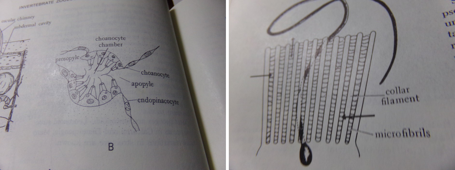
from one pseudopodium to adjacent ones and mucus secretion complete the collar. The flagellum pulls water through the interstices of the collar filtering out particles and its are carried to the base of the collar to be ingested in food vacuoles [1].
In calcareous sponge with large choanocytes, the food is digested here (Figure 1).
(a) (b) Figure 1: Scheme of a section through a choanocyte chamber. b. Details of the collar of a choanocyte as revealed by electron microscopy (a-b) from 1.
Nematocysts
The mother cell call cnidoblast is localized in different zones of the animal, such as epithelium (ectoderm or endoderm).
The above mentioned cell for a process call cnidogenesis give a cell called nematocysts. During this process there is a metamorphosis process into the mother cell where will built different structures like capsule, filament and shaft and when the filament is so long it is coiled into the capsule. The filament has enzymes of different molecular weight and these include lipolytic and proteolytic proteins that catabolized prey tissues.
When the cnidarians inject the nematocysts content or venom that initiates one toxic and immunological reaction in the envenomated organism because this venoms contain the enzymes mentioned. Certain cnidarians venoms contain amines such as serotonin, histamine, bunodosine and caissarone [2].
The nematocysts is very important on taxonomy of cnidarians, especially in the taxonomy of order Actiniaria (sea anemones), notwithstanding they are employed too in taxonomy of Hydra, and medusae too.
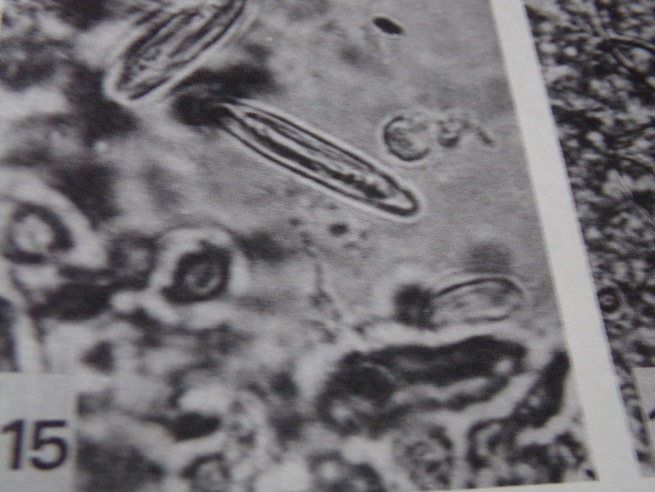
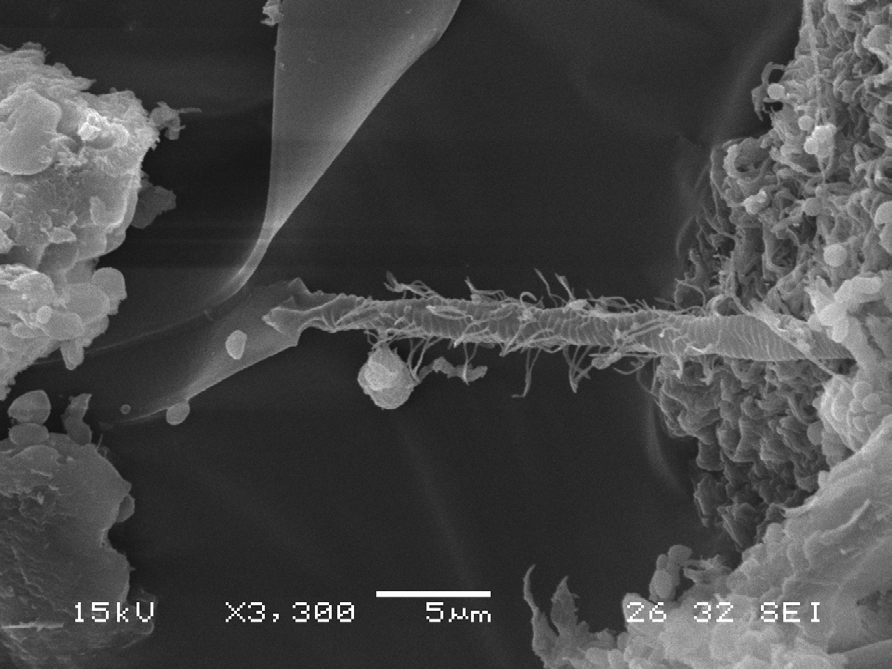
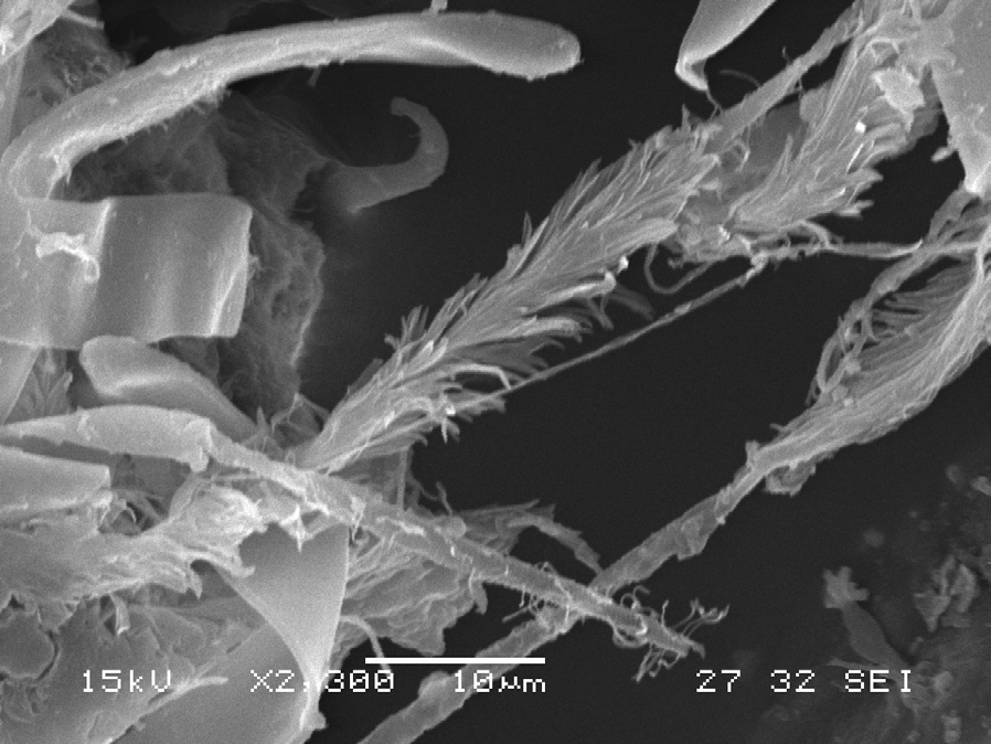
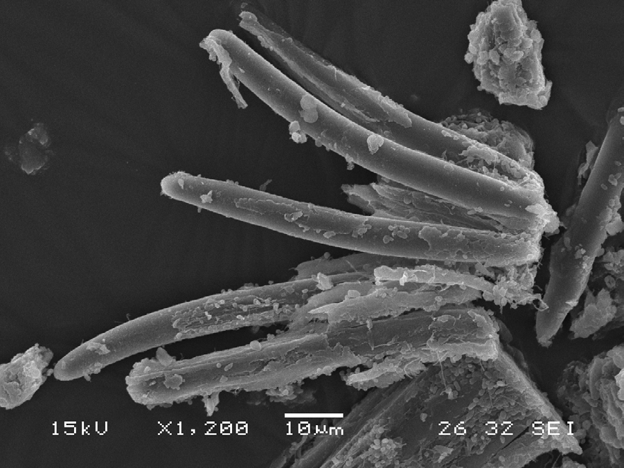
The nematocysts are so important that there is a classification of them on based their morphology, so one can read in some paper of taxonomy of cnidarians names such as eurytele, macrobasic p-mastigophore, microbasic b-mastigophore, holotrichous, basitrichous, etc. For example it can see with electronic microscopy three nematocysts type microbasic from acontium (a) and here it can see details of filament and other with optic microscopy coming from lips of gastric peduncle of medusae (b).
The acontium is the end free portion of mesentery and it is not present in all sea anemone. They are present in acontiarian and mesomyarian sea anemone. This acontium is lying on wall of mesentery and it is floating to gastric cavity when some stimulus produce their activity (Figure 2).
From point ecology view, this cells are so important because they are employed by catching press and in some cnidarians they are used for settled the planulae larvae to the substratum, and another functions like defense, aiding digestion and supporting structure [3, 4, 5, 6].
When some organism are swimming near of cnidarian or it is near there is a stimulus and it origin discharge of filament of nematocysts and like this filament has enzimes of high molecular weight, it produce paralysis of prey or death of it.
Colloblasts
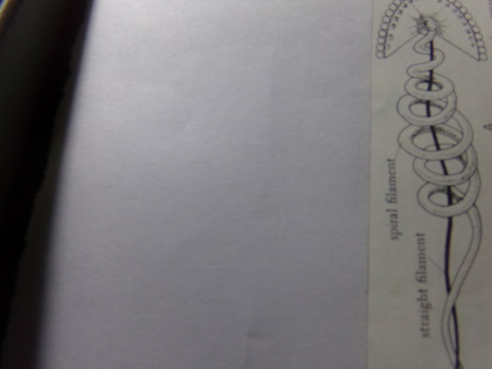
The colloblasts is a highly specialized cell but lacking normal cell structures; they are on tentacles and carry the food to mouth (Figure 3).
It is covered by sticky material and tethered by a straight filament and if it is pulled out by struggling prey, the colloblasts is retracted by a spiral contractile filament.
Discussion
The main axis of this contribution is the functional relationship between choanocite cells, nematocysts cells and colloblasts cells.
These cells types have in common the capture of food and each one developed functional morphology for this activity. It will be success and the final propose will be sure getting food for the species.
The evolution line from Porifera through Cnidarian to Ctenophora according to appearances has a direct line in relation to capture food, so different species from these phyla have food resource sure.
It is very important because always the study of evolution has a view to morphological forms, to an ecological niche, to animal distribution, but it is very scarce or nothing about the evolution to sure food a how it can get through specialized structure like above mentioned.
The three studied structures have their degree of complexity and each one has a structure different one to another and this complexity is out of influence of environmental, because sponge are benthic and benthic cnidarians (sea anemone, polyps hydrozoa) have different apparatus for catching food, meanwhile ctenophore and cnidarians like medusae are neritic and oceanic habitats and both of them have different structure for catching food too. So this fact shows here how the evolution is independent from medium and the final propose is to make sure the food for the individual that in last instance is the species.
Acknowledgments
The author thanks to Dr. Agustín Garese (Biology Laboratory of Cnidaria (LABIC). Faculty of Exacts and Natural Sciences. National University of Mar del Plata) for the picture of nematocysts from electronic microscopy.
References
-
Meglitsch PA (1972) Invertebrates Zoology. In: 2nd (Edn.), Oxford University Press pp: 834.
-
Jouaei M, Yamagihara AA, Madio B, Nevalainen TJ, Alevooa PF, et al. (2015) Ancient venom system: a review on Cnidarian toxin. Toxin 7(6): 2251-2271.
-
Zamponi MO (1990) The larva of Olindias sambaquiensis Muller, 1861 (Cnidaria: Limnomedusae): swimming behavior and substrate selection. Physis (Buenos Aires) Secc A 48(114-115): 21-24.
-
Werner B (1965) The nettle capsules of the Cnidaria with special consideration of the Hydroida.I. Classification and significance for systematics and evolution. Helgolander wiss Meeresunters.
-
Chatton E (1938) Taxonomic and cytological research on free-living dinoflagellates of the genus Polykrikos, Butschli, their cnidocysts and cnidogenesis. In: Scientific Treatises and Works (1906-1932). F Sottano Printing Company, Brazil, pp: 63-75.
-
Garese A (2013) Study of the cnidoma of sea anemones (Cnidaria: Actiniaria and Corallimorpharia): composition, abundance and biometry. Ciencias Marinas. UNMdP. Doctoral Thesis, pp: 1-137.
- Mitochondrial Bio-Logistics: Steering Co-Enzyme Q10 and Lycopene Synergies within the Science 4.0 Bio-OS Framework
- Hymenoptera Specimens from the Caño Negro Wetland, of the National Museum Collection, Costa Rica
- Science 4.0: Comprehensive Architecture of the Biological Operating System (Bio-OS) A Framework for Systemic Resilience and Industrialized Bio-Governance
- Rabbit on, or Hare Back? Understanding Climate Change
- Clinical Validation of Science 4.0: Flow Steering and Epigenetic Drift Inversion on a 76-Year-Old Hybrid System
- Seeds Planted by another Mind