Radiographic Imaging and Osteometric Analysis Os Penis in Red Fox (Vulpes vulpes)
The aim of this study was to determine the macroscopic features of the os penis of the Red Fox (Vulpes vulpes), as well as radiographic images and morphometric measurements using a digital electronic caliper. In this study, 4 adult male red fox os penises, which were previously brought dead from nature by students and whose skeletons were exhibited in our anatomy museum, were used. Macroscopic and morphometric measurements were taken and anatomical structures were analysed by radiographic imaging. Os penis is thicker at the proximal end and tapers towards the distal end, terminating in a cartilaginous tip. There are three margins, one dorsal and two ventrolateral to the sulcus urethiralis. The dorsal edge forms a crista on the median of the bone, extending from proximal to distal. The crista becomes shallow at the distal 1/3 of the bone and disappears completely at the cartilaginous end. Sulcus urethralis is found on the ventral side Os penis of the Red Fox measured as, length of the os penis 56.70mm, Distal diameter 2.96mm, Proximal diameter 3.85mm, length of the sulcus urethralis 35.13mm, width of sulcus urethralis 2.95mm, length of the fibrous cartilaginous tissue 5.70, width of the fibrous cartilaginous tissue 1.77mm. As a result, it has been observed that the os penis of the red fox, which is a part of wildlife, is similar to the other members of the team, the dogs, and radiographic images have been combined with new techniques and a literary contribution has been made. Wild animal anatomy has been supported by more detailed examination of these species. It is thought that the data obtained in the study can be used in future morphometric, zoo-archaeological and taxonomic studies.
Abbreviations
LOP: Length of the Os Penis; LSU: Length of the Sulcus Urethralis; WSU: Width of Sulcus Urethralis; LFCT: Length of the Fibrous Cartilaginous Tissue, WFCT: Width of the Fibrous Cartilaginous Tissue; DDOP: Distal Diameter Os Penis, PDOP:
Proximal Diameter Os Penis.
Introduction
The Red Fox (Vulpes vulpes) was the most geographically spread species of the Carnivores and represents as one of the most abundant carnivore in Egypt. It was taxonomy under Kingdom: Animalia, Phylum: Chordata, Class: Mammalia, Order: Carnivora, Family: Canidae, Tribe: Vulpini [1]. The extremely adaptable and most numerous representative of the Vulpes genus is the red fox (Vulpes vulpes Linnaeus, 1758). It is present in the steppe biomes and mixed forests of Eurasia within the temperate climate zone [1, 2]. The red fox has adapted to life in different types of habitats and climates and can be seen in urban and suburban areas, which has led to partial changes in behavior and in its manner and type of nutrition [1, 3]. Vulpes vulpes belongs to the order of Carnivora; however, unlike dogs and cats, it is actually an undomesticated canid, between a large cat and a small dog in size [3]. However, as the largest species of the genus Vulpes, its body size ranges between 3 and 14 kg [4].
The OS penis is a bony structure located in the corpus cavernosum penis. It is located in the distal part of the penis. It is shaped like a corrugated probe and its opening faces ventrally and the urethra runs in this opening. This groove is called sulcus urethralis. In the German Shepherd and Turkish Shepherd Dogs, the sulcus urethralis is more closed and flat at the proximal end, while it widens and becomes shallower towards the distal end and disappears completely in the distal l/3. The dorsoventral diameter is thicker than the laterolateral diameter. The distal part of the bone ends in a hyaline, fibrous or fibrocartilaginous structure [5, 6].
Radiographic diagnosis method still maintains its gold standard value in the diagnosis of diseases in veterinary medicine. It is the most preferred diagnostic method due to its affordable cost and easy applicability in the diagnosis of diseases. It is understood by studies that radiography is frequently used especially in bone-related problems [7, 8, 9, 10]. However, a good quality radiograph is needed for a good evaluation. For this purpose, the desired region should be taken in the appropriate position, at the appropriate dose and the radiographic anatomy of the region should be well known in order to evaluate the radiograph.
This study was carried out to determine the anatomical features of the red fox os penis. It is expected to contribute to the literature by knowing the radiographic anatomy for the radiographic examination method, which is an important method especially in disease diagnoses today, and by detailing the anatomical structures in wild animal species.
Materials and Methods
In this study, 4 adult male red fox os penises, which were previously brought dead from nature by students (with the permission of the General Directorate of Nature Conservation and National Parks of the Ministry of Agriculture and Forestry with the number E-21264211-288.04-14713474) and whose skeletons were exhibited in our anatomy museum, were used. The dead animals were firstly subjected to maceration procedures by removing the skin and muscles, which are routine dissection procedures, and then made ready for craniometric measurements. Digital callipers were used for craniometric measurements (Figure 1). Surfaces of the penis bone were examined for macroanatomical observations. Nomina Anatomica Veterinaria [11] was used in the spelling of anatomic terminology.
For morphometric measurement points, the greatest length of the os penis and the diameter of the proximal/distal part, the length and width of the fibrocartilaginous part, the length and width of the sulcus urethralis were calculated with digital callipers.
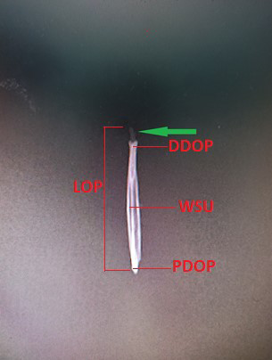
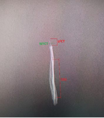
Radiography device (Varian medical systeme X-RAY PRODUCTS, RAD-14) in Fırat University Animal Hospital was used for radiographic images. Radiographic images of the penis were taken. The images were transferred to the computer and the necessary anatomical parts were marked on the image.
Results
Os penis of Red Fox is thicker at the proximal end and tapers towards the distal end, terminating in a cartilaginous tip. There are three margins, one dorsal and two ventrolateral to the sulcus urethiralis. The dorsal edge forms a crista on the median of the bone, extending from proximal to distal. The crista becomes shallow at the distal 1/3 of the bone and disappears completely at the cartilaginous end. Sulcus urethralis is found on the ventral side. Sulcus urethralis is wider in the proximal side but it shows a shallower course towards the distal side. The distal end shows a concavity towards the ventral side. This part, which is composed of cartilage, is developed and spoon-like. In the distal part of the bone, a fibrous cartilaginous tissue with a length of 5.70mm mm and a thickness of 1.77 mm was detected.
Os penis of the Red Fox measured as, length of the os penis 56.70mm, Distal diameter 2.96mm, Proximal diameter 3.85mm, length of the sulcus urethralis 35.13mm, width of sulcus urethralis 2.95mm, length of the fibrous cartilaginous tissue 5.70, width of the fibrous cartilaginous tissue 1.77mm (Table 1).
| Parameters | Morphometric measurement (Average of 4 red foxes) |
|---|---|
| Length of the os penis (LOP) | 56.70mm |
| Distal diameter os penis (DDOP) | 2.96mm |
| Proximal diameter os penis (PDOP) | 3.85mm |
| length of the sulcus urethralis (LSU) | 35.13mm |
| width of sulcus urethralis (WSU) | 2.95mm |
| Length of the fibrous cartilaginous tissue (LFCT) | 5.70mm |
| Width of the fibrous cartilaginous tissue (WFCT) | 1.77mm |
Table 1: Findings measured on the os penis by classical morphometry (mm).
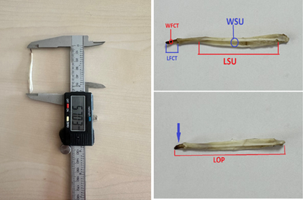
Os penis of the red fox was visualized by radiographic imaging. Dorsoventral and laterolateral position images were taken and transferred to the computer. Morphometric measurement points were also marked on the radiographic images (Figures 1 & 2).
Discussion
The fox os penis is thicker proximally and distally it has a structure resembling a corrugated probe with a sulcus urethralis on the ventral side, which gradually tapers towards the end [12]. With this shape, it shows exactly the characteristics reported for the dog in classical books [5, 6, 7, 8, 9, 10, 11, 12, 13]. Although Yilmaz O, et al. [14] reported that the sulcus urethralis terminated at distal of the os penis and the crista on the dorsal margin terminated at distal, this ratio was found to be for both formations in the fox. The dorsoventral diameter was found to be greater than the laterolateral diameter throughout the length of the bone as reported by Miller [6].
In the study of Canady A [15], it was stated that a cartilaginous structure was not observed on the distal tip of os penis belonging to the fox. The absence of cartilaginous structure might be attributed to the fact that the materials were brought from a museum. In our study, cartilaginous structure was found to be present. According to the study of Gultiken ME, et al. [12], it was determined that the proximal part of the os penis of the fox is bone and the distal part is cartilage. Our study was similar to this study and cartilaginous structure was found in the distal part.
Canady A [15] measured with digital calliper as length of the os penis 47.01-63.41 mm, dorsoventral thickness 2.64-4.60 mm, length of the sulcus urethralis 31.02-46.41 mm, width of sulcus urethralis 1.70-3.59 mm, in red fox. In the study by Haligur A, et al. [16] os penis was measured from 3D reconstruction images, length of it 65.32-67.53 mm, widht of it 5.71-6.25 mm, and dorsoventral thickness 3.48-3.74 mm.
The sulcus urethralis was measured from 3D reconstruction images, length of it 2.97-3.37 mm, width of it 43.86-45.59 mm. Our study is similar to these studies measured as Length of the os penis 56.70mm, Distal diameter 2.96mm, Proximal diameter 3.85mm, length of the sulcus urethralis 35.13mm, width of sulcus urethralis 2.95mm, Length of the fibrous cartilaginous tissue 5.70mm.
Schwery O, et al. [17] and Canady A [15] showed that a lower variability in the length of the penis bone and a greater variability in its thickness can play an important role in increasing strength of the bone and preventing its possible fracture during copulation. Some data on the length of the penis in several species of Canidae were shown by Burt WH [18], Larivière S, et al. [19] and Dixson AF, et al. [20]. When we consider the data in our study, it was observed that the results were similar to other studies and were similar to canidae species.
As a result, it has been observed that the os penis of the red fox, which is a part of wildlife, is similar to the other members of the team, the dogs, and radiographic images have been combined with new techniques and a literary contribution has been made. Wild animal anatomy has been supported by more detailed examination of these species. It is thought that the data obtained in the study can be used in future morphometric, zoo-archaeological and taxonomic studies.
Declaration of Conflicting Interests
The author(s) declared no potential conflicts of interest with respect to the research, authorship, and/or publication of this article.
Data Availability Statement: The data that support the findings of this study are available from the corresponding author upon reasonable request
References
-
Porobic JM (2017) Geometric-Morphometric Analyses of Golden Jackal (Canis aureus) and Red Fox (Vulpes vulpes) Skulls from the Territory of Serbia: Biogeographical Aspects of Morphological Variability. University of Belgrade, Serbia.
-
Lloyd HG (1980) The Red Fox. Batsford, UK.
-
Gloor S, Bontadina F, Hegglin D, Deplazes P, Breitenmoser U (2001) The rise of urban fox populations in Switzerland. Mamm Biol 66: 155-164.
-
Nowak RM (1999) Walker’s Mammals of the World. 6th (Edn.), The Johns Hopkins University Press: Baltimore, USA.
-
Getty R (1975) Sisson and Grossman’s the Arıatomy of the Domestic Animals. In: 5th (Edn.), Evans HE, Christensen GC (Eds.), Philadelphia.
-
(2019) Miller’s anatomy of the dog. In: 2nd (Edn.), pp: 1181.
-
Şen Y, Bumin A (2015) Radiological Anatomy of Bovine Feet. Turkish Clinics J Vet Sci Surg 1(1): 8-12.
-
Arslan OF, Altindag A (2023) Temporomandibular Joint Anatomy Disorders and Imaging Methods. Health & Science pp: 117-145.
-
Okkesim A, Yilmaz B, Yilmaz S (2017) Initial Intervention and Radiographic Imaging in Maxillofacial Trauma. ADO Journal of Clinical Sciences 8(1): 1553-1562.
-
Koc S (2023) Radiological Evaluation of Extremity Long Bone Fracture Cases Encountered in Cats and Dogs Applied to Our Clinic. Aydin Adnan Menderes University, Institute of Health Sciences.
-
Nomina Anatomica Veterinaria (2017) International Committee on Veterinary Gross Anatomical Nomenclature: General Assembly of the World Association of Veterinary Anatomists. In: 6th (Edn.), Gent.
-
Gultiken ME, Yildiz D, Bolat D (2004) The anatomy of os penis in red fox (Vulpes vulpes). Ankara Universitesi Veteriner Fakültesi Dergisi 51: 71-73.
-
Nickel R, Schummer A, Seiferle E, Frewein J, Wilkens H, et al. (1986) The anatomy of the domestic animals. Volume 1. The locomotor system of the domestic mammals J Anat 150: 289.
-
Yilmaz O, Yildi H, Bahadir A, Yilmaz B, Serbest A (1993) A Comparative Study on Os Penis of Turkish Shepherd and German Shepherd Dogs. Bursa Uludag University, Research Information System 12(2): 62-67.
-
Canady A (2013) Variability of the baculum in the red fox (Vulpes vulpes) from Slovakia. Zoology and Ecology 23(3): 165-170.
-
Haligur A, Ozkadif S (2019) Male genital organs in the red fox (Vulpes vulpes); Macroanatomic and Three- dimentional Reconstruction Aspect. Mehmet Akif Ersoy University Journal of Health Sciences Institute 7(2): 89- 98.
-
Schwery O, Kohnemann BA, Michler FU, Brinkmann W (2011) Morphometrical characterisation of a raccoon (Procyon lotor L.) population from Müritz National Park (Germany) by means of the Os baculum. Beiträge zur Jagd & Wild forschung 36: 605-617.
-
Burt WH (1960) Bacula of North American mammals. University of Michigan Miscellaneous Publications of the Museum of Zoology 113: 1-75.
-
Lariviere S, Ferguson SH (2002) On the evolution of the mammalian baculum: vaginal friction, prolonged intromission or induced ovulation. Mammal Review 32(4): 283-294.
-
Dixson AF, Anderson M (2004) Sexual behavior, reproductive physiology and sperm competition in male mammals. Physiology and Behavior 83(2): 361-371.
- Mitochondrial Bio-Logistics: Steering Co-Enzyme Q10 and Lycopene Synergies within the Science 4.0 Bio-OS Framework
- Hymenoptera Specimens from the Caño Negro Wetland, of the National Museum Collection, Costa Rica
- Science 4.0: Comprehensive Architecture of the Biological Operating System (Bio-OS) A Framework for Systemic Resilience and Industrialized Bio-Governance
- Rabbit on, or Hare Back? Understanding Climate Change
- Clinical Validation of Science 4.0: Flow Steering and Epigenetic Drift Inversion on a 76-Year-Old Hybrid System
- Seeds Planted by another Mind