Exposure Keratopathy Secondary to Palpebral Paralysis in Cavia porcellus: A Case Report
Background: Ulcerative keratitis in Cavia porcellus may be secondary to eyelid paralysis caused by otitis media. Diagnoses performed by professionals with specific knowledge, and supported by diagnostic tests are essential for proper management and treatment. Case Description: A 2-year-old guinea pig, exhibiting persistent dryness of the ocular surface and ulcerative keratitis, was referred to veterinary ophthalmology attributed to a lack of positive response to previous treatment. Laboratory and imaging tests were performed, including X-ray and computed tomography of the skull, were conducted. Additionally, specific ophthalmic tests were performed, revealing detectable alterations. The results showed notable leukocytosis and increased alkaline phosphatase. Additionally, there were indications of right otitis media and internal otitis. The ocular examination identified eyelid ptosis and ulcerative keratitis attributed to exposure. A new therapeutic protocol was implemented, incorporating novel medications and involving acupuncture as an integrative method. The response to treatment was satisfactory, leading to the complete improvement of ulcerative keratitis and recovery of eyelid movement.
Introduction
Guinea pigs (Cavia porcellus) have been used as laboratory animals for a long time. However, their increasing popularity as an unconventional pet has spurred a growing interest in studies aimed at their overall health, including research in the field of ophthalmology [1].
Owing to the unique features discovered in studies involving these animals, it has been observed that they exhibit low corneal sensitivity and possess a vestigial third eyelid. This is in addition to blinking infrequently, approximately 2 to 5 times every 20 min, which underscores the importance of ensuring proper spreading of the tear film for the maintenance of the corneal surface [2]. Moreover, the cornea of the guinea pig (PDI) is soft, transparent, shiny, and wide; histologically, it is composed of epithelium, Bowman’s layer (found in primates and some other species), stroma, Descemet’s membrane, and endothelium [3].
Systemic disorders, secondary inflammatory processes, trauma, and other conditions can culminate in corneal degeneration [4]. Orbital diseases and facial nerve paralysis are the common causes of keratitis in these animals [5].
Several diseases and abnormalities have been observed in this species even in healthy individuals. Of these occurrences, some were species-specific, and others were common to the other animals evaluated. In anatomical observations, the path of the facial nerve gains significant relevance when accessing the middle and inner ear, particularly before reaching rostral structures. This is crucial because otitis, often of bacterial etiology, can settle and systemically compromise the patient. Furthermore, may affect nearby organs, according to its extent [6].
Case Presentation
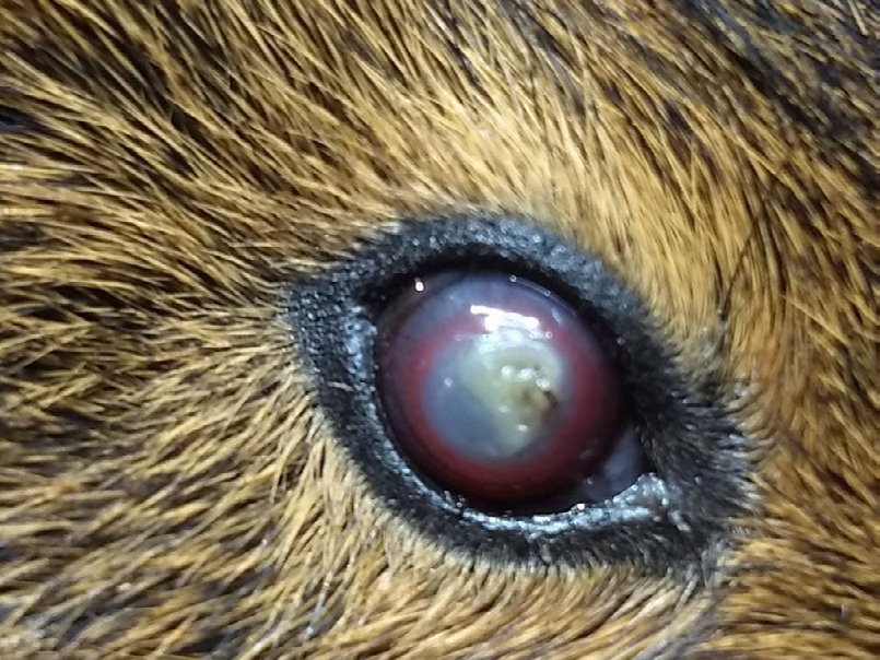
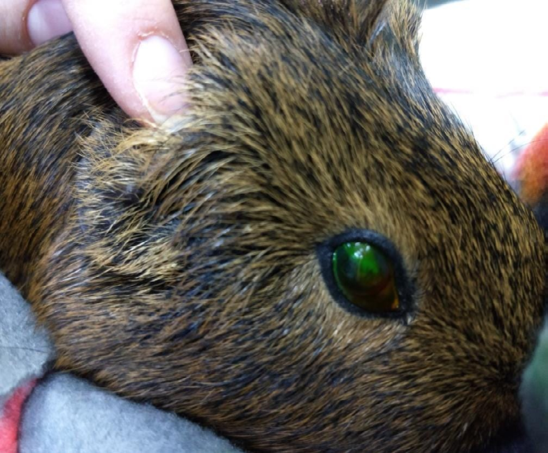
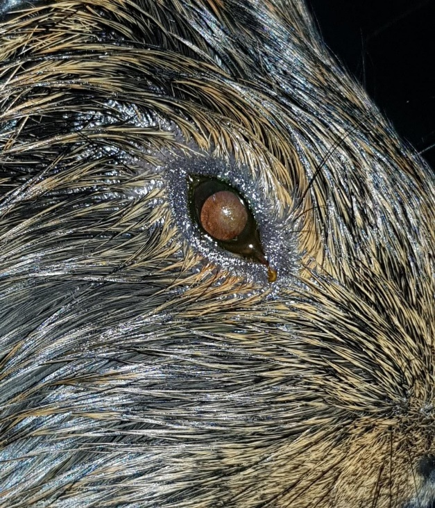
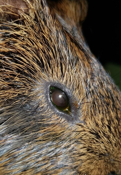
A 2-year-old male guinea pig of the species Cavia porcellus was referred for ophthalmologic care owing to a 15-day history of complaining about a corneal ulcer in the right eye (Figure 1). This was initially treated with Tobrex® (tobramycin 3 mg/ml) and Lacrima plus® (dextran 1 mg/ ml + hypromellose: 3 mg/ml), both eye drops being applied twice a day. However, there was limited satisfactory clinical improvement observed with this treatment. Upon ophthalmic examination, the patient exhibited an intraocular pressure of 13 mmHg in both eyes. Positive fluorescein test results were noted in the right eye, and negative fluorescein test results in the left eye. In the right eye, the patient presented with eyelid paralysis, lower eyelid ptosis, dryness of the corneal surface (3+), corneal opacity (1+), neovascularization, and extensive corneal continuity (Figure 2). The initiated ocular treatment included Regencel® (retinol acetate 10,000 IU/g, amino acids 25 mg/g, methionine 5 mg/g and chloramphenicol 5 mg/g) 5 times a day, Epitegel® (dexpanthenol 50 mg/g) in the intervals and acupuncture for facial paralysis. In addition to ophthalmic alterations, the patient also experienced weight loss and hyporexia, which were monitored and treated by a clinical veterinarian. Blood counts, transaminase, alkaline phosphatase, creatinine, urea, abdominal ultrasonography, skull radiography, and cranial computed tomography were performed. The complete blood count showed significant leukocytosis (2,750 – 14,732/μl) attributed to heterophilia, monocytosis and eosinophilia and increased AF (55 – 108 U/L). Furthermore, abdominal ultrasonography revealed cystic enlargement of the seminal vesicles, thickening and opacity in both tympanic bullae, opacity of the right tympanic cavity, osteolysis and bone proliferation of the ipsilateral temporal bone, suggestive of chronic otitis media and internal bone, and osteomyelitis in the right temporal bone. Seven days after the consultation, improvement in the ocular condition was observed with a negative fluorescein test, a shiny surface, and a decrease in corneal neovascularization (Figure 3). The treatment was maintained, and the importance of acupuncture, which had not yet been initiated, was reinforced.
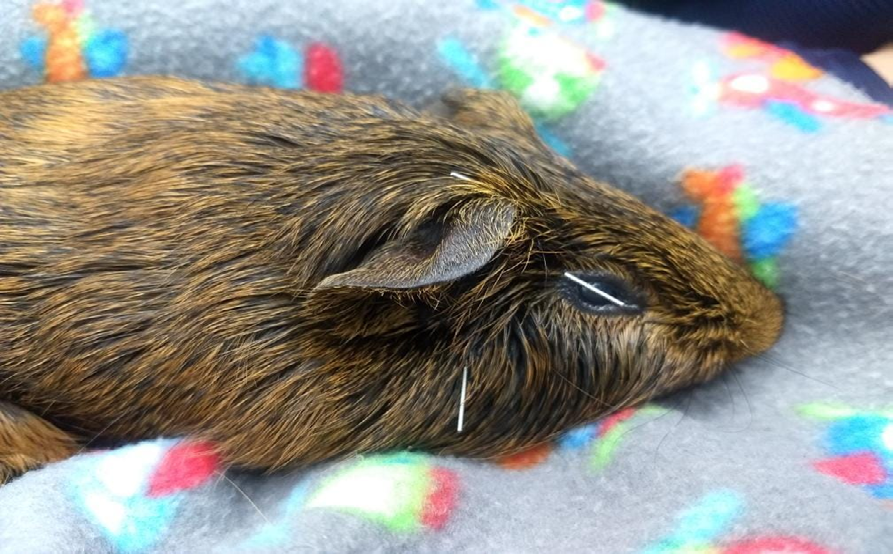
Fifteen days later, at the beginning of the acupuncture sessions (Figure 4), the patient presented with eyelid movements, a tear meniscus, and a shiny surface (Figure 5).
Discussion
There are two forms of neurogenic keratitis: neurotrophic, which is associated with a lack of sensory innervation (trigeminal nerve); and neuroparalytic, which is attributed to the lack of motor innervation (facial nerve) to the orbicularis muscle of the eyelids, resulting in facial paralysis. Neuroparalytic keratitis, which is the one in the reported case, results in ulcerative keratitis owing to severe exposure and initially presents with epithelial degeneration and stromal edema, with advancement, dryness, vascularization, and opacification of the cornea, which in some cases can reach deeper layers and even cause perforation of the corneal structure [3]. Knowledge about these particularities and their occurrence in the species is a great diagnostic differential, including the choice of tests to elucidate the general picture more quickly.
Once a diagnosis is made, the initial treatment is usually symptomatic, with topical lubricants and antibiotics to prevent secondary bacterial infections. In cases of dryness owing to exposure, the use of an ointment is indicated because of its longer contact time and good protective action, infection control, and corneal lubrication [3].
Corneal lesions in guinea pigs attributed to facial nerve palsy, for which the response to clinical treatment is unsatisfactory, may require a surgical procedure, such as temporary tarsorrhaphy, to assist in the recovery process and local protection [5, 6].
The prey characteristics and behavior, which were originally classified in nature, confer high resistance in demonstrating symptoms in the most diverse conditions to which they are subjected, which can cause a delay in detecting the disease and seeking veterinary support [7]. Additionally, animals with low corneal sensitivity, such as guinea pig, tend to have delayed responses to discomfort when presenting with eye disorders [3].
Similar to other species, the use of integrative auxiliary therapies, such as acupuncture, has demonstrated effectiveness in the present case. It stands as a valuable tool in the treatment of eyelid paralysis [8].
Conclusions and Case Relevance
The diagnosis of corneal ulcer goes beyond its detection, it is necessary to investigate its origin and cause so that the appropriate treatment can be implemented. Attention, knowledge, and rapid diagnosis of corneal ulcerative processes are essential for recovery and good prognosis of the case.
References
-
Williams D, Sullivan A (2010) Ocular disease in the guinea pig (Cavia porcellus): a survey of 1000 animals. Veterinary Ophthalmology 13(S1): 54-62.
-
Seyed MP, Maneli AM, Reza S (2016) Intraocular pressure, tear production, and ocular Echobiometry in guinea pigs (Cavia porcellus). JAALAS (Journal of the American Association for Laboratory Animal Science) 55: 475-479.
-
Safatle AMV, Galera PD (2023) Clinical and surgical veterinary ophthalmology. In: Payá (Ed.), Brazil, pp: 800.
-
Kirk NG (1991) The metabolic basis of ophthalmic diseases in animals. Animal Eye Res 101(2): 1-16.
-
Montiani FF, Moore BA, Shlomo GB (2022) Wild and exotic animal ophthalmology. Mammals 2.
-
James K, Version AMG (2016) Rabbit head diseases: description of clinical cases with imaging diagnosis. Master’s thesis. Lisbon: Lusófona University of Humanities and Technology.
-
Cubas ZS, Silva JCR, Dias JLC (2014) Wild animals treaty. In: 2nd (Edn.), 1: 2014.
-
Cavalcanti, Portela VAB, Coelho MCOC (2013) Electroacupuncture in the treatment of idiopathic facial nerve palsy in dogs. Case Report. Journal of Continuing Education in Veterinary Medicine and Animal Science of CRMV-SP (MV&Z Journal) 11n: 3.
- Mitochondrial Bio-Logistics: Steering Co-Enzyme Q10 and Lycopene Synergies within the Science 4.0 Bio-OS Framework
- Hymenoptera Specimens from the Caño Negro Wetland, of the National Museum Collection, Costa Rica
- Science 4.0: Comprehensive Architecture of the Biological Operating System (Bio-OS) A Framework for Systemic Resilience and Industrialized Bio-Governance
- Rabbit on, or Hare Back? Understanding Climate Change
- Clinical Validation of Science 4.0: Flow Steering and Epigenetic Drift Inversion on a 76-Year-Old Hybrid System
- Seeds Planted by another Mind