A Case Report of Feline Cryptococcosis Diagnosed by Cerebrospinal Fluid Analysis
Cryptococcosis is an infectious, cutaneous and/or systemic, chronic fungal disease with global distribution, caused by fungi of Cryptococcus genus that can infect humans and domestic animals, including felines. Two species of the genus Cryptococcus are involved in the disease in cats, Cryptococcus neoformans and Cryptococcus gattii. The C. gattii, in addition to affecting immunosuppressed hosts, can also cause disease in immunocompetent individuals. This report presents the clinical findings of a case of pneumonia, ocular and neurological alterations in a three-year-old male, mixed-breed feline due to Cryptococcus neoformans with diagnosis confirmed through analysis of cerebrospinal fluid with identification of the fungus using the Indian ink dye and its cultivation and growth in Sabouraud dextrose agar from a sample of cerebrospinal fluid.
Introduction
Cryptococcosis is a systemic fungal disease that affects humans and domestic animals, including felines [1], caused by fungi of Blastomycetes class, Cryptococcaceae family, Cryptococcus genus, and Cryptococcus neoformans and Cryptococcus gattii species [1, 2]. Immunocompromised humans and animals can be affected by C. neoformans, although C. gattii affects both immunosuppressed and immunocompetent hosts [2]. The C. neoformans has a cosmopolitan distribution and can be isolated from samples of bird droppings, mainly pigeons (Columba livia), soil, and decaying fruits and vegetables [3].
Animals acquire the infection from organic material present in the environment contaminated by fungus, which disperses its spores through the air, and acquire disease by inhaling blastoconidia of Cryptococcus spp.. In the respiratory system, the primary infection occurs, affecting the nasal cavity and lungs. From this primary focus, the fungus can spread to various organs, including the central nervous system [4].
Pathogenesis of cryptococcosis is determined by the host’s immune status, strain virulence, and inoculum size [5]. In pets such as dogs and cats, states of malnutrition, immunosuppression caused by drugs, viral or bacterial infections and neoplasias predispose to infection, which can determine greater severity or worse prognosis [6].
Cryptococcosis presents four important clinical syndromes; respiratory, nervous, cutaneous and ocular, and in felines the respiratory form with nasal involvement is the most frequent [1, 2].
In respiratory conditions, animals present dyspnea, sneezing, respiratory rales, unilateral or bilateral nasal discharge, purulent, serous or bloody mucus. Firm nodular masses may be observed in the subcutaneous tissue, mainly in the nasal plane or mirror. Lung lesions may or may not result in clinical manifestations, although they can be detected using chest radiographs, computed tomography scans or observed in autopsy findings [7].
Neurological picture of cryptococcosis can be evidenced histologically as diffuse or focal meningoencephalomyelitis with formation of granulomas. Here, animals may present depression, disorientation, cervicalgia, ataxia, paresis, paraplegia, convulsions, vocalization, decreased consciousness, spasticity, walking in circles, anisocoria, mydriasis, visual deficit, blindness, deafness and loss of smell [8, 9].
The diagnosis is initially established by data obtained during anamnesis and physical examination findings.
Hematological and biochemical findings are usually not specific, and radiographic examination may or may not show changes [2]. The confirmation of cryptococcosis diagnosis can be obtained using different laboratory procedures such as cytofungal examination, serology, histopathology, culture and fungal isolation [2, 6, 10]. The prognosis of felines affected by cryptococcosis, in which there was no involvement of the central nervous system, is reserved; and those that presented neurological involvement is reserved to poor [11].
Case Report
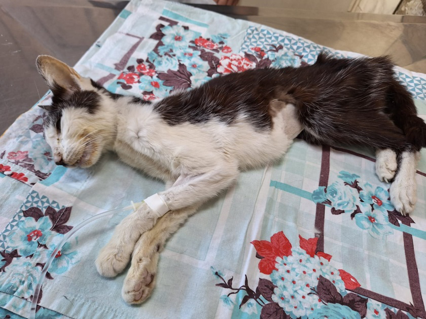
In September 2024, a three-year-old male, castrated and vaccinated feline, without a defined breed, was attended and presented with dyspnea, tetraparesis and weight loss. The owner informed him that the feline had access to street and contact with pigeons. During clinical examination, the animal presented dyspnea, tetraparesis, mydriasis, anisocoria, absence of deep pain in locomotor limbs, generalized muscular weakness, remaining in lateral decubitus throughout the clinical examination (Figure 1).
A complete blood count, measurement of alanine aminotransferase (ALT), alkaline phosphatase (ALP), glucose, thyroxine (free T4), TSH, type I urine, abdominal ultrasound and chest radiography were performed. In the blood count, a smaller number of erythrocytes (4.5 million/ mm3; normal values=5.0-10.0 million/mm3) and leukocytes (5.2 thousand/mm3; normal values=5.5-19, 5 mil/mm3) and hematocrit (23%; normal=24.0-45.0%) was observed. The dosages of ALT (108 U/L; normal values=20-107 U/L), ALP (128; normal values=23-107 U/L), and glucose (142 mg/dL; normal=60-132 mg/ dL) were slightly increased. Type I urinalysis was performed by collecting urine through a urethral probe, and the biochemical examination showed the absence of glucose, ketone bodies and occult blood. In sedimentoscopy, erythrocytes, leukocytes and bacteria were not observed. A rapid test for feline immunodeficiency vírus (FIV) and feline leukemia vírus (FeLV) was performed, and the serum sample was reactive for FIV.
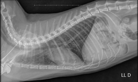
Abdominal ultrasound not revealed any alteration, and the chest radiography showed opacification of the lung fields with a mixed pattern, interstitial and bronchial (Figure 2).
Based on the history, clinical signs and chest radiographic findings suggestive of cryptococcosis, the animal was anesthetized to perform a puncture of cisterna magna to collect cerebrospinal fluid. In the cerebrospinal fluid sample, pleocytosis was observed with the presence of a large number of neutrophils (Figure 3A). In the examination of the cerebrospinal fluid stained with Gram, rounded shapes with clear cytoplasm were observed, suggestive of Cryptococcus spp. (Figure 3B), which was confirmed as Cryptococcus neoformans through the use of India ink, where the fungus with its capsule and the bud formation are evident (Figure 3C).
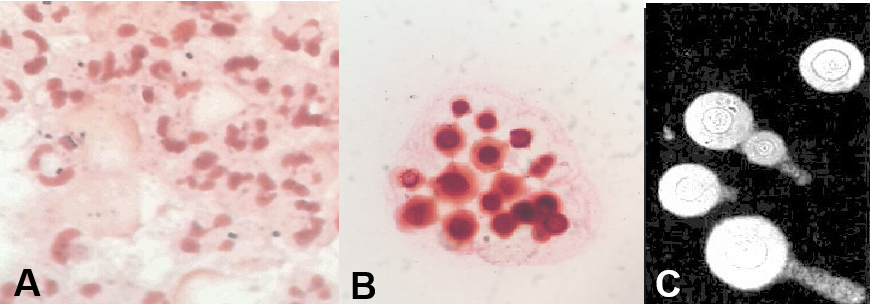
- Pleiocytosis with large numbers of neutrophils in cerebrospinal fluid.
- Round Cryptococcus spp. cells with a light lilac cytoplasmic background or as Gram-negative lipoid bodies.
- Identification of Cryptococcus neoformans yeasts using India ink.
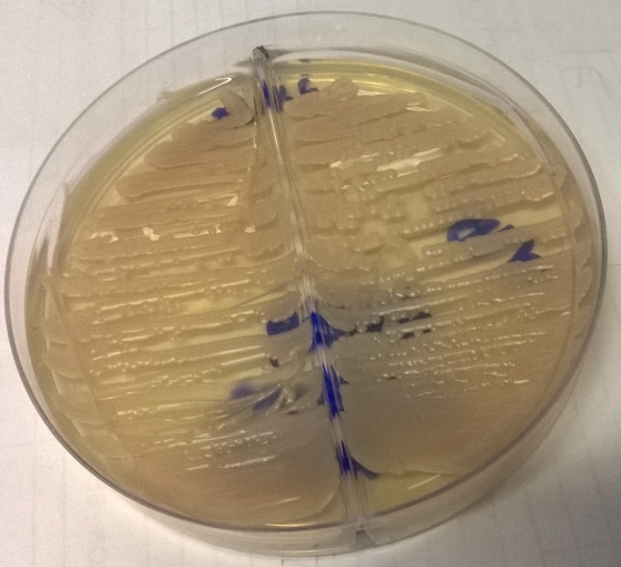
Part of the cerebrospinal fluid sample was plated on Sabouraud dextrose agar, where whitish mucoid colonies grew within 3 days (Figure 4). Due to the weakened clinical condition and reserved prognosis, the owner opted to euthanize of cat. Due to the owner’s lack of authorization, a necropsy was not performed.
Discussion
Feline cryptococcosis is caused by systemic fungi of the genus Cryptococcus, which occurs after the inhalation of blastoconidia, which develop in bird droppings and fallen vegetation on the ground [1, 2, 3]. Clinical signs may vary from localized or systemic lesions, which may appear in isolation or together, causing respiratory, neurological, ocular and cutaneous syndrome [1, 7, 8, 9]. The most common forms are cutaneous and involvement of upper respiratory tract, the clinical signs of which manifest in the form of sneezing, runny nose, nasal secretion, nodules and swelling of the nasal plane. In this case report, the classic form of feline cryptococcosis was not observed, which typically presents rostral involvement of the nasal cavity; nasal secretion, unilateral or bilateral, and distortion of the nasal plane [2, 7].
Central nervous system involvement occurs in 25% of cats affected by cryptococcosis, producing seizures, paresis, ataxia, depression, blindness, and retinitis when there is ocular involvement [8, 9, 12]. Considering that in the context of neurocryptococcosis the airways are the main entry point for Cryptococcus spp., the clinical manifestations are diverse, presenting as an asymptomatic infection to severe pneumonia [2]. Here, the clinical manifestations were relatively nonspecific and the pulmonary lesions, with subsequent neurological changes, were responsible for the symptoms presented.
Evidence of upper respiratory tract infection occurs in 50% to 60% of cases; ocular and neurological signs occur in 15% and 25% of cases, respectively [2, 13]. Lower respiratory tract clinical signs are not very common and weight loss and anorexia are more common in chronic cases [14]. Here, the clinical signs such as weight loss, ocular and neurological changes presented by the animal were compatible with those described in literature.
In humans, the main predisposing condition for fungal infections involves diseases that cause immunodeficiency, particularly the human immunodeficiency virus/acquired immunodeficiency syndrome (HIV/AIDS) [15, 16, 17]. In cats, it has been postulated that FIV and/or FeLV infection may predispose to Cryptococcus infection [18, 19, 20], although literature findings are not unanimous [21]. In this case report, the presence of retroviral disease caused by FIV was serologically determined. The C. neoformans infection in immunocompromised patients usually occurs with few symptoms or even asymptomatically. Infection of the lungs can result in its spread through the bloodstream, and due to its affinity for the central nervous system, the fungus tends to cross the blood-brain barrier, establishing itself and causing fungal meningitis. Because fungal infections of the central nervous system in dogs and cats are uncommon, antemortem diagnosis can be difficult and is definitively made by cytology, biopsy, or culture of an affected organ or cerebrospinal fluid. In cases of neurocryptococcosis, it is recommended to test for the fungus in the cerebrospinal fluid [2, 22]. In cases of cryptococcosis, the cerebrospinal fluid is commonly found with hyperproteinorrhachia, hypoglycorrhachia, and pleocytosis, which was described in this case report. Diagnosis of the fungus in cerebrospinal fluid samples can be performed by direct visualization of fungal structures (thick capsule) by gram staining, although the use of Indian ink is more suitable for diagnosing cryptococcal meningitis [23]. In this case report, yeast-like structures were observed in the cerebrospinal fluid using Gram staining, and the presence of C. neoformans was confirmed using India ink staining.
Fungal culture is considered the gold standard method for diagnosing cryptococcosis, and has a sensitivity of 98 to 100% using cerebrospinal fluid samples. To perform the culture, samples are plated on Sabouraud dextrose agar, CHROMagar Candida, Niger agar, or minimal medium agar containing L-DOPA [24, 25, 26, 27]. When cultivated on Sabouraud dextrose agar at 37°C, Cryptococcus species produce whitish mucoid colonies in 2 to 3 days [24], what was described in this case report. Another method of diagnosing cryptococcosis using a cerebrospinal fluid sample would be to perform the polymerase chain reaction [28, 29]. Although available in Brazil, this test is little known and used in the diagnosis of cryptococcosis in cats.
Conclusion
Cats that roam free in urban areas are potentially exposed to Cryptococcus, since they may come into contact with pigeon feces. The presence of pigeon feces and some decomposing vegetation substrates can be considered a risk factor for domestic felines. When cryptococcosis is suspected, knowledge of the clinical and diagnostic aspects is essential for recognizing and diagnosing the disease and choosing the appropriate treatment, informing the owner about the risks to human health and alternatives for preventing the disease.
References
-
Maziarz EK, Perfect JR (2016) Cryptococcosis. Infect Dis Clin North Am 3091: 179-206.
-
Pennisi MG, Hartmann K, Lloret A, Ferrer L, Addie D, et al. (2013) Cryptococcosis in cats; ABCD guidelines on prevention and management. J Feline Med Surg 1597: 611-618.
-
Bahn YS, Sun S, Heitman J, Lin X (2020) Microbe profile: Cryptococcus neoformans species complex. Microbiology 166(9): 797-799.
-
Yamamura D, Xu J (2021) Update on pulmonary cryptococcosis. Mycopathologia 186(5): 717-728.
-
Alspaugh JA (2015) Virulence mechanisms and Cryptococcus neoformans pathogenesis. Fungal Genet Biol 78: 55-58.
-
Castellá G, Abarca ML, Cabañes FJ (2008) Cryptococcosis and pets. Rev Iberoam Micol 25(1): S19-S24.
-
Rodrigues TO, Godoy JR, Malandrim P, Sossai V, de Souza MT (2020) Cryptococcosis in a feline - case report. Revista MV&Z 18(3): 1-7.
-
Sykes JE, Sturges BK, Cannon MS, Gericota B, Higgins RJ, et al. (2010) Clinical signs, imaging features, neuropathology, and outcomes in cats and dogs with central nervous system cryptococcosis from California. J Vet Intern Med 24(6): 1427-1438.
-
Huang CH, Cehn KS, Chia MY, Chiou HY, Shia WY (2023) Cryptococcal granuloma of basal ganglia due to Cryptococcus neoformans in a cat: a case report and literature review. J Vet Med Sci 85(4): 412-416.
-
Barrs VR, Beczkowski PM, Talbot JJ, Hobi S, Teoh SN (2024) Invasive fungal infections and oomycoses in cats: 1. Diagnostic approach. J Feline Med Surg 26(1): 1-22.
-
Barrs VR, Hobi S, Wong A, Sandy J, Shubitz LF, et al. (2024) Invasive fungal infections and oomycoses in cats: 2. Antifungal therapy. J Feline Med Surg 26(1): 1-16.
-
Wronski JG, de Cecco BS, Raiter J, Henker LC, de Lorenzo C, et al. (2023) Ophtalmic and immunopathological characterization of systemic infectious disease in cats. Vet Pathol 60(3): 352-359.
-
Coelho HE, Moura LR, Orpinelli SRT, Kock GP, Machado FME (2009) Feline meningitis associated with Cryptococcus neoformans in the Municipality of Uberaba-MG - Case report Vet Not 159(1): 29-34.
-
Franco DQS, de Oliveira GBM, Luiz ACS, dos Reis PSB, Pulz LH (2019) Cryptococcal pneumonia and leptomeningitis in feline: a case report. Revista MV&Z 17(2): 14-22.
-
Gushiken AC, Saharia KK, Baddley JW (2021) Cryptococcosis. Infect Dis Clin North Am 35(2): 493-514.
-
Tugume L, Ssebambulidde K, Kasibante J, Ellis J, Wake RM, et al. (2023) Cryptococcal meningitis. Nat Rev Dis Primers 9(1): 62.
-
Gifford A, Jayawardena N, Carlesse F, Lizararo J, McMullan B, et al. (2024) Pediatric cryptococcosis. Pediatr Infect Dis J 43(4): 307-312.
-
Mancianti F, Giannelli C, Bendinelli M, Polli A (1992) Mycological findings in feline immunodeficiency virus- infected cats. J Med Vet Mycol 30(3): 257-259.
-
Jacobs GJ, Medleau L, Calvert C, Brown J (1997) Cryptococcal infection in cats: factors influencing treatment outcomes, and results of sequential serum antigen titers in 35 cats. J Vet Intern Med 119(1): 1-4.
-
Barrs VR, Martin P, Nicoll RG, Beatty JÁ, Malik R (2000) Pulmonary cryptococcosis and Capillaria aerophila infection in na FIV-positive cat. Aust Vet J 78(3): 154- 158.
-
O´Brien CR, Krockenberger MB, Wigney DI, Martin P, Malik R (2004) Retrospective study of feline and canine cryptococcosis in Australia from 1981 to 2001; 195 cases. Med Mycol 42(5): 449-460.
-
Lavely J, Lipsitz D (2005) Fungal infections of the central nervous system in the dog and cat. Clin Tech Small Anim Pract 20(4): 212-219.
-
Dorko E, Kmetová M, Dorko F, Bracoková I, Danko J, et al. (2000) Prevalence of Cryptococcus neoformans in clinical specimens. Folia Microbbiol (Praha) 45(4): 369- 372.
-
Hasimoto e Souza LK, Costa CR, Fernandes Ode F, Abraão FY, Silva TC, et al. (2013) Clinical and microbiological features of cryptococcal meningitis. Rev Soc Bras Med Trop 46(3): 343-347.
-
Guelfand L, Grisolía P, Bozzano C, Kaufman S (2003) Comparison of methods for the identification of the most common yeasts in the clinical microbiology laboratory. Rev Argent Microbiol 35(1): 49-53.
-
Sukroongreung S, Lim S, Tantimavanich S, Eampokalap B, Carter D, et al. (2001) Phenotypic switching and genetic diversity of Cryptococcus neoformans. J Clin Microbiol 39(6): 2060-2064.
-
Brilhante RSN, España JDA, de Alencar LP, Pereira VS, Castelo-Branco DSCM, et al. (2017) An alternative method for the analysis of melanina production in Cryptococcus neoformans sensu lato and Cryptococcus gatii sensu lato. Mycoses 60(10): 697-702.
-
Kano R, Fujino Y, Takamoto N, Tsujimoto H, Hasegawa A (2001) PCR detection of the Cryptococcus neoformans CAP59 gene from a biopsy specimen from a case of feline cryptococcosis. J Vet Diagn Invest 13: 439-442.
-
Okabayashi K, Kano R, Watanabe T, Hasegawa A (2006) Serotypes and mating types of clinical isolates from feline cryptococcosis in Japan. J Vet Med Sci 68: 91-94.
- Mitochondrial Bio-Logistics: Steering Co-Enzyme Q10 and Lycopene Synergies within the Science 4.0 Bio-OS Framework
- Hymenoptera Specimens from the Caño Negro Wetland, of the National Museum Collection, Costa Rica
- Science 4.0: Comprehensive Architecture of the Biological Operating System (Bio-OS) A Framework for Systemic Resilience and Industrialized Bio-Governance
- Rabbit on, or Hare Back? Understanding Climate Change
- Clinical Validation of Science 4.0: Flow Steering and Epigenetic Drift Inversion on a 76-Year-Old Hybrid System
- Seeds Planted by another Mind