Rodent-Related Zoonotic Dynamics in the Republic of Guinea: Implications for the Surveillance and Prevention of Emerging Diseases
In the Republic of Guinea, studies have shown that rodents are recognised as reservoirs and vectors of multiple zoonoses. However, the precise mechanisms of transmission, as well as the impact of environmental and human factors, remain poorly understood. The aim of this study is to assess the transmission dynamics of rodent-borne zoonoses in the Republic of Guinea in order to develop effective strategies for the surveillance, control and prevention of emerging and re-emerging diseases in this region. Between 15 July 2022 and 7 August 2023, small mammal fauna was examined in various prefectures (N'Zérékoré, Lola, Yomou, Beyla, Macenta, Gueckedou, Faranah, Kindia, Kankan, Siguiri, Labé, Dalaba and Dubréka) to identify the presence of zoonotic pathogens. A total of 1,855 rodents of 18 species (including Rattus rattus, Mus musculus and Mastomys natalensis) were captured. Capture methods were based on the use of Sherman and Formizon traps. The captured specimens were subjected to in-depth biometric and pathological analyses. Blood and tissue samples were used to detect viral and bacterial pathogens using PCR techniques: centrifugation and molecular sequencing. The tests revealed the presence of Lassa virus RNA in Mastomys natalensis. Coxiella burnettii infections were identified in Xerus erythropus, indicating a risk for humans. Borrelia spp, Leptospira spp, Anaplasma spp and Ehrlichia were also detected, indicating active circulation of these pathogens among the rodents studied. This study highlights the importance of carefully monitoring the proliferation of small mammals in Guinea in order to prevent the transmission of zoonotic diseases to humans. It calls for heightened vigilance and precautionary measures when handling wildlife, especially where interaction with human communities is frequent
Introduction
Rodents are the reservoirs of around 40% of known zoonoses, and have been responsible for major epidemics and pandemics throughout history. Zoonoses associated with rodents can be viral (such as smallpox), bacterial (such as plague), helminthic (such as schistosomiasis) or due to protozoa (such as toxoplasmosis). Some of these diseases can evolve into human-to-human transmission, causing more than 400 million illnesses worldwide every year. What’s more, rodents are undoubtedly carriers of as yet unsuspected parasites that could be the source of new emerging diseases [1].
Their strong affinity for urban environments, their phylogenetic proximity to humans, their anthropophilic nature and the subsequent dissemination via global trade of certain invasive exotic species - such as the house mouse and the black rat - make rodents crucial players in the multiscale spread of zoonoses. Recent work in several West African countries (such as Benin, Niger and Senegal) has showń that rodents carry zoonotic pathogens (e.g. the Lassa virus, leptospires, plague bacilli, the infectious agent of typhus), responsible for particularly deleterious epidemic episodes, and unfortunately very often neglected in their anticipation their consideration and/or management [1].
Lassa fever is a viral haemorrhagic fever with a rodent host, Mastomysnatalensis, and is endemic in certain regions of West Africa. There have been repeated epidemics of Lassa fever in Sierra Leone, Nigeria and Liberia, and even in Guinea [2].
The disease is transmitted mainly to humans through direct or indirect contact with rodent body fluids such as urine, faeces, saliva and blood. Secondary human-to-human transmission occurs through contact with body fluids or objects in the home or during healthcaré [3]. Human-to- human transmission can also occur through the secretion of aerosols in the form of sneezes, sputum, faeces, urine, blood and fluid [4]. There is no vaccine and prevention is recommended through improved hygiene practices, including food storage, rodent control measures and infection control practices [2].
The latest report on human infectious diseases shows that there are almost 1407 pathogens affecting humans worldwide. Of these, 800 or 58% are caused by zoonotic pathogens transmitted to people by animals. Another study identified 335 human infectious diseases that have emerged in the last 6 decades. This represents 25% of all human infectious diseases. Of these 335 recently emerging human diseases, 202 (60%) are caused by zoonotic pathogens and 144 (43%) are caused by pathogens whose main source is wildlife. The rate of disease emergence has increased over the last six decades [5].
Rodents are both pets and laboratory animals. In these areas, they are appreciated for their qualities. On the other hand, as wild rodents, they have an importance in public health, which is why they sometimes, and rightly so, have a bad reputation. They are particularly hated for carrying serious diseases such as the plague [6].
Infectious diseases (zoonoses), the subject of our study, are the consequence of damage caused to their host by microparasites. These organisms include viruses, bacteria, fungi and protists, as well as prions1 [6].
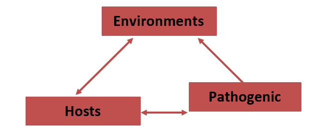
Every time we try to control human and animal diseases in order to limit their socio-economic and ecological impact, we act by modifying certain aspects of their ecology. Disease ecology is therefore a crucial area of science for those involved in disease management and control. This concept is often illustrated by a triangle of interactions.
The aim of this study is therefore to prove the zoonotic importance of rodents in contact with humans, in order to demonstrate the need to find new ways of combating rodents that carry zoonoses.
General Objective
Analyse the zoonotic dynamics of rodents in Guinea in order to assess their impact on public health and develop effective prevention strategies.
Specific Objectives
- Identify the rodent species present in different regions.
- Carry out analyses to detect viruses, bacteria or parasites potentially transmissible to humans.
- Analyse the transmission mechanisms of pathogens between rodents and humans.
- Develop recommendations for limiting contact between humans and rodents.
Materials and Methods
Study Environment
In view of the diversity of ecosystems in our country, we chose prefectures by natural region where diversity would be reasonable.
Materials
Biomaterial: The biomaterial consisted of rodents captured in the various study areas. Capture campaigns were carried out between 15 July 2022 and 7 August 2023.
The biomaterial was collected using a snowball effect. From the first sample found, we collected others according to availability.
Study Framework
The Centre International de Recherche des Infections Tropicales en Guinée (CIRIT-Guinée) was our study setting. It is located in the prefecture of N’Zérékoré, within the N’Zérékoré Regional Hospital, and has a staff of ten, including trainees. The infrastructure comprises 4 offices and 4 analysis laboratories.
Working Methods
Small mammals were captured using Sherman and Formizon traps, baited with dried fish. The traps were set directly in homes, warehouses, barns, vegetable gardens and agricultural fields. At these locations, we marked the spot where the traps would be set. The size of a site is 72 by 135 metres. The traps on this site were placed at exactly the same distance from each other. All the traps at each site were of the same type and were numbered.
Trapping at each site was carried out 4 times a year: Dry season (early and late) October April and Rainy season (early and late) June September.
Each trapping session lasted approximately 8 days. The traps were checked twice a day, in the morning and in the evening, at 12-hour intervals.
The captured rodents were placed in breathable cotton bags that had been moistened beforehand to prevent dehydration. All rodents used in this study were handled in accordance with international animal welfare guidelines, including the Guide for the Care and Use of Laboratory Animals and the AVMA Guidelines for the Euthanasia of Animals [7].
Molecular Study
The blood samples were centrifuged for 10 minutes at 800 rpm to precipitate the red blood cells and obtain serum. The serum obtained was then centrifuged for 10 minutes at 14,000 rpm to precipitate the white blood cells. Total RNA was extracted from the precipitated white blood cells and, separately, from 100 to 500 μl of blood plasma.
The tissue samples were homogenised in 500 µl of saline. Next, nucleic acids were extracted from 100 µl of 10% homogenate using the Amplisense Ribo Prep kit.
For some animal species, species identification was confirmed by sequencing the cytochrome oxidase (cox1) gene region. For this purpose, amplification was performed using primers described in [11].
The sequences obtained were compared using the blast algorithm with the sequences deposited in GenBank.
In addition, animal tissue samples were examined by PCR for the presence of RNA from Lassa virus, Ebola virus, Marburg virus, Jingmen tick virus, Crimean-Congo haemorrhagic fever virus, Flavivirus, Orthobunyavirus, Hantavirus. We also tested for the presence of DNA from Borrelia SPP, Rickettsia spp, Anaplasma spp, Ehrlichia spp and Coxiella burnetti.
Of the animals captured, some were released on the spot after analysis and those transported to the laboratory were all euthanised.
Animals were euthanised by isoflurane inhalation followed by a confirmatory physical method. Specifically, each animal was individually placed in an induction chamber containing an isoflurane buffer (>5%) until deep anaesthesia was achieved (absence of pinch reflex). Death was then induced by rapid cervical dislocation, in accordance with current ethical standards.
A post-mortem examination preceded by weighing, measuring the animal, determining its sex, place of capture, animal identifier, trap number and species identification, followed by the collection of internal organ tissues (brain, lungs, liver, kidneys, spleen, blood and urine). The carcasses were disposed of via the establishment’s secure biosecurity circuit [8, 9, 10].
Statistical Analysis
The data were processed statistically using the following
Results
software: SPSS (20.0) and statistica (version 8.0) (Tables 1-4).
Number of patients
$$ P r e v e l e n c e = \frac {N u m b e r o f p a t i e n t s}{T o t a l P o p u l a t i o n} $$
| Order | Families | Species |
|---|---|---|
| Rodentia | Muridae | Mus musculus(Linnaeus, 1758) ou spp. |
| Rodentia | Muridae | Mus spp.(Linné, 1758) |
| Rodentia | Muridae | Mus setulosus(Peters, 1876) |
| Rodentia | Muridae | Rattus rattus(Linnaeus, 1758) |
| Rodentia | Muridae | Lemniscomys zebra(Heuglin, 1864) |
| Rodentia | Muridae | Mastomys natalensis(Smith, 1834) |
| Rodentia | Muridae | Praomys daltoni(Thomas, 1892) |
| Rodentia | Soricidae | Praomys rostratus(Miller, 1900) |
| Rodentia | Soricidae | Dasymys rufulus(Meunier, 1900) |
| Rodentia | Soricidae | Lemniscomys striatus(Linnaeus, 1758) |
| Rodentia | Soricidae | Lophuromys sikapusi(Temminck, 1853) |
| Rodentia | Soricidae | Crocidura spp.(Walger, 1832) |
| Rodentia | Nesomyidae | Cricetomys gambianus(Waterhouse, 1840) |
| Rodentia | Sciuridae | Xerus erytropus(Desmarest, 1817) |
| Rodentia | Sciuridae | Heliosciurus gambianus(Ogilby, 1835) |
| Rodentia | Sciuridae | Funisciurus pyrropus(Cuvier, 1833) |
| Rodentia | Sciuridae | Epixerus ebii(Temminck, 1853) |
| Rodentia | Gliridae | Graphiurus kelleni(Reuvens, 1890) |
Table 1: Inventory of rodents caught.
In the course of our research, we identified five (5) families: Muridae, Soricidae, Nesomyidae, Sciuridae and Gliridae, divided into (14) genera: Mus, Rattus, Lemniscomys, Mastomys, Praomys, Dasymys, Lophuromys, Crocidura, Cricetomys, Xerus, Heliosciurus, Funisciurus, Epixerus, and Graphiurus kelleni, as well as 18 species. The Muridae family was the most represented, accounting for 11 of the 18 species identified. On the other hand, the Sciuridae family accounted for 4 species, while the least represented families were the Soricidae, Nesomyidae and Gliridae, each represented by just one species.
| Prefectures, Pathogens | N’Zéré- koré | Kindia | Lola | Yomou | Beyla | Macenta | Guéc- kédou | Kankan | Siguiri | Labé | Dalaba | Dubréka | Faranah | Total |
|---|---|---|---|---|---|---|---|---|---|---|---|---|---|---|
| Mus musculus | 24 | 47 | 5 | 3 | 13 | 10 | 20 | 12 | 13 | 14 | 7 | 39 | 23 | 230 |
| Mus spp. | 13 | 18 | 6 | 5 | 11 | 7 | 12 | 9 | 9 | 7 | 6 | 17 | 14 | 134 |
| Rattus rattus | 48 | 57 | 49 | 17 | 39 | 45 | 42 | 40 | 43 | 35 | 52 | 46 | 43 | 556 |
| Mastomys natalensis | 12 | 13 | 5 | 4 | 9 | 12 | 8 | 8 | 8 | 6 | 4 | 12 | 15 | 116 |
| Crocidura spp. | 7 | 18 | 5 | 2 | 6 | 7 | 8 | 3 | 3 | 13 | 5 | 13 | 11 | 101 |
| Lophuromys sikapusi | 10 | 13 | 7 | 6 | 12 | 8 | 0 | 9 | 9 | 5 | 7 | 13 | 16 | 115 |
| Dasymys rufulus | 15 | 15 | 4 | 8 | 20 | 14 | 0 | 5 | 4 | 7 | 5 | 15 | 11 | 123 |
| Lemniscomys striatus | 5 | 16 | 3 | 4 | 13 | 5 | 0 | 10 | 0 | 3 | 4 | 11 | 12 | 86 |
| Mus setulosis | 0 | 5 | 3 | 1 | 5 | 4 | 0 | 0 | 3 | 2 | 6 | 3 | 7 | 39 |
| Cricetomys gambianus | 6 | 18 | 11 | 12 | 14 | 16 | 4 | 10 | 10 | 17 | 12 | 15 | 12 | 157 |
| Xérus erytropus | 2 | 11 | 1 | 5 | 9 | 7 | 2 | 5 | 0 | 13 | 3 | 12 | 15 | 85 |
| Lemniscomys zébra | 5 | 3 | 3 | 3 | 8 | 6 | 0 | 2 | 1 | 7 | 0 | 0 | 11 | 49 |
| Praomys daltoni | 3 | 3 | 0 | 0 | 0 | 1 | 0 | 1 | 0 | 2 | 0 | 0 | 0 | 10 |
| Praomys rostratus | 2 | 2 | 0 | 0 | 0 | 0 | 0 | 0 | 0 | 1 | 0 | 0 | 2 | 7 |
| Funisciurus pyrropus | 0 | 0 | 0 | 0 | 1 | 0 | 0 | 0 | 0 | 0 | 0 | 0 | 4 | 5 |
| Heliosciurus gambianus | 0 | 0 | 0 | 0 | 3 | 1 | 0 | 0 | 0 | 3 | 0 | 0 | 5 | 12 |
| Epixerus ebii | 1 | 0 | 0 | 2 | 1 | 3 | 0 | 0 | 0 | 4 | 0 | 0 | 7 | 18 |
| Graphiurus kelleni | 1 | 0 | 0 | 1 | 2 | 1 | 0 | 0 | 0 | 5 | 0 | 0 | 2 | 12 |
| Total | 154 | 239 | 102 | 73 | 166 | 147 | 96 | 114 | 103 | 144 | 111 | 196 | 210 | 1855 |
Table 2: Summary of rodent species caught by prefecture.
Analysis of the table shows that the prefecture of Kindia recorded the highest number of rodents, with a total of 239 specimens, followed by Faranah and Dubréka, with 210 and 196 specimens respectively. The most abundant species was Rattus rattus, with a total of 556 individuals, followed by Mus musculus (230 individuals) and Cricetomys gambianus (157 individuals). On the other hand, the least represented species, six in number, were: Epixerus ebii (18 individuals), Graphiurus kelleni (12), Heliosciurus gambianus (12), Funisciurus pyrropus (5), Praomys daltoni (10) and Praomys rostratus (7).
| Prefectures/pathogenes | Borrelia spp. | Mamarenavirus lassa | Anaplasma spp. | Leptospira spp. | Ehrlichia spp. | Coxiella burnettii | Total |
|---|---|---|---|---|---|---|---|
| Mus musculus | 12 | 0 | 0 | 0 | 0 | 0 | 12 |
| Mus spp. | 9 | 0 | 0 | 0 | 0 | 0 | 9 |
| Rattus rattus | 15 | 0 | 0 | 0 | 0 | 0 | 15 |
| Mastomys natalensis | 2 | 2 | 0 | 0 | 0 | 0 | 4 |
| Crocidura spp. | 0 | 0 | 3 | 0 | 0 | 0 | 3 |
| Lophuromys sikapusi | 0 | 0 | 0 | 0 | 0 | 0 | 0 |
| Dasymys rufulus | 2 | 0 | 0 | 3 | 0 | 0 | 5 |
| Lemniscomys striatus | 0 | 0 | 2 | 1 | 0 | 0 | 3 |
| Mus setulosis | 0 | 0 | 0 | 0 | 0 | 0 | 0 |
| Cricetomys gambianus | 0 | 0 | 0 | 0 | 4 | 0 | 4 |
| Xérus erytropus | 0 | 0 | 0 | 0 | 0 | 1 | 1 |
| Lemniscomys zébra | 2 | 0 | 0 | 0 | 0 | 0 | 2 |
| Praomys daltoni | 0 | 0 | 0 | 0 | 0 | 0 | 0 |
| Praomys rostratus | 0 | 0 | 0 | 0 | 0 | 0 | 0 |
| Funisciurus pyrropus | 0 | 0 | 0 | 0 | 0 | 0 | 0 |
| Heliosciurus gambianus | 0 | 0 | 0 | 0 | 0 | 0 | 0 |
| Epixerus ebii | 0 | 0 | 0 | 0 | 0 | 0 | 0 |
| Graphiurus kelleni | 0 | 0 | 0 | 0 | 0 | 0 | 0 |
| Total | 42 | 2 | 5 | 4 | 4 | 1 | 58 |
Table 3: Distribution of pathogens by rodent species.
Molecular analysis of the results showed the presence of DNA from Borrelia spp. bacteria, with infections in various rodents caught in different prefectures. The rodent species infected were: Mus musculus, Mus spp, Rattus rattus, Mastomys natalensis, Dasymys rufulus, Lemniscomys zébra. 42 positive samples were identified, indicating potential circulation of Borrelia spp. among rodents in the Republic of Guinea.
Further studies, such as sequencing and microagglutination reaction, are needed to assess the pathogenicity of this bacterium for humans. In addition, blood sediment samples were analysed showing the presence of DNA from Anaplasma spp, Leptospira spp, and Ehrlichia spp. 4 (four) cases indicating the presence of Ehrlichia spp. were detected in Cricetomys gambianus, 4 positive cases were identified for Leptospira spp, distributed as follows: 1 Lemniscomys striatus and 3 Dasymys rufulus, while for Anaplasma spp. 5 cases were identified in Lemniscomys striatus and Crocidura spp.
Analysis of brain tissue samples from Mastomys natalensis detected the presence of Lassa virus RNA in samples from 2 Mastomys natalensis.
| Prefectures/ pathogenes | N’Zéré- koré | Kindia | Lola | Yomou | Beyla | Macenta | Guéc- kédou | Kankan | Siguiri | Labé | Dalaba | Dubréka | Faranah | Total |
|---|---|---|---|---|---|---|---|---|---|---|---|---|---|---|
| Borrelia spp. | 9 | 8 | 2 | 1 | 0 | 2 | 1 | 5 | 3 | 4 | 2 | 5 | 0 | 42 |
| Mamarenavirus lassa | 2 | 0 | 0 | 0 | 0 | 0 | 0 | 0 | 0 | 0 | 0 | 0 | 0 | 2 |
| Anaplasma spp. | 2 | 1 | 0 | 0 | 0 | 0 | 1 | 1 | 0 | 0 | 0 | 0 | 0 | 5 |
| Leptospira spp. | 1 | 1 | 0 | 0 | 0 | 0 | 0 | 0 | 0 | 0 | 0 | 2 | 0 | 4 |
| Ehrlichia spp. | 2 | 1 | 0 | 0 | 0 | 0 | 0 | 1 | 0 | 0 | 0 | 0 | 0 | 4 |
| Coxiella burnettii | 1 | 0 | 0 | 0 | 0 | 0 | 0 | 0 | 0 | 0 | 0 | 0 | 1 | |
| Total | 17 | 11 | 2 | 1 | 0 | 2 | 2 | 7 | 3 | 4 | 2 | 7 | 0 | 58 |
Table 4: Breakdown of agents by Prefecture.
Analysis of the samples revealed 58 positive cases out of the 1 855 samples taken from the various rodent species captured.
Brain tissue samples from Mastomys natalensis were used to detect the presence of Lassa virus RNA in samples from 2 Mastomys natalensis captured in N’Zérékoré. In addition, blood plasma samples from all rodents tested negative for the presence of hantavirus RNA, while blood sediment samples revealed the presence of Coxiella burnettii bacterial DNA in Xerus erythropus in N’Zérékoré.
In addition, blood sediment samples were analysed for the presence of DNA from the bacteria Anaplasma spp. and Ehrlichia spp. Ehrlichia spp. were detected in Cricetomys gambianus in Kindia and Kankan, while DNA from Anaplasma spp, was identified in Lemniscomys striatus in N’Zérékoré and Crocidura spp. in Gueckedou, and the presence of DNA from the bacterium Borrelia spp. was detected in various rodents captured in different prefectures. Finally, blood sediment samples taken from all the rodents were tested for the presence of Leptospira spp. bacterial DNA, and four positive samples were identified, all from N’Zérékoré (Samoé), indicating a potential circulation of Leptospira spp. among rodents there (Figures 1-3).
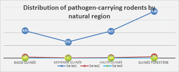
Forest Guinea recorded the highest number of captures, totaling 738 specimens out of 1855, followed by Lower Guinea and Upper Guinea, with 435 and 427 specimens captured respectively. The lowest catches were observed in Middle Guinea, with 255 specimens. In terms of rodents carrying pathogens, Forest Guinea had the highest number of cases, with 24 infected individuals (1.29%), while Lower Guinea had 18 (0.97%).
The results highlight a higher zoonotic risk in Forest Guinea, requiring increased surveillance and targeted efforts to limit pathogen transmission.
In Lower Guinea, although the prevalence is slightly lower, the total number of cases remains worrying because of the large number of rodents caught.
Analysis of our results justifies the importance of adapting zoonotic disease prevention and control strategies to the specific ecological and epidemiological features of each region.
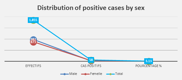
Analysis of brain tissue, blood sediment and organ samples identified 58 pathogens, or 3.13%, of which 38 (2.05%) were found in females and 20 (1.08) in males. These results indicate a higher prevalence of pathogens in females than in males.
These data highlight the importance of differentiating between the sexes in the study of zoonoses, as females could play a more important role in the transmission dynamics of pathogens. An in-depth analysis of the causes of this difference could guide surveillance and prevention strategies, taking account of ecological and behavioural specificities.
Discussion
The composition of the populations in the different study sites during the 2 years of research was as follows: 18 species found compared with 11 species listed by Inapogui, P. et al. in 2000. The greatest species richness was obtained in the Kindia region, where we captured 16 different species against 12 species inventoried by Kéita, N. in 2021 in Kindia [12, 13].
In 2000, Inapogui, P. et al. noted that the rodent population in the selected areas was largely polyspecific and representative of the middle zones of Guinea. The main families characteristic of West Africa are represented. They recorded 11 species, including 8 species of Muridae; one species of Crocidura spp. (Soricidae) and one species of Tatera kempi (Gerbillidae) made up the bulk of the rodent fauna. All these species are likely to play an epidemiological role in the transmission of diseases or as predators of crops and harvests.
Among the true emerging diseases, we were mainly interested in zoonoses linked to wild animals, as there is relatively little data describing or reporting the emergence of these diseases [14].
Our results, following a PCR carried out on blood and organ samples, enabled us to detect 2 cases of Mammarenavirus lassa. This result confirms the declarations by the Guinean health authorities of a fatal case of lassa haemorrhagic fever and more than 30 contacts recorded in the prefecture of Yomou during the month of May 2021, and which was also detected in a 17-year-old patient from the sub-prefecture of Kassadou, prefecture of Guéckédou on 20 April 2022. All this shows that rodents are reservoirs of pathogens and a source of infection for ectoparasites and humans [15].
Hantavirus
Blood plasma samples from all rodents were tested for the presence of hantavirus RNA using genus-specific primers. The result was negative - no hantavirus RNA was detected.
Coxiella burnettii
Blood sediment samples from all the rodents were analysed for the presence of Coxiella burnetti_i bacterial DNA. The pathogen was also detected in tissue samples from _Xerus erythropus killed by a hunter in the N’Zérékoré prefecture. This pathogen can cause serious human illness, so care should be taken when cutting up and preparing infected animals.
Anaplasma spp. and Ehrlichia spp.
Blood sediment samples from all rodents were analysed for the presence of DNA from Anaplasma spp. and Ehrlichia spp. Ehrlichia spp. were detected in samples of Cricetomys gambianus caught in Kindia and Kankan. Anaplasma spp. DNA was detected in two animals: Lemniscomys striatus N’Zérékoré (Samoé) and Crocidura spp. in Gueckedou.
Borrelia
Blood sediment samples taken from all rodents were tested for the presence of Borrelia spp. DNA. Borrelia spp. were detected in animals caught in Labé, Dubréka, N’Zérékoré (Samoé) and Kankan. Three animals Dasymus rufulus, Lemniscomys Zebra, Mus spp. from Samoé were infected with Borrelia spp. In Labé, Dubréka and Kankan, synanthropic rodents Rattus rattus, Mastomys natalensis and Mus musculus were infected. Sequencing is required to genotype Borrelia species.
Leptospira
Blood sediment samples taken from all rodents were tested for the presence of bacterial DNA of Leptospira spp. Four positive samples were identified, all taken in N’Zérékoré (Samoé). Leptospires were detected in Dasymus rufulus and Lemniscomys striatus. This result indicates that Leptospira spp. are circulating among rodents in Samoé. Further studies (sequencing, microagglutination reaction) are required to determine its pathogenicity for humans.
Rodents are an example of a reservoir of diseases that can be transmitted to humans. Other more common diseases, such as arboviroses, and in particular those transmitted by mosquitoes (dengue, chikungunya, etc.), are also closely linked to climatic and environmental conditions. The conditions in which they spread reflect human activities, disrupting ecosystems and eroding biodiversity.
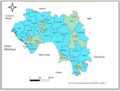
Analysis of the results of our work shows that the areas at risk are the N’Zérékoré and Kindia regions (where the survey was carried out and where rodents are abundant).
Conclusion
At the end of our research work in the four natural regions of the Republic of Guinea on Rodent-related zoonotic dynamics in the Republic of Guinea: implications for the surveillance and prevention of emerging diseases, several specimens were caught and divided into different families and species.
We tested brain tissue samples from Mastomys natalensis for the presence of Lassa virus RNA. A total of 1 855 animals captured in 13 prefectures were tested. Viral RNA was detected in tissue samples from 2 Mastomys natalensis captured in N’Zérékoré.
Blood plasma samples from all rodents were tested for the presence of hantavirus RNA using genus-specific primers. The result was negative - no hantavirus RNA was detected.
Blood sediment samples from all the rodents were analysed for the presence of Coxiella burnettii bacterial DNA. The pathogen was also detected in tissue samples from Xerus erythropus killed by a hunter in the Lola prefecture. This pathogen can cause serious human illness, so care should be taken when cutting up and preparing infected animals.
Blood sediment samples from all the rodents were analysed for the presence of DNA from Anaplasma spp. and Ehrlichia spp. Ehrlichia spp. were detected in samples of Cricetomys gambianus caught in Kindia and Kankan. Anaplasma spp. DNA was detected in two animals: Lemniscomys striatus N’Zérékoré (Samoé) and Crocidura spp. in Gueckedou.
Blood sediment samples taken from all small mammals were tested for the presence of Borrelia spp. DNA. Borrelia spp. were detected in animals captured in Labé, Dubréka, N’Zérékoré (Samoé) and Kankan. Three animals: Dasymus rufulus, Lemniscomys Zebra, Mus spp. from N’Zérékoré (Samoé) were infected with Borrelia spp. In Labé, Dubréka and Kankan, synanthropic rodents Rattus rattus, Mastomys natalensis and Mus musculus were infected. Sequencing is required to genotype Borrelia species.
Blood sediment samples taken from all rodents were tested for the presence of bacterial DNA of Leptospira spp. Four positive samples were identified, all taken in N’Zérékoré (Samoé). Leptospires were detected in Dasymus rufulus and Lemniscomys striatus. This result indicates that Leptospira spp. are circulating among rodents in Samoé. Further studies (sequencing, microagglutination reaction) are required to determine its pathogenicity for humans.
PCR and RT-PCR analyses, combined with bacteriological examinations of rodent tissues and organs, have enabled us to draw up a health status report, while confirming the role of the reservoir for certain zoonoses.
Based on our observations, the general health situation appears to be of little concern, although the presence of zoonoses that are potentially dangerous to humans, particularly professionals, cannot be overlooked.
These diseases are transmitted from host to host under favourable conditions, reflecting their dependence on ecosystem disturbances.
As a result, these diseases and their vectors are likely to circulate within a borderless zone, increasing the risk of contamination for humans. Since wild animals are a major reservoir of zoonotic pathogens, it is vital to identify and understand the transmission routes between wildlife and humans.
Recommendations
Pathogens affecting animals can also cause serious illness in humans. In the absence of a vaccine, the only way to reduce the risk of human infection is to make people and all those involved in healthcare aware of the risk factors, and to recommend measures to limit exposure to the virus. These measures include:
1. Human Community Health • Rat control: It is essential to control the rat population near inhabited areas to reduce the risk of disease transmission. Rat control should be carried out using poisoned bait, but with extreme caution, as rat poison can be harmful to humans. Individual traps and other alternative methods should also be used. • Epidemic management: In the event of an epidemic disease, such as plague, it is crucial to start by disinsectising homes rather than immediately killing the rats. This prevents disease-carrying fleas from moving on to other hosts such as humans. • Careful handling of infected animals: Pathogens can cause serious illness in humans, so extreme caution is required when cutting up and preparing potentially infected animals.
2. Animal health • Surveillance and prevention: It is essential to integrate disease prevention, surveillance and response into government services, taking into account the health of domestic animals, wildlife and the environment. • Strengthening surveillance by the OIE: To better prevent zoonotic diseases, the OIE must consolidate the surveillance and notification of pathogenic micro- organisms, particularly those affecting wildlife.
3. Governmental and institutional involvement • Integration of government services: Public health, animal health and environmental management services must be integrated to enable a coordinated response to epidemics and other health threats. This approach requires the adoption of the ‘One World, One Health’ model, which encourages inter-institutional collaboration. • Collaboration and communication: This new approach requires a high degree of collaboration and communication between different government agencies and institutions, which traditionally have had little contact. • Surveillance and research: The OIE must step up its efforts to monitor pathogens and major epidemiological events. It is also essential to promote interdisciplinary research, breaking down barriers between disciplines and institutions, to bring health services and eco- epidemiology specialists closer together.
Conflicts of Interest
The authors declare no conflict of interest in this article.
Contribution of the Authors
• Conceptualisation: Bonaventure KOLIÉ, Alpha Oumar Sily DIALLO, Boubacar Sidy Sily BAH, Mariama BAH. • Data curation: Bonaventure KOLIÉ, Alpha Oumar Sily DIALLO, Boubacar Sidy Sily BAH. • Formal analysis: Bonaventure KOLIÉ, Mariam BAH, DIALLO Souleymane. • Fund acquisition: Vasily G. Akimkin, Lyudmila S. Karan. • Investigation: Bonaventure KOLIÉ, Alpha Oumar Sily DIALLO, Boubacar Sidy Sily BAH, DIALLO Souleymane, Youssouf CONDE, Amara CISSE, Noumouny SACKO, Raphael TOLNO, Marat T. MAKENOV, Sanaba BOUMBALY. • Methodology: Bonaventure KOLIÉ, Alpha Oumar Sily DIALLO, Boubacar Sidy Sily BAH, Souleymane DIALLO. • Project administration: Sanaba BOUMBALY, Alpha Oumar Sily DIALLO, Boubacar Sidy Sily BAH. • Supervision: Sanaba BOUMBALY, Alpha Oumar Sily DIALLO, Boubacar Sidy Sily BAH • Validation: Bonaventure KOLIÉ, Alpha Oumar Sily DIALLO, Boubacar Sidy Sily BAH, Souleymane DIALLO. • Visualisation: Bonaventure KOLIÉ • Editing original project: Bonaventure KOLIÉ, Alpha Oumar Sily DIALLO, Boubacar Sidy Sily BAH, Souleymane DIALLO. • Writing revision and editing: Bonaventure KOLIÉ, Aly Nènè MANSARÉ, Boubacar Sidy Sily BAH, Alpha Oumar Sily DIALLO, Mariama BAH, Souleymane DIALLO.
Acknowledgements
I would like to thank the Guinean government, in particular the Ministry of Higher Education, Scientific Research and Innovation, the University of Kindia (UK), the Institut Supérieur des Sciences et de Médecine Vétérinaire de Dalaba (ISSMV/Dalaba), the Institut de Recherche en Biologie Appliquée de Guinée (IRBAG/Kindia), the Centre International de Recherche sur les Infections Tropicales en Guinée (CIRIT-GUINEE) and the Moscow-Russia Epidemiology Research Institute.
References
-
Christophe AD (2023) Zoonoses et urbanisation durable au Sud: comprendre les risques pour mieux les prévenir. Dangles, Olivier; Sabrié, Marie-Lise. Sciences de la durabilité: comprendre, co- construire, transformer (2)2: 28-31.
-
Bonwitt J, Kelly AH, Ansumana R, Agbla S, Sahr F, et al. (2016) Rat- atouille: Une étude de méthode mixte pour caractériser la chasse et la consommation de rongeurs dans le contexte de la fièvre de Lassa. EcoSanté 13: 234- 247.
-
Dzingirai V, Bukachi S, Leach M, Mangwanya L, Scoones I, et al. (2017) Facteurs structurels de vulnérabilité aux zoonoses en Afrique. Transactions philosophiques de la Société royale pp: 372.
-
Inegbenebor U, Okosun J, Inegbenebor J (2010) Prévention de la fièvre Lassa au Nigeria... Transactions de la Société Royale de Médecine et d’Hygiène Tropicales 104: 51-54.
-
Organisation mondiale de la santé animale (OIE) (2010) Atelier de formation des Points Focaux Nationaux de l’OIE pour la faune sauvage pp: 1-42.
-
Toma B, Thiry E (2003) Qu’est-ce qu’une maladie émergente ?. Epidemiol. et santé anim No 44: 1-11.
-
Wilson DE, Reeder DM (1993) Mammal Species of the World: A Taxonomic and Geographic Reference. Washington, Smithsonian Institution Press pp: 1206.
-
Rosevear DR (1969) Rodents of West Africa. Trustees of the Birtish Museum (Nat. Hist.), London, UK.
-
(2011) Guide for the Care and Use of Laboratory Animals pp: 246 pages.
-
Underwood, Wendy A (2020) AVMA guidelines for the euthanasia of animals pp: 2020-2021.
-
Karan G (2016) Sexage et phylogénie, à partir des gènes CHD (-Z et -W) et COX-1, des oiseaux de proie du Québec et de perroquets d’attrait vétérinaire.
-
Inapogui AP, Bausch DB, Demby AH, Dieng BM, Koulibaly MJ (2000) Activités du virus Lassa chez les petits Mammifères captures en guinée. Rapport Scientifique, Laboratoire de Mammalogie de L’IRBAG, pp: 28-34.
-
Namory K (2019) Dynamique saisonnière des rongeurs (muridae et soricidae) dans la préfecture de Kindia (Basse Guinée), République de Guinée.
-
Cleaveland S, Laurenson MK, Taylor LH (2001) Diseases of humans and their domestic mammals: pathogen characteristics, host range and the risk of emergence. Phil Trans R Soc Lond B 356: 991-999.
-
French X (2022) Guinée : un cas de fièvre hémorragique à virus Lassa signaler dans le sud.
- Mitochondrial Bio-Logistics: Steering Co-Enzyme Q10 and Lycopene Synergies within the Science 4.0 Bio-OS Framework
- Hymenoptera Specimens from the Caño Negro Wetland, of the National Museum Collection, Costa Rica
- Science 4.0: Comprehensive Architecture of the Biological Operating System (Bio-OS) A Framework for Systemic Resilience and Industrialized Bio-Governance
- Rabbit on, or Hare Back? Understanding Climate Change
- Clinical Validation of Science 4.0: Flow Steering and Epigenetic Drift Inversion on a 76-Year-Old Hybrid System
- Seeds Planted by another Mind