The Functional Outcome of Four-in-One Technique: Dorsal Closing-Wedge & Shortening Osteotomy, Debridement, Micro-Fracture in the Treatment of Freiberg’s Disease
Introduction: Osteochondritis dissecans of the femoral condyles is a subchondral bone necrosis, rare, more or less extended. The osteochondral fragmentation that results from it is responsible for very invalid pain and joint dysfunction. The aim of this work is to evaluate the clinical, radiological and evolutionary aspects of patients operated for osteochondritis dissecans of the femoral condyles by the technique of mosaicplasty at Order of Malta’s Hospital Center in Dakar CHOM. Patients and Method: This is a retrospective, single-center study over a period of 67 months, involving 8 patients (6 men and 2 women). The average age was 25 years and the average BMI 22.93. Standard radiography has been performed in all our patients. 2 patients had MRI and 1 patient did a CT. The collected data were the time of care, the IKDC score, the ICRS score and the Hughston / SFA score. The following characteristics of the lesion - the surface and the location - were also studied. Intraoperatively, the number and diameter of the pads, the location of the donor site and the associated operative procedures were recorded. At last follow-up, patients were assessed according to the Hughston functional and radiological scores, and IKDC. All the patients were immobilized for 6 weeks and benefited from the re-education sessions. Results: At an average follow-up of 36.5 months, the subjective results of the patients were very satisfactory in 62.5% of the cases and the clinical results according to the IKDC were excellent. Healing of osteochondral lesions was achieved in all our patients and 75% were in stage IV according to the radiological Hughston score.
Introduction
Freiberg [1], in 1914, first described what now stands to be the fourth most common osteochondrosis. It is five times more common in females and presents in adolescence. The magnitude of the problem can be appreciated when considering that apart from causing pain, the condition can restrict mobility, limit daily as well as recreational activities and prevent females from wearing fashionable footwear, a major psychological burden. The primary defect involves an interrupted vascular supply to the subchondral bone at the maturing epiphysis of the affected Meta-Tarso-Phalangeal joint (MTP) [2], resulting in bone necrosis and inevitably growth disturbances to the epiphysis or apophysis. Any of the MTP joints may be affected, however, 68% of cases have been found to affect the 2nd MTP joint [3]. It has been suggested that the aetiology is multi-factorial; however, it is unclear whether the initial insult is traumatic or vascular. Conservative measures reducing the load and stress on the metatarsal are used in treating Freiberg’s disease [4, 5, 6], as the first line management. However, failure of conservative management is dealt with operative techniques including debridement [7], osteotomy (dorsal closing-wedge [8] or shortening [9], osteochondral plug transplantation, resection arthroplasty [10, 11]. We proposed a multipronged approach to the management of Freiberg disease. A combined dorsal closing wedge with shortening of the metatarsal and joint debridement and micro-fracture for treating the Freiberg infarction was done. The hypothesis is that dorsal closing wedge and shortening alone cannot help cartilage regeneration. Addition of micro-fracture aids in cartilage regeneration and debridement clears the joint of debris which might further perpetuate damage if left unattended. The aim of our study is to evaluate the functional outcome of Freiberg’s disease patients who were surgically treated with this four in one procedure.
Materials and Methods
Between January 2004 and April 2009, a cohort of 15 symptomatic patients with Freiberg’s disease was surgically treated with the novel four in one procedure. The study was registered with the institutional ethical committee. All patients complained of persistent pain in the affected metatarsal despite intensive conservative treatment. They indicated a reduced range of motion and on clinical examination presented with tenderness over the affected metatarsal head. Patients who failed a trial of conservative management were included. We classified the patients in to various stages of Smillie [12] based on preoperative radiographic and intraoperative findings of the metatarsal head (Table 1). According to this classification system, 1 patient had type I, 6 patients had type II, 4 patients had type III and 2 patients had type IV osteonecrosis. None of our patients showed evidence of type V osteonecrosis. However all stages underwent the four in one procedure in our study. It was the observation of the senior author that cartilage regeneration procedures irrespective of the stage of the disease would help in early healing of the diseased dorsal segment of head.
| Stage I | Subtle fracture of subchondral epiphysis |
|---|---|
| Stage II | Central collapse and flattening of the metatarsa head |
| Stage III | Depression and further flattening of metatarsal head |
| Stage IV | Loose body and separation of fragment |
| Stage V | End-stage degenerative joint disease, marked flattening and widening |
Table 1: Smillie Classification (1967).
Operative Technique
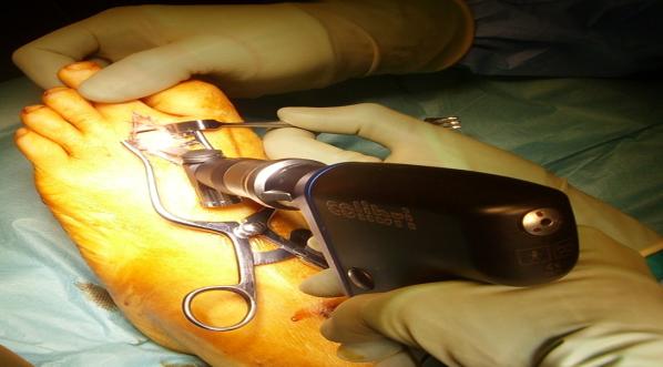
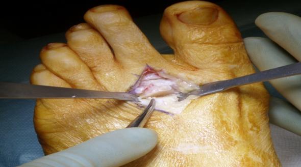
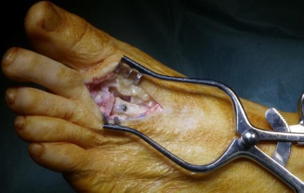
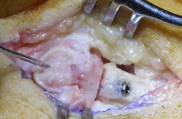
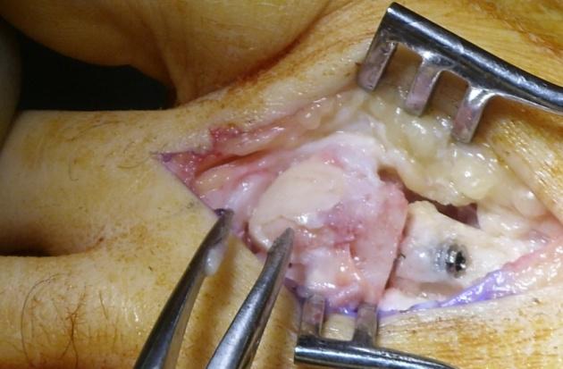
Tourniquet was used for all cases. A dorsal longitudinal incision was made and extensor tendon was laterally retracted. The Meta-Tarso-Phalangeal (MTP) joint as well as the distal metaphysis of the metatarsal bone was exposed. All patients underwent debridement of the MTP joint with removal of loose bodies and synovetomy if needed. A long oblique dorsal wedge osteotomy was performed with an oscillating saw offloading the diseased dorsal cartilage allowing shortening (Figure 1). The metatarsal head was rotated, brining the intact plantar aspect of the articular cartilage into articulation with the proximal phalanx and fixed with a single Barouk headless compression screw (Figure 2). The degenerate articular cartilage was debrided (Figure 3). Micro fractures were created using k-wire (Figure 4). The incision was closed with vicryl sutures and 20ml 0.5% Chirocaine was administered as an ankle block. If deemed appropriate, they were offered physiotherapy to optimise mobilisation exercises. All operations were undertaken by one orthopaedic surgeon to eliminate any intra-operative technique variability.
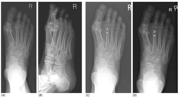
Postoperatively patients were mobilized as able in a heel weight bearing shoe for 6 weeks. Once radiographic evidence of union was noticed normal shoe wear was gradually resumed. Clinical and radiological assessments were performed at 6 weeks after surgery and periodically at three, six and twelve months and then yearly. All patient details including clinical and radiological records were reviewed. They were assessed by subjective patient satisfaction scores with regard to the operation. Pre- and post-operative pain scores were recorded based on visual Analogue Scale (VAS) on a scale of 0 to 10. American Orthopaedics Foot and Ankle Society (AOFAS) scores a standardized method of scoring for the lesser metatarsophalangeal joint was used to assess the functional outcome. This scoring system slots in numerical scales to assess function, alignment and pain. They were scored according to their severity of pain, limitation of daily and recreational activities, ability to wear fashionable footwear, restriction of movement and alignment of their toes. Complications were reviewed from the clinical records. Post-operative metatarsal shortening was evaluated on the dorsoplantar weight-bearing radiographs with a method modified from Jones, et al. [13]. (Figure 5) The continuous data were expressed as mean and range. Wilcoxon signed rank test and Mann Whitney U test were used for assessing the difference in functional outcome measures. The null hypothesis was rejected if the p value was <0.05.
Results
There were 13 female and 2 male patients with a mean age of 36.6 years (range 15-61). The mean follow up period was 15.7 months (range 9-36). 12 patients indicated that the operation provided complete pain relief (p<0.05). 3 patients suffered mild to moderate post- operative pain [14]. Patients had disease of the 2nd metatarsal head and 1 patient had disease of the 3rd metatarsal head. X-rays showed an average healing time of 12.7 weeks (range 6-24 weeks). Patient satisfaction was excellent at an average of 9.3 (range 6-10) (Figure 6).
The mean AOFAS scores were 54 pre-operatively and 82 post-operatively. The mean metatarsal head shortening was 2.18mm (range 0.02-5.51). 1 patient developed a haematoma over the wound site within a week of surgery, which resolved spontaneously. No other complications were reported and there was no neurovascular deficit evident in any patients.
comfortable footwear but required the use of a shoe insert. 11 patients indicated that they did not experience any limitations in their daily or recreational activities after the operation, but 4 patients experienced varying degrees of limitations. 14 of the patients did not report any post-operative complications such as tenderness over the osteotomy site, signs of infection or non-union. 1 patient complained of mild tenderness over the osteotomy site. None of the patients were restricted to wearing modified shoes or braces and none were dependent on a walking aid. None experienced residual limitations of quality of life. None complained of transfer metatarsalgia in adjacent metatarsals (Table 2).
| h | Osteotomy | e | ||||||||||||||||||||||||||||||
|---|---|---|---|---|---|---|---|---|---|---|---|---|---|---|---|---|---|---|---|---|---|---|---|---|---|---|---|---|---|---|---|---|
| S | tud | y | Affected | Stage | P | reoperativ | e | p | Post op | e | Patient | Follow-up in | ||||||||||||||||||||
| Sex | A | g | e | Sid | e | ealing tim | ||||||||||||||||||||||||||
| no | metatarsal | (Smillie) | pain score | ain scor | satisfaction | months | ||||||||||||||||||||||||||
| months | ||||||||||||||||||||||||||||||||
| 1 | F | 25 | R | Third | III | 9 | 0 | 9 | 11 | 15 | ||||||||||||||||||||||
| 2 | F | 15 | L | Second | II | 9 | 0 | 9 | 7 | 14 | ||||||||||||||||||||||
| 3 | F | 22 | L | Second | IV | 9 | 0 | 9 | 23 | 20 | ||||||||||||||||||||||
| 4 | M | 50 | R | Second | II | 8 | 6 | 6 | 24 | 12 | ||||||||||||||||||||||
| 5 | F | 39 | R | Second | I | 9 | 4 | 10 | 8 | 12 | ||||||||||||||||||||||
| 6 | F | 61 | L | Second | IV | 8 | 0 | 10 | 18 | 11 | ||||||||||||||||||||||
| 7 | F | 41 | R | Second | III | 9 | 0 | 9 | 24 | 6 | ||||||||||||||||||||||
| 8 | F | 38 | R | Second | II | 9 | 0 | 9 | 11 | 12 | ||||||||||||||||||||||
| 9 | F | 40 | R | Second | III | 10 | 0 | 10 | 8 | 36 | ||||||||||||||||||||||
| 10 | M | 23 | R | Second | III | 9 | 0 | 9 | 12 | 10 | ||||||||||||||||||||||
| 11 | F | 43 | L | Second | II | 8 | 0 | 10 | 6 | 9 |
Table 2: Details of the fifteen patients who underwent four in one procedure.
| 12 | F | 42 | R | Second | II | 7 | 0 | 10 | 10 | 17 |
|---|---|---|---|---|---|---|---|---|---|---|
| 13 | F | 41 | R | Second | II | 9 | 0 | 10 | 10 | 12 |
| 14 | F | 33 | R | Second | II | 9 | 1 | 10 | 8 | 26 |
| 15 | F | 37 | R | Second | II | 10 | 0 | 10 | 11 | 24 |
Table 3: Details of the fifteen patients who underwent four in one procedure.
Discussion
Over the years, researchers have described various methods of surgically treating Freiberg’s disease.9-12,18- 20,22 Certain patients may improve with conservative treatment alone 13, however, surgical intervention becomes necessary if symptoms are uncontrolled. Surgical debridement produces favourable results (Freiberg’s monogram); however, it does not alter the anatomic and physical conditions initially precipitating Freiberg’s disease [15]. Metatarsal head excision has produced satisfactory results. Ihedioha, et al. [11] reported that 8 out of 9 patients treated with excision arthroplasty reported complete pain relief and continued to wear commercially available footwear. They did not require any walking aids or insoles and were able to walk reasonable distances without pain. However, associated complications such as progressive hallux valgus, transfer metatarsalgia, gait disorders [16] and cosmetic derangement are permanent [17]. A long 2nd metatarsal is often the cause of abnormal overloading and so shortening osteotomy seems a logical surgical choice in such circumstances. Smith et al noted complete pain relief in 15 out of 16 feet; however 7 patients were left with residual stiffness and certain functional limitations, such as an inability to flatten their toes while standing [9]. Miyamoto, et al. investigated the success of osteochondral plug transplantation in 4 patients with late-stage Freiberg’s disease patients. The bone was harvested from the femoral condyle at the knee. The plug transplants all healed within 12 months of surgery and the AOFAS score improved from 70.8 pre-operatively to 97.5 post-operatively 10. However, there is an inherent risk of donor site morbidity. Our technique avoids donor site morbidity. Dorsal closing-wedge osteotomy has been reported to give stable results and good post-operative functionality. There is a reduction in symptoms by joint debridement and also elimination of stress on the metatarsal head to allow healing. No long-term complications have been noted in the literature [15]. A residual loss of flexion and extension is apparent but does not limit the patients’ ability to walk or run. Initially, the procedure was carried out via an intra-articular route but an extra-articular approach has the advantages of increased stability by use of a compression screw, and affords a degree of metatarsal shortening and joint decompression [18, 19]. We used a single compression screw as our fixation method. The disadvantage of using K-wires is the need to remove them before any weight-bearing activities can be commenced [20]. K-wires are usually cut long to allow removal but this irritates adjacent soft tissues causing capsular or tendon adhesion, limits early motion and encourages infection. Plates and screws can lead to stress concentration at the implant and thus bone weakening, but this was not noted in any of our patients. It has been suggested that metallic fixation methods require further implant removal [21]; however, this is not routinely warranted unless there are complications. None of our patients were re-operated for implant removal. Absorbable pin fixation has been reported as an alternative method and allows placement of the base of the wedge in the necrotic dorsal portion with sufficient stability, minimising shortening and elevation of the metatarsal [22]. Lee, et al. [22] reported that in all 12 patients treated with this technique, the osteotomies healed within a few months and pain measurement on a visual analogue scale improved from 8.0 to 2.3. The pins can be cut short allowing free range of motion and smooth gliding of tendons. Because they decompose gradually, stress shielding can be prevented [22, 23]. Patients operated by this method reported increased MTP joint dorsiflexion and it was hypothesised that this may be due to the absence of protruding fixation materials. However, inflammatory foreign body reaction has been the primary drawback to absorbable implants [21]. Our method employs a simple technique without the use of complex equipment, allowing ease of reproducibility. It also brings the plantar intact articular surface to articulate with the proximal phalanx and allows shortening of the metatarsal. Both of these help off- loading the dorsal diseased cartilage. Debridement and micro fractures help regenerating the cartilage through subchondral stem cells.
There are two weaknesses to this study design. It is a retrospective study with inherent bias and a small cohort were observed. However, the relative rarity of the condition makes this a widely accepted reality. We are therefore proposing a prospective multicentre trial. We conclude that the dorsal closing-wedge shortening osteotomy combined with surgical debridement and microfracture is a simple and reliable four-in-one method of treating Freiberg’s disease with no major complications.
References
-
Freiberg AH (1926) The so-called infarction of the second metatarsal bone. J Bone Joint Surg 8: 257-261.
-
Stanley D, Betts R, Rowley D (1990) Assessment of etiological factors in the development of Freiberg’s disease. J Foot Surg 29: 444-447.
-
Binek R, Levinsohn E, Bersani F, Rubenstein H (1988) Freiberg disease complicating unrelated trauma. Orthopaedics 11: 753-757.
-
Trott A (1991) Developmental disorders. In: Jahss M (Ed.), Disorders of the foot and ankle. 2nd (Edn.), WB Saunders, Philadelphia, pp: 613-614.
-
Wu K (1986) Surgery of the metatarsal region. In: Surgery of the foot, Lea & Febiger, Philadelphia, pp: 143-146.
-
Tachdijan M (1990) Freiberg’s infarction. In: Paediatric orthopaedics. 2nd (Edn.), WB Saunders, Philadelphia, pp: 1006-1010.
-
Freiberg A (1914) Infraction of the second metatarsal bone-a typical injury. Surg Gynecol Obstet 19: 191- 193.
-
Gauthier G, Elbaz R (1979) Freiberg’s Infarction: A subchondral bone fatigue fracture. Clin Orthop 142: 93-95.
-
Smith TWD, Stanley D, Rowley DI (1991) Treatment of Freiberg’s disease. J Bone Joint Surg 73(1): 129- 130.
-
Miyamoto W, Takao M, Uchio Y, Kono T, Ochi M (2008) Late-stage Freiberg disease treated by osteochondral plug transplantation: a case series. Foot ankle International 29(9): 950-955.
-
Ihedioha U, Sinha S, Campbell AC (2003) Surgery for symptomatic Freiberg’s disease: exicision arthroplasty in eight patients. The foot 13(3): 143- 145.
-
Simillie IS (1967) Treatment of Freiberg’s infarction. Proc R Soc Med 60(1): 29-31.
-
Jones S, Al Hussainy HA, Ali F, Betts RP, Flowers MF (2004) Scarf osteotomy for hallux valgus. A prospective clinical and pedobarographic study. J Bone Joint Surg 86B: 830-836.
-
Hoskinson J (1974) Freiberg’s disease: A review of the long term results. Proc R Soc Med 67(2): 106-107.
-
Katcherian DA (1994) Treatment of Freiberg’s disease. Foot and ankle injuries in sport 25(1): 69-81
-
Viladot A, Viladot A (1991) Osteochondroses: Aseptic necrosis of the foot. In: Jahss M (Ed.), Disorders of the foot and ankle, 2nd (Edn.), WB Saunders, Philadelphia, pp: 617-638.
-
Thompson F, Hamilton W (1987) Problems of the second metatarsophalangeal joint. Orthopaedics 10(1): 83-89.
-
James M, Rowley D, Betts P (1985) An operation for Freiberg’s disease. J Bone Joint Surg 67B: 671.
-
Botek G, Anderson MA, Balis G (2007) Dorsiflexory wedge osteotomy to treat Freiberg’s Infarction of the second metatarsal head. Podiatry Internet Journal 2(9): 1.
-
Capar B, Kutluay E, Mujde S (2007) Dorsal closing wedge osteotomy in the treatment of Freiberg’s disease. Acta Orthop Traumatol Turc 41(2): 136-139.
-
Bostman OM (1994) Economic considerations on avoiding implant removals after fracture fixation by using absorbable devices. Scand I Soc Med 22: 41-15.
-
Lee SK, Chung MS, Baek GH, Oh JH, Lee YH, et al. (2007) Treatment of Freiberg disease with intra- articular dorsal wedge osteotomy and absorbable pin fixation. Foot and ankle International 28(1): 43-48.
-
Ashammakhi N, Rokkanen P (1997) Absorbable polyglycolide devices in trauma and bone surgery. Biomaterials 18(1): 3-9.
- Return to Work Among Manual Workers After the Latarjet Procedure: A Cohort Study of 43 Patients
- Refractory Pelvic Collection Following Modified Stoppa Approach for Both-Column Acetabular Fracture Fixation: A Case Report
- Comparative Study of Dynamic Knee Phenotypes Under Loaded and Unloaded Conditions: Clinical Impact
- Locked Intramedullary Nailing of the Tibia Using a Humeral Nail: A Care Case Report
- Subtalar Dislocation: About a Case Report
- Surgical Site Infection in Orthopedics in a Country with LimitedResources: Indications, Treatment and Results