Posterior Interosseous Nerve Syndrome Caused by Intramuscular Lipoma: A Rare Case of Radial Neuropathy
Lipomas are common benign soft tissue neoplasms that occur usually in subcutaneous tissue. In rare instances they can occur in the deep soft tissue such as intramuscular and parosteal sites. When an intramuscular lipoma occurring in the proximal forearm or adjacent to the proximal radius they can compress the posterior interosseous branch of radial nerve and cause paralysis of posterior interosseous nerve. In this report we describe an unusual case of a 55-year-old man with posterior interosseous nerve syndrome caused by quite small intramuscular lipoma.
Introduction
The radial nerve originates from the posterior cord of the brachial plexus and it divides into two main branches at the level of the lateral epicondyle. One of these branches is the superficial branch, it’s provides sensory innervation to the skin on the dorsolateral surface of the hand. The other branch becomes the posterior interosseous nerve (PIN), a pure motor nerve. PIN injury is most commonly seen after trauma such as Monteggia fractures [1]. Inflammatory disease, neuralgic amyotrophy and space-occupying lesions are the other non traumatic causes of PIN syndrome (PINS) [1, 2].
Lipomas are benign soft tissue tumors that occur commonly in subcutaneous tissue. Rarely do they present in the deep soft tissue [3]. When intramuscular lipoma occurring in the proximal forearm they can compress the posterior branch of radial nerve and may cause PIN syndrome which characterized by the pure extensor muscle weakness of digital extension that is insidious in onset [4].
In this report we present a new case of PIN syndrome due to quiet small intramuscular lipoma unlike the known cases.
A 55-year-old man was admitted to our clinic with a one-year history of pain and parenthesis in his 2nd, 3rd and 4th fingers, followed by the inability to extend his left index finger. He was unable to perform his domestic activities because of weakness and paresis of the hand. There was no history of trauma or other illness. There was absence of extension of the left index finger and weakness of wrist extension when compared with left (Figure 1). There was no sensory deficit. In nerve conduction studies of the left upper extremity, reduced muscle action potentials of the radial nerve was detected and radial nerve motor conduction velocity was prolonged. In electromyography motor unit loss were recorded in the extensor carpi ulnaris and extensor indicis muscle. With these findings he was diagnosed as having PIN syndrome. In the physical examination there was no palpable mass of left forearm below the elbow. Magnetic resonance imaging (MRI) was performed and revealed a small mass lesion, measuring 1.5 cm at its largest diameter in the anterolateral aspect of the left forearm just below the elbow (Figure 2). Surgical exploration and PIN decompression was planned. General anesthesia was administered and curvilinear incision was made in the proximal forearm. The supinator muscle exposed a fatty encapsulated mass and dissection of the mass revealed it is compressing the PIN. The nerves were carefully released and a lipoma was excised totally. Gross examination of the tumor demonstrated a well circumscribed, 1.5 x 1.5 cm yellow mass clinically consistent with a lipoma (Figure 3). Histological examination of the tumor confirmed it to a benign lipoma. In the early postoperative period, a dynamic splint was applied and, a physiotherapy program was started 2 weeks after surgery. 45 days after operation, there were no changes on electroneuromyography (ENMG) findings. At a six month follow-up the patient neurological examination was the same at at the beginning. No local recurrences were detected.
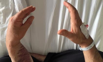
Figure 3: A) The brachioradialis muscle is reflected to expose a fatty encapsulated 1.5 x 1.4 cm mass. The dissection of the mass revealed that it is compressing the PIN. B) The nerves were carefully released and after excision of the mass, the superficial radial nerve and the PIN were preserved. C) Well circumscribed 1.5 x 1.5 cm yellow fatty mass.
Discussion
Symptomatic radial nerve compression caused by lipoma is relatively uncommon. Lipomas over the radial nerve are rare causes of chronic entrapment of the PIN [3]. While lipomas themselves occur commonly in subcutaneous tissue, it is rare to find them in deeper sites as in our case. Other causes of non-traumatic paralysis of the PIN compression are impingement of the nerve at the supinator muscle, rheumatoid arthritis and neuralgic amyothrophy [2, 4, 5].
The diagnosis of PINS is based through clinical history, physical and neurological examination and is confirmed by electrophysiological studies. Patients presenting with lipoma compression will typically describe weakness of digital extension that is insidious in onset [1, 4]. It causes paresis and paralysis of finger extensors without sensory loss. To diagnose PIN, as seen in our patient, electromyographic findings of brachioradialis must be normal. If there is any suspicion a mass as the causative factor of PINS, MRI scan is the method of choice for evaluating their presence and extent [3]. In our case report, there was no palpable mass on the forearm and no trauma history of patient.
Treatment of PIN palsy caused by lipoma is surgical excision [3, 4]. Anterior approach involves dissection between brachioradialis and brachialis muscle and allows for seperation of the PIN under direct vision [4]. The prognosis of PINS relates to the duration of symptoms, early diagnosis and surgical excision [2, 3]. Therefore clinical presentation of PINS must be kept in mind, a comprehensive physical examination should be performed, electrophysiological studies must be revealed and the location of compression must be defined. When the cause of this syndrome cannot be clarified, MRI should be performed and the presence of an space-occupying lesion should be investigated. The prognosis after surgical excision of a lipoma is good. Malignant transformation has not been reported. The longest reported duration of symptoms with full healing postoperatively being 18 months [3, 6]. Jurgens and Haupt [7] revealed 20 patients with PIN paralysis, and they reported that prognosis after surgical excision depended on the duration of symptoms. Thus, long-term paralysis reduces the possibility of reinnervation.
In conclusion early diagnosis and immediate surgical excision are very important in the treatment of PINS. Especially in non-traumatic paralysis, tumoral masses should be considered. It should be kept in mind that the masses may be quite small and non-palpable, as in our case.
References
-
Fitezgerald A, Anderson W, Hooper G (2002) Posterior interosseous nerve palsy due to parosteal lipoma. J Hand Surg Br 27(6): 535-537.
-
Ekinci AS, Çiftçi Ş, Aydoğdu İ (2017) A rare case of focal neuropathy: posterior interosseous neuropathy due to lipoma. Turk J Neurol 23(1): 29-31
-
Allagui M, Maghrebi S, Touati B, Koubaa M, Hadhri R, et al. (2014) Posterior interosseous nerve syndrome due to intramuscular lipoma. Eur Orthop Traumatol 5(1): 75-79.
-
Murphy A, Williams J (2009) Posterior interosseous nerve palsy caused by lipoma: A case report. Can J Plast Surg 17(4): 42-44.
-
Ganapathy K, Winston T, Seshadri V (2006) Posterior interosseous nerve palsy due to intramuscular lipoma. Surg Neurol 65(5): 495-496.
-
Berry JB, Richard MH (1973) Parosteal lipoma producing paralysis of the deep radial nerve. South Med J 66(11): 1298-1300.
-
Jürgens R, Haupt WF (1987) The supinator syndrome: study of the course in 20 patients and therapeutic recommendations. Nervenarzt 58(1): 30-32.
- Return to Work Among Manual Workers After the Latarjet Procedure: A Cohort Study of 43 Patients
- Refractory Pelvic Collection Following Modified Stoppa Approach for Both-Column Acetabular Fracture Fixation: A Case Report
- Comparative Study of Dynamic Knee Phenotypes Under Loaded and Unloaded Conditions: Clinical Impact
- Locked Intramedullary Nailing of the Tibia Using a Humeral Nail: A Care Case Report
- Subtalar Dislocation: About a Case Report
- Surgical Site Infection in Orthopedics in a Country with LimitedResources: Indications, Treatment and Results