Preliminary Phytochemical Screening, Pharmacognostic and Physicochemical Evaluation of Leaf of Argyreia Pilosa Wight & ArnA
<p>Objective: To analyze the pharmacognostic characteristics and physicochemical parameters of the leaves of Argyreia pilosa Wight & Arn (A. pilosa Wight & Arn). Methods: Microscopic characters and powder analysis had been carried out with the help of a microscope. The physiochemical properties such as loss on drying, total ash value, acid insoluble ash value, water soluble ash value, extractive values and fluorescence of A. pilosa had been performed. Results: Macroscopically, the leaves are simple, ovate in shape, acute apex with an entire margin, petioles (1-3 cm) long. Microscopically, the leaf showed the presence of epidermal cells with uniseriate multicellular covering trichomes and anomocytic stomata, followed by 3-5 layered collenchymatous cells and 10-15 numbered Conjoint, collateral closed vascular bundles are some of the diagnostic characteristics observed from the anatomical study. Powder microscopy of leaf revealed the presence of uniseriate multicellular covering trichomes, lignified xylem vessels, and epidermis with anomocytic stomata. The investigations also included leaf surface data i.e., quantitative leaf microscopy and fluorescence analysis. Physiochemical parameters such as loss of drying, extractive values, and ash values were also determined. Preliminary phytochemical screening showed the presence of flavonoids, alkaloids, tannins, steroids, carbohydrates, glycosides, amino acids and proteins. Conclusion: The morphological, microscopical and physicochemical parameter results provided in this paper may be utilized as a basis for the preparation of a monograph on A. pilosa leaves. </p>
Materials and Methods
Plant Collection and Authentication
The plant obtained from Tirupati, Chittoor district of Andhra Pradesh, India during the month of March 2016 and authenticated by Dr. K. Madhava chetty, Taxonomist at Sri Venkateswara University Tirupati, India. Voucher specimen No. 1922 was deposited at the herbarium for future reference. One portion of the leaf is preserved in Formalin: Acetic acid: Alcohol mixture for histological studies and the remaining portion was shade dried, powdered and sieved through 20 mesh and kept in an air tight container for future use.
Chemicals
All analytical grade chemicals were utilized in this study were procured from E. Merck, Germany, absolute alcohol, Phloroglucinol, acetic acid, chloral hydrate, H2SO4, NaOH, HNO3, FeCl3, distilled water, Conc. HCl and chloroform.
Pharmacognostic Evaluation
Morphological evaluation: Organoleptic evaluation of A. pilosa leaves has been carried out in accordance the color, Journal of Natural & Ayurvedic Medicine
size, odor, shape, and taste as per WHO Quality Control methods of herbal medicine [15].
Microscopic Evaluation
Preparation of sections: Microscopic studies had been done by preparing thin hand section of the leaf with the help of sharp cutting edge of the blade, then cleared with chloral hydrate solution, stained with phloroglucinol- hydrochloric acid (1:1) and mounted in glycerin. Powdered microscopy: The powder microscopy was carried out in accordance with the procedure described in Khandelwal [16].
Quantitative Analysis
The quantitative examinations including stomatal number, stomatal index, vein islet number and vein termination number were studied using standard method [2]. Preparation of extracts and preliminary phytochemical analysis: The powdered material had been extracted with various solvents according to its polarity i.e., Petroleum ether, chloroform, ethyl acetate and methanol. 5g leaf powder was extracted with 20 ml of the respective solvent by maceration at room temperature for 24 hours. Then, filtered through what man filter paper and collect the filtrate, concentrated with roto-evaporator. Then, the extracts had been subjected to preliminary phytochemical screening according to standard methods [16, 17].
Physicochemical Analysis
Physicochemical parameters such as ash value, moisture content and extractive values were determined according to the procedures mentioned in WHO quality control methods for herbal materials [15]. Determination of loss on drying: About 10 g of powder drug (without preliminary drying) was measured and positioned in a tared evaporating dish and was dried out at 105°C. The drying, as well as weighing, was being executed at 1 h time intervals until the difference among two successive weighing was not been more than 0.25%. A consistent weight was purported to reach whenever two successive weighing after drying for 30 min and cooling for 30 min in a desiccator, demonstrated not more than 0.01 g difference [15].
Determination of total ash: About 2.0 g of the powder drug was measured properly and incinerated in a silica crucible at a temperature not beyond 450°C till free of carbon. The resulting ash was cooled after which measured. The procedure was repeated to acquire a constant weight. The percentage of total ash with regards to the air‑dried drug was eventually determined [15]. Water soluble ash: The ash was acquired based on the procedure specified above and boiled for 5 minutes together with 25 ml of water, strained and the insoluble matter was acquired on an ash less filter paper. It was furthermore washed together with hot water and inflamed approximately 15 minutes at a temperature not beyond 450°C and weighed. The difference in weight of inflamed and total ash signifies the water-soluble ash. The percentage of water soluble ash was determined with regards to the air dried drug [15]. Determination of acid insoluble ash: The ash was acquired based on the procedure described above and boiled for 5 minutes with 25 ml of 2 M hydrochloric acid, filtered and insoluble matter was amassed on an ash less filter paper. It was even more rinsed with hot water and ignited for 15 minutes at a temperature not exceeding 450°C and weighed. The percentage of acid insoluble ash was determined with regards to the air dried drug [15].
Determination of Extractive Value
Water soluble extractive value: 4 gm of the air dried powder drug had been macerated with 100 ml of water in a closed flask for 24 hours, and shaken frequently during first 6 hours then permitted to stand for 18 hours. It was filtered; 25 ml of filtrate was evaporated in a flat shallow dish, and dried at 105°C and weighed. Percentage of water-soluble extractive value was determined with regards to air-dried drug [15]. Methanol soluble extractive: 4 gm of the air dried powder drug was macerated with 100ml of methanol in a closed flask for 24 hours, and shaken frequently during first 6 hours and permitted to stand for 18 hours. Then it was filtered, during filtration, precaution was taken against loss of ethanol; 25 ml of filtrate was evaporated in a flat shallow dish, and dried at 105°and weighed. Percentage of Ethanol soluble extractive value was calculated with regards to air-dried drug [15].
Journal of Natural & Ayurvedic Medicine
Fluorescence analysis: Various reagents were utilized to check the fluorescence activity. In this, 0.1 g of leaf powder was blended with 1.5 ml of respective reagent (Table 4). The mixture was placed on a slide for a minute and observed under visible light, short ultra-violet light (254 nm) and long ultraviolet light (365 nm) [11, 12].
Thin Layer Chromatographic Profile
TLC was carried out to examine the variance in bioactive chemical constituents. Readymade TLC plates (coated with silica gel 60 F254 on aluminum sheets) obtained from Merck Germany were used. The TLC profile of the methanolic extract was analyzed using various solvent systems. TLC plates were developed in TLC chamber. The chromatograms were then observed under UV-254 nm and UV-365 nm light. Spraying reagent vanillin-sulphuric acid is used. Pictures were taken with Sony camera (DSC-WX200) and the Rf values were calculated with the following formula.
Rf = Distance travelled by Solute
________ Distance travelled by Solvent
Results
Morphological Characteristics
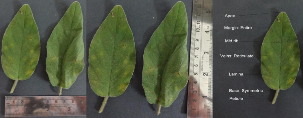
The morphological characteristics of A. pilosa leaves were described in figure 1 and Table 1.
| Morphological | |||||
|---|---|---|---|---|---|
| Observation | |||||
| characters | |||||
| Leaf | |||||
| Size | Length: 8 – 14cm (Avg) Width: 2- 5cm (Avg) | ||||
| Shape | Ovate | ||||
| Apex | Acute | ||||
| Base | Symmetric | ||||
| Venation | Reticular | ||||
| Surface | Pubescent | ||||
| Margin | Entire | ||||
| Petiole | Medium | ||||
| Colour | Green (Dorsal Surface) | ||||
| Pale green (Ventral surface) | |||||
| Odour | Characteristic | ||||
| Taste | Slightly astringent |
Table 1: Morphological characteristics of _Argyreia_ _pilosa_ Wight & Arn. Leaf.
Anatomical Description
Leaf: The transverse section of leaf passing through midrib is convexly protruding at the lower side slightly with more prominent ridged on the upper side (figure 2a), showed uniseriate epidermal cells on both surfaces of the leaf, which was covered by thick cuticle. The epidermis is composed of rectangular shaped cells and contains an anomocytic type of stomata (figure 2b). There is uniseriate multicellular covering trichomes (figure 2c) on the adaxial and abaxial surface of epidermal cells, relatively more on abaxial surface. The epidermal cells followed by 3 -5 layered collenchymatous cells beneath the upper epidermis, and 2-3 layered collenchymatous cells above lower epidermal cells in the midrib region (figure 2d). The cells of collenchyma were thick walled and round in shape showing small intercellular spaces, followed by broad parenchymatous ground cells with intercellular spaces (figure 2e). Conjoint, collateral closed vascular bundles 10-15 were present in ground tissue Journal of Natural & Ayurvedic Medicine
(figure 2f). The phloem consists of companion cells and sieve tubes and xylem consists of spiral annular thickened vessels, tracheids, fibers and xylem parenchyma. T. S lamina shows single layered palisade cells beneath the upper epidermis and 2-3 layered spongy parenchyma above lower epidermis (figure 2g).
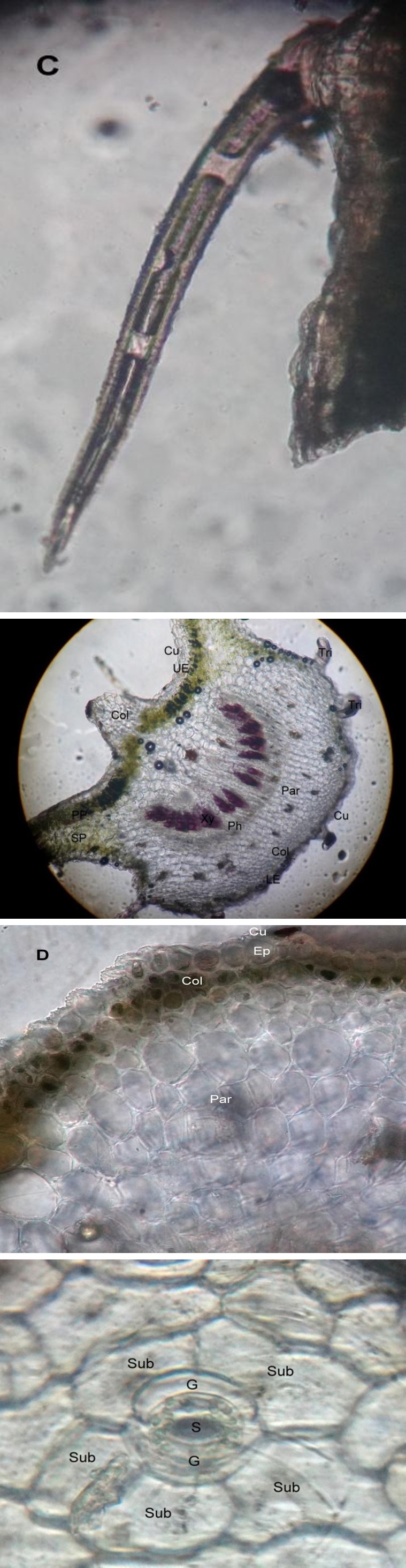
Figure 2(a): Transverse section of Midrib portion of Argyreia pilosa Wight & Arn. Cu: Cuticle; UE: Upper epidermis; Col: Collenchyma cells; PP: Palisade Parenchyma; SP: Spongy Parenchyma; Xy: Xylem; Ph: Phloem; Par: Parenchyma cells; LE: Lower epidermis; Tri: Trichomes.
Figure 2(b): Epidermal cells showed Anomocytic stomata. S: Stoma; G: Guard cells; Sub: Subsidiary cells.
Figure 2(c): Epidermal cells showed uniseriate multicellular covering trichomes.
Figure 2(d): Detailed TS of midrib of leaf showed upper epidermis, collenchyma and parenchymatous cells Cu: Cuticle; Ep: Epidermis; Col: Collenchyma cells; Par: Parenchyma.
Journal of Natural & Ayurvedic Medicine
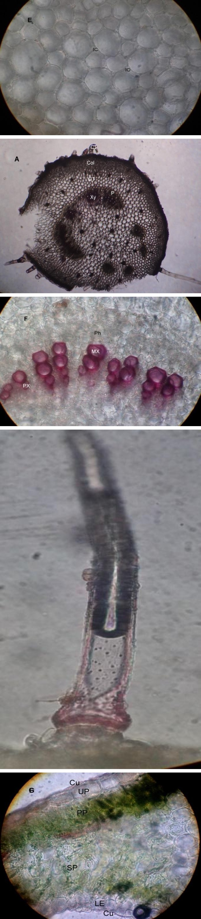
Figure 2(e): Ground tissue of leaf midrib showed Intercellular spaces IC: Intercellular spaces.
Figure 2(f): T.S of Midrib portion of Argyreia pilosa showed Vascular Bundles Ph: Phloem; MX: Metaxylem; PX: Protoxylem.
Figure 2(g): T. S of Lamina of Argyreia pilosa Wight & Arn. Leaf. Cu: Cuticle; UP: Upper Epidermis; PP: Palisade Parenchyma cells; SP: Spongy Parenchyma cells; LE: Lower epidermis.
Petiole: Circular shaped petiole was observed in T.S (figure 3a), showing a layer of thick walled epidermis with uniseriate multicellular covering trichomes (figure 3b). Followed by 4–6 layers of collenchymatous cells were present beneath the epidermal layer (figure 3c). Various sized parenchymatous cells form the ground tissue with intercellular spaces. Vascular bundles are open, bicolateral and arranged in a ring, which was present at the center of the petiole and nature is similar to that of the leaf (figure 3d).
Figure 3(a): Transverse section of Petiole of Argyreia pilosa Wight & Arn. CT: Covering Trichomes; Col: Collenchyma cells; GT: Ground Tissue; Xy: Xylem; P: Phloem.
Figure 3(b): Transverse section of petiole of Argyreia pilosa Wight & Arn. showed uniseriate multicellular covering trichomes.
Journal of Natural & Ayurvedic Medicine
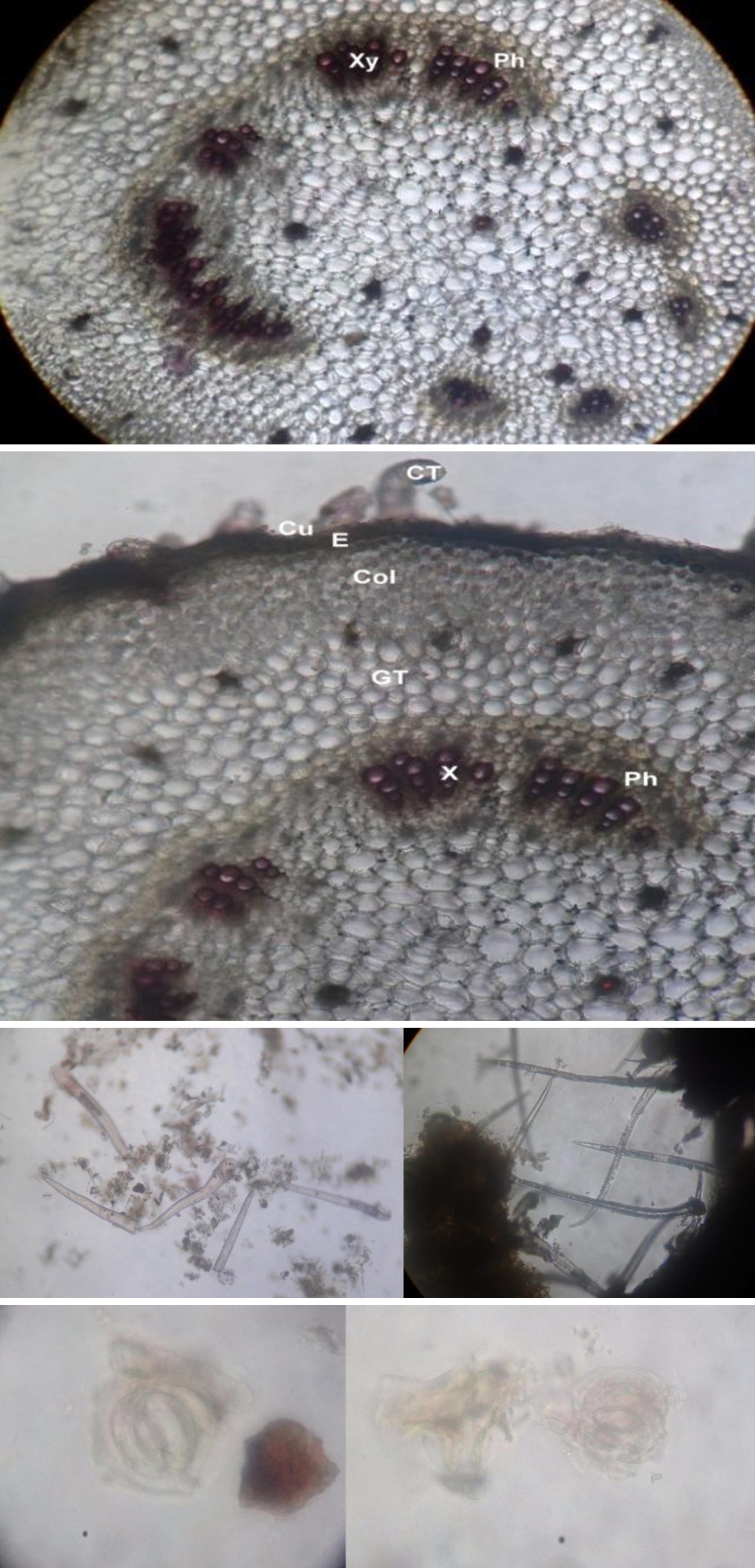
Figure 3(c): Detailed T.S of petiole showed the epidermis, collenchyma, ground tissue and vascular Bundles. CT: Covering Trichome; Cu: Cuticle; E: Epidermis; Col: Collenchyma cells; GT: Ground Tissue; X: Xylem; Ph: Phloem.
Figure 3(d): T.S of petiole showed the arrangement of vascular bundles. Xy: Xylem; Ph: Phloem. Powder Microscopy: The crude powder of leaf was green in color with characteristic odor and taste. Microscopic study of the powder showed revealed different characters such as anomocytic stomata, covering trichomes and xylem vessels (Figure 4).
Figure 4(a): Powder microscopy of leaf showed the presence of uniseriate multicellular covering trichomes.
Figure 4(b): Powder microscopy of leaf showed the presence of Anomocytic Stomata.
Journal of Natural & Ayurvedic Medicine
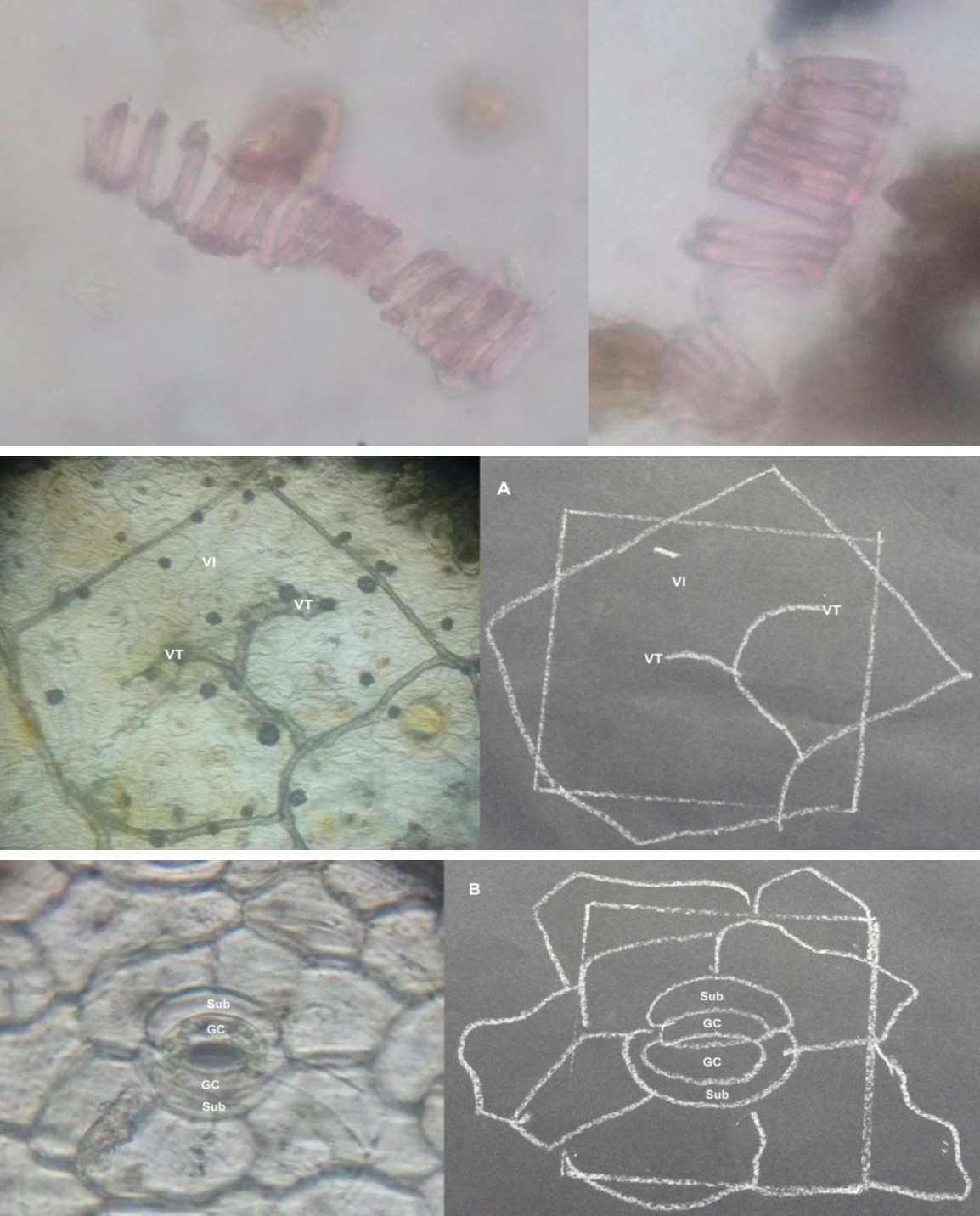
Figure 4(c): Powder microscopy of leaf showed the presence of lignified Xylem Vessels.
Leaf Constants
Leaf venation was reticulate with 4 - 5 pairs of alternate lateral veins. Vein islet number is 12 ± 6 and vein termination number is 15 ± 5.5 (figure 5a). The stomatal number and stomatal index for lower epidermis are 22.2 ± 6.4 and 25 per sq. mm respectively, for upper epidermis 11.1 ± 7.8 and 33.33 per sq. mm, respectively (Figure 5b).
Figure 5(a): Photograph showed the presence of Vein Islet and Vein Termination in surface view of Leaf. VI: Vein Islet; VT: vein terminal.
Figure 5(b): Photograph showed the presence of Anomocytic stomata in surface view of Leaf. GC: Guard cell; Sub: Subsidiary cell.
Journal of Natural & Ayurvedic Medicine
Preliminary Phytochemical Analysis
The results of qualitative phytochemical analysis of crude powder of A. pilosa leaf are shown in Table 2.
| Parameters | Values %w/w | |||
|---|---|---|---|---|
| Moisture content (Loss ]on drying) | 7.5 ± 0.15 | |||
| Total ash | 4.6 ± 0.36 | |||
| Acid insoluble ash | 1.92 ± 0.12 | |||
| Water soluble ash | 2.83 ± 0.23 | |||
| Petroleum ether soluble extractive value | 0.63 ± 0.05 | |||
| Chloroform soluble extractive value | 1.34 ± 0.03 | |||
| Ethyl acetate soluble extractive value | 2.67 ± 0.05 | |||
| Alcohol soluble extractive value | 8.78 ± 0.02 | |||
| Water soluble extractive value | 12.32 ± 0.05 |
Table 2: Physicochemical Parameters of leaf powder of Argyreia pilosa. Wight & Arn.
Physicochemical Parameters
The results attained from various determinations of physicochemical analysis are produced in Table 3.
| Pet. ether | Ethyl acetate | Chloroform | Methanol extract | |||||||||||||
| Phytoconstituents | Method | |||||||||||||||
| extract | extract | extract | ||||||||||||||
| Flavonoids | Shinoda Test | - | + | - | + | |||||||||||
| Zn. Hydrochloride test | - | + | - | + | ||||||||||||
| Lead acetate Test | - | + | - | + | ||||||||||||
| Volatile oil | Stain test | - | - | - | - | |||||||||||
| Alkaloids | Wagner Test | - | - | + | + | |||||||||||
| Hager’s Test | - | - | + | + | ||||||||||||
| Tannins & phenols | FeCl Test 3 | - | - | - | + | |||||||||||
| Potassium dichromate test | - | + | - | + | ||||||||||||
| Saponins | Foaming Test | - | - | - | - | |||||||||||
| Steroids | Salkowski test | + | - | + | + | |||||||||||
| Fixed oils and fats | Spot test | + | - | - | - | |||||||||||
| Carbohydrates | Molish test | - | - | - | + | |||||||||||
| Acid compounds | Litmus test | - | - | - | + | |||||||||||
| Glycoside | Keller-Killani Test | - | - | - | + | |||||||||||
| Amino acids | Ninhydrin test | - | - | - | + | |||||||||||
| Proteins | Biuret | - | - | - | + |
Table 3: Phytochemical analysis of various extracts of _Argyreia_ _pilosa_ Wight & Arn. Leaves. + Present – Absent Fluorescence A
Table 3: Phytochemical analysis of various extracts of Argyreia pilosa Wight & Arn. Leaves. + Present – Absent Fluorescence Analysis: Fluorescence analysis of leaf powder was performed out after treating with different solvents. Fluorescence was observed at 254 and 365 nm comparing its change of color in the visible light. The observations are presented in Table 4 shows the variation in color.
Journal of Natural & Ayurvedic Medicine
| UV light | |||||||||||
|---|---|---|---|---|---|---|---|---|---|---|---|
| Solvent used | Visible light | ||||||||||
| At short (254nm) | At Long (365nm | ) | |||||||||
| Distilled water | Green | Green | Black | ||||||||
| Methanol | Greyish white | Dark green | Greenish black | ||||||||
| 1N Hcl | Green | Black | Black | ||||||||
| 50% HNO 3 | Pale green | Greenish white | Red | ||||||||
| FeCl 3 | Pale yellow | Dark blue | Black | ||||||||
| CHCl 3 | Pale green | Buff | Buff | ||||||||
| Picric acid | Yellowish white | Dark blue | Black | ||||||||
| Ethyl acetate | Green | Buff | Greenish black |
Table 4: Fluorescence analysis of _Argyreia_ _pilosa_. Wight & Arn leaf powder. Thin Layer Chromatography: The methanolic extract
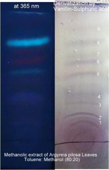
Adsorbent Mobile Phase Spraying
No. of Spots Rf Values
Reagent
0.08 0.15 0.28 0.34 0.39 0.52 0.56 0.63 0.69 Table 5: TLC Chromatographic profile of Methanolic extract of A. pilosa Wight & Arn.
Toluene : Methanol
Vanillin Sulphuric
Silica gel
9 (90:10) acid
Discussion
Indian systems of medicine utilize the majority of the crude drugs which are of plant origin. It is important that standards need to be set down to control and check the identity of the plant and confirm its quality before use. Hence a detailed pharmacognostic assessment is an extremely an important prerequisite. In accordance with World Health Organization (WHO), the organoleptic and histological description of a medicinal plant could be the first step towards establishing its identity and purity and should be performed before to any tests tend to be undertaken [18]. A.pilosa, extensively utilized in conventional medicines has tremendous therapeutical potential due to its various biological activities. The prominent diagnostic characteristics of leaf were uniseriate multicellular covering trichomes, anomocytic stomata, and lignified xylem vessels. These characters can be utilized for standardization of drugs as well as useful for preparation of plant monograph and also reduces the possibilities of adulteration, when the drug is available in the powdered form Studies of physicochemical parameters can serve as an important source to judge the purity and quality of crude drugs. Ash values are utilized to establish the quality and purity of the crude drug. It implies the existence of various impurities like carbonate, oxalate, and silicate. The water soluble ash is water soluble part of total ash, employed to calculate the amount of inorganic substances found in the drugs. The acid insoluble ash comprises mostly silica and indicates contamination with earthy matter. The moisture content of drugs might be at a minimum level in order to suppress the growth of microorganisms like bacteria, yeast or fungi during storage. The extractive values are helpful to judge the chemical constituents present in the crude drug and also assist in the evaluation of particular constituents soluble Journal of Natural & Ayurvedic Medicine
in a specific solvent. Total ash and acid insoluble ash are essential indices to illustrate the quality and purity of the herbal medicine. Total ash consists of physiological ash, which is derived from plant tissue itself, and nonphysiological ash that is usually from atmosphere contaminations includes sand and soil. Total ash content alone is not adequate to indicate the quality of herbal medicine because the plant materials usually contain a significant level of physiological ash, calcium oxalate in particular. Therefore, the acid insoluble ash content is another index to indicate the quality of herbal medicine [19, 20, 21]. The phytochemical analysis of extracts viz., petroleum ether, chloroform, ethyl acetate and methanol were analyzed and it indicates the presence of tannins, flavonoids, steroids, glycosides, volatile oil, amino acids, proteins, and alkaloids.
Conclusion
Standardization of herbal drugs is very much crucial because they are produced from heterogeneous sources which could result in variations. These kinds of variations can cause spurious results in various pharmacological and phytochemical studies. Argyreia pilosa Wight & Arn. leaves are recognized for many therapeutical properties, therefore, the current study might be beneficial to supplement the information in respect to its identification, authentication, and standardization; no such information is available for the same till date.
References
-
Akbar S, Hanif U, Ali J, Ishtiaq S (2014) Pharmacognostic studies of stem, roots and leaves of _Malva parviflora_ L. Asian Pac J Trop Biomed 4(5): 410-415.
-
Amponsah IK, Mensah AY, Otoo A, Mensah MLK, Jonathan J (2014) Pharmacognostic standardisation of _Hilleria latifolia_ (Lam.) H. Walt. (Phytolaccaceae). Asian Pac J Trop Biomed 4(12): 941-946.
-
Traiperm P, Staples GW (2014) A new endemic thai species of _Argyreia_ (Convolvulaceae). Phytotaxa 164(4): 281-285.
-
Chetty KM, Shivaji K, Rao KT (2008) Flowering plants of chittoor district 4th (Edn) Student offset’s printers, Tirupathi,Andhra pradesh, India. _5._ Ambasta SP (2006) The useful plants of India 5th (Edn) Publications and Information Directorate, CSIR
-
New Delhi, India. **7.** Manjunatha BK, Krishna V, Pullaiah T (2004) Flora of Davanagere District, Regency publications, Karnataka, New Delhi, India. **8.** Matthew KM (1995) An Excursion Flora of Central Tamilnadu, CRC press, India.
-
Galani VJ, Patel BG, Patel NB (2010) _Argyreia_ _speciosa_ (Linn. f.) sweet: A comprehensive review. Pharmacogn Rev 4(8): 172-178.
-
Lalan BK, Hiray RS, Ghongane BB (2015) Evaluation of Analgesic and Anti-Inflammatory Activity of Extract of _Holoptelea_ _Integrifolia_ and _Argyreia_ _Speciosa_ in Animal Models. J Clin Diagn Res 9(7): FF01-4.
-
Yadav KS, Yadav NP, Rawat B, Rai VK, Shanker K, et al. (2014) An assessment of wound healing potential of _Argyreia_ _speciosa_ leaves. Scientific World J 2014: 1- 6.
-
Galani VJ, Patel BG (2011) Psychotropic activity of _Argyreia_ _speciosa_ roots in experimental animals. Ayu 32(3): 380-384.
-
Motawi TK, Hamed MA, Hashem RM, Shabana MH, Ahmed YR (2012) Protective and therapeutic effects of _Argyreia_ _speciosa_ against ethanol-induced gastric ulcer in rats. Z Naturforsch C 67(1-2): 47-57.
-
Jhade D, Ahirwar D, Jain R, Sharma NK, Gupta S (2011) Pharmacognostic Standardization, Physico- and Phytochemical Evaluation of _Amaranthus_ _Spinosus_ Linn. Root. J Young Pharm 3(3): 221-225.
-
Ghorpade P, Siddiqui A, Patil MJ, Rukhsana AR (2012) Pharmacognostic and phytochemical evaluation of _Celosia argentea._ Phcog J 4(33): 7-15.
-
Anonymous (1998) Quality Control Methods for Medicinal Plant Materials. Geneva: Office of the Publications, World Health Organization.
-
Khandelwal KR (20012) Practical pharmacognosy techniques and experiments 19th (Edn) Nirali Prakashan, New Delhi.
-
Harborne JB (1984) Phytochemical methods A guide to modern techniques of plant analysis, 2nd (Edn) Springer, USA Journal of Natural & Ayurvedic Medicine
-
Rakholiya K, Chanda S (2012) Pharmacognostic, Physicochemical and Phytochemical Investigation of _Mangifera indica_ L. var. Kesar leaf. Asian Pac J Trop Biomed 2(2): S680-S684.
-
Dave R, Nagani K, Chanda S (2010) Pharmacognostic studies and physicochemical properties of the _Polyalthia_ _longifolia_ var. pendula leaf. Phcog J 2(3): 572-576.
-
Vaghasiya Y, Nair R, Chanda S (2008) Antibacterial and preliminary phytochemical and physico - chemical analysis of _Eucalyptus_ _citriodora_ Hk leaf. Nat Product Res 22: 754-762.
-
Prasanth D S N B K, Rao AS, Yejella RP (2016) Assessment of Pharmacognostic, Phytochemical and Physicochemical Standards of _Aralia racemosa_ (L.) root. Ind J Pharm Edu Res 50(3s): S225-S231.
- Management of Ear Keloid with Ksharsutra: A Case Study
- Yoga and Global Sustainability: A Holistic Path to One Earth, One Health
- Autoimmune Diseases in Ayurveda: A Narrative Review with Classical and Modern Perspectives
- Management of Cluster Headache Associated with Pituitary Apophysitis by CERT (Chakrasiddh Energy Release Technique): A Case Report on Energy Rebalancing
- Zygophyllum Geslini Coss : Biochemicals and Antioxidant Activity
- Observations of a Beginner Vaidya