Identification of Bioactive Chemical Constituents Presents in the Aqueous Extract of Telfairia Occidentalis and Its in vitro Antioxidant Activities
Medicinal plants are utilized as complementary and alternative medicine in different kinds of diseases. The present study was designed to evaluate phytochemical components, of aqueous leaf extract of Telfairia occidentalis and its antioxidant potentials against free radicals. The phytochemical component of the extract was determined by standard methods while the functional groups of the phytochemicals were carried out using FTIR techniques. The antioxidant abilities of aqueous leaf extract of Telfairia occidentalis were evaluated by various antioxidant assays, including 1,1-diphenyl-2-picrylhydrazyl (DPPH), hydrogen peroxide, hydroxyl radicals, scavenging activities. These various antioxidant activities were compared with standard antioxidants such as ascorbic acid and Gallic acid. Qualitative analysis of the extract revealed that the extract is rich in phenol, coumarins, saponin, tannin, anthraquinone, betacyanin, glycosides, oils and resin, quinones, flavonoids, alkaloids, sterols, vitamin C, terpenoids, sterols and phytosterols. Moreover, the results of FTIR analysis confirmed the presence of aromatic CH bend, C-O stretch, phenol, aromatic ring stretch, alkenyl stretch, hydroxyl group (alcohol) in the extract. The extract exhibited high reduction capability and powerful free radical scavenging, especially against DPPH radical and hydrogen peroxide. The extract shows higher inhibition activities than both ascorbic and Gallic acid. The results obtained in the present study clearly established the antioxidant potency of aqueous leaf extract of Telfairia occidentalis, which may account for some of the medical claims attributed to this plant.
Introduction
Medicinal plants house many secondary metabolites such as flavonoids, alkaloids, phenols, tannins, phlobotanins, saponins, glycosides steroids which enhance some physiological activities in human [1]. World Health Organization (WHO) indicated that medicinal plants contain chemical substances that can be used for the synthesis of useful drugs [2]. From this, the knowledge of these metabolites are essential, the constituents of a plant aid its usage in the synthesis of drug to fight against some diseases. These secondary metabolites are also known as phytochemicals, which are plant chemicals that protect plant cell walls from any form of environmental pollution, also from being attacked by any pathogens like; bacteria, virus, etc.
Telfairia occidentalis is is a medicinal plant with a number of claimed pharmacological activities. It is classified in the tribe Joliffieae of the subfamily Cucurbitaceae. It is called fluted pumpkin, ugwu. Telfairia occidentalis occurs in the forest zone of West and Central Africa and they are found more in Benin, Nigeria and Cameroon. Kayode and kayode [3] reported that Telfairia occidentalis is a well- known vegetable all over Nigeria and constituted the main vegetable food. Previous experimental studies from our laboratory documented the modulatory effects of Telfairia occidentalis extract on chemically induced electrolyte imbalance, pancytopenia, oxidative stress and hepatorenal damage [4, 5, 6].
Similarly, some researchers have reported so many findings on Telfairia occidentalis, for instance, Alada [7] and Dino, et al. [8] documented that herbal preparation of the plant has been used in the treatment of anaemia, chronic fatigue and diabetes. It has been used to manage cholesterolemia, liver problems and impaired defense immune systems [9]. The plant has been used in soup and folk medicine preparation for the management of various diseases like diabetics, anaemia and gastrointestinal disorder [10]. From all these usage, it is of great importance to know the exact constituents of Telfairia occidentalis that has the ability to cure all these diseases. Thus, this study was undertaken to identify the phytochemical components of aqueous leaf extract of Telfairia occidentalis and to examine the extract’s free radical scavenging and antioxidant abilities.
Materials And Methods
Chemicals and Reagents
Folin–Ciocalteu’s reagent, HCl, methanol, gallic acid, H2SO4, Na2CO3, aluminium chloride, potassium acetate, potassium persulphate, sodium nitroprusside, hydrogen peroxide, sulphanilic acid, glacial acetic acid, naphthylethylenediamine dichloride, NADH were all purchased from Merck, USA. DPPH (1,1-diphenyl-1,2-picryl hydrazyl), TPTZ (2,4,6,-tripyridyl-s-triazine), Ferrozine, Deoxyribose Sigma (St Louis, MO, USA). Trichloroacetic acid (TCA), L-Ascorbic acid, and all other chemicals and reagents used were of analytical grade.
Plants Collection
Fresh samples of Telfairia occidentalis leaves were harvested in a garden at Obada area of Kings University, Ode-Omu, Osun State, Nigeria. The plant was identified and deposited at the herbarium of the Department of Biological Sciences, Osun State University, Osogbo, Osun State, Nigeria. This research was conducted between November, 2019 and February 2020.
Preparation of Extract
The fresh samples of Telfairia occidentalis leaves were air dried at room temperature to constant weight after which they were pulverized into powder using an electrical blender.
The powdered leaf materials were cold-macerated with 6 volumes of 80% distilled water for 72 hours. Crude extract was obtained through filtration and then concentrated. The paste was then freeze dried.
Qualitative Phytochemical Analysis
Qualitative screening of aqueous leaf extract of Telfairia occidentalis was carried out to identify the active phytochemicals like phenols, flavonoids, saponins, tannins, coumarins, alkaloids, terpenoids, anthraquinones and anthocyanins.
Test for anthraquinones: 2% diluted hydrochloric acid was added to 1 mg of aqueous leaf extract of Telfairia occidentalis. The appearance of red colour was interpreted as the presence of anthraquinone [11].
Test for phenols: Assessment of phenol was determined following the method described by Harborne [11]. Briefly, 2 ml of distilled water was added to 1 mg of aqueous leaf extract of Telfairia occidentalis and 10% ferric chloride was added in it. The confirmation sign of phenols presence was formation of green or blue color.
Test for coumarins: 1 mg of aqueous leaf extract of Telfairia occidentalis was reacted with 1 ml of 10% sodium hydroxide. Formation of yellow colour in test sample was an indication of the presence of coumarins [11].
Test for flavonoids
Alkaline reagent test: This test was carried out using the procedure described by Trease and Evans [12]. 1 ml of 2 N sodium hydroxide was added to 1 mg of aqueous leaf extract of Telfairia occidentalis. Formation of yellow colour was interpreted as the presence of flavonoids.
FeCl3 test: Few drops of FeCl3 solution were added in 1 ml of the extract. Existence of flavonoids was indicated by formation of blackish red precipitate [13].
Test for saponins: Froth formation with distilled water: 2 mg of the extract was mixed with 2 ml of distilled water in the test tube. After this accumulation, the test sample was mixed vigorously for almost 15 min. The formation of a soapy layer indicated the presence of saponins in test samples [11].
Test for alkaloids
Mayer’s test: 2 ml of the extract was reacted with concentrated HCl and a special reagent named Mayer’s reagent. Formation of white precipitates or appearance of green colour was indication of alkaloids presence [12].
Hager’s test: Few drops of Hager’s (Saturated picric acid solution) reagent were added to 2 ml of the extract. Formation
of bright yellow precipitates specified the manifestation of alkaloids [13].
Test for anthocyanin and betacyanins: 1 mg of the extract was taken in the test tube and followed by the addition of 2 ml of 1 N sodium hydroxide. The test sample was boiled at 100 °C for about 10 min. Anthocyanin presence was indicated by the formation of bluish green colour while yellow colour formation was indicative of betacyanin presence [12].
Test for sterols
Salkowski test: To 2 ml of the extract, 5 ml of chloroform was added and then 1 ml concentrated H2SO4 was carefully dispensed along the walls of the tube. The appearance of reddish colour in the lower layer indicated the existence of sterols [13].
Test for vitamin C: Dinitrophenyl hydrazine was dissolved in concentrated sulphuric acid and allowed to react with 1 ml of the extract. Appearance of yellow precipitates indicated the presence of vitamin C in test samples.
Test for proteins
Xanthoproteic test: According to this procedure, 1 ml of extract was treated with few drops of conc. nitric acid. Presence of proteins in test samples was indicated by the formation of yellow colour.
Biuret test: 0.5 mg of extract was taken and equal volume of sodium hydroxide solution (40%) was added to it. After that few drops of 1% CuSO4 solution was added. Appearance of violet colour in test samples manifested protein presence.
Emulsion test with olive oil: 1 ml of extract was poured in test tubes followed by addition of 5–6 drops of olive oil and shaken vigorously to form a stable froth. Formation of an emulsion was the confirmatory sign of saponin presence [13].
Test for tannins
FeCl3 test: To 1 mg of extract, 2 ml of 5% ferric chloride was added. Appearance of greenish black or dark blue colour was the indication of tannins presence [12].
Alkaline reagent test: 2 ml of 1 N NaOH solution was added in 2 ml of the extract. Appearance of yellow to red colour showed the presence of tannins [13].
Test for triterpenoids: 1 ml of Libermann-Buchard Reagent (conc. H2SO4 + acetic anhydride) was added in 1.5 ml extract. Triterpenoids were determined by the appearance of bluish- green color in the test samples.
Test for terpenoids: 0.5 mg of extract was taken in the test tube and 2 ml of each chloroform and concentrated sulphuric acid was added to plant sample. Presence of terpenoids was indicated by the formation of brown layer in the middle of other two layers [12].
Test for glycosides (Keller Killanis’ test): To 1 ml of extract, 1 ml glacial acetic acid was added and left to cool down. After cooling two drops of FeCl3 were added and 2 ml of concentrated H2SO4 along the walls of test tube was dispensed carefully. Development of reddish brown coloured ring at the intersection of two layers indicated the presence of glycosides [13].
Test for oils and resins (Filter paper test): Extract was applied on filter paper and checked for the establishment of transparent appearance which was a positive sign for the presence of oils and resins in respective test sample.
Test for steroids and phytosteroids: 1 ml of chloroform and few drops of concentrated sulphuric acid were added to 1 ml of the extract. Formation of brown-colored ring indicated steroids presence whereas appearance of bluish- brown coloured ring marked the presence of phytosteroids in the test samples.
Test for phlobotannins: To 1 ml of the extract, few drops of 10% ammonia solution were added. Formation of pink- coloured precipitates showed the existence of phlobatannins in samples.
Test for quinones: A volume of 1 ml of extract was allowed to react with 1 ml concentrated sulphuric acid. Appearance of red colour manifested the occurrence of quinones.
FT-IR Spectroscopic Analysis
Fourier Transform Infrared Spectrophotometer (FTIR) is currently the best technique and equipment to evaluate types of chemical bonds/functional groups present in natural products or phytochemicals. The wavelength of light absorbed is the salient feature of the chemical bonds seen in the annotated spectrum. By interpreting the infrared absorption spectrum, the chemical bonds in a compound can be determined. Dried powder of aqueous leaf extract of Telfairia occidentalis was used for FTIR analysis. 10mg of the dried extract powder was encapsulated in 100mg of KBr pellet, in order to prepare translucent sample discs. The powdered sample of the extract was loaded in FTIR spectroscope (Shimadzu, Japan), with a Scan range from 400 to 4000 cm-1 with a resolution of 4 cm-1.
Hydrogen Peroxide Scavenging Assay
The ability of the extract to scavenge hydrogen peroxide was determined according to the method of Ilhami, et al. [14]. A solution of hydrogen peroxide (40 mM) was prepared in phosphate buffer (pH 7.4). Different concentrations of plant extract were added to a hydrogen peroxide solution (0.6
ml, 40 mM). The absorbance of hydrogen peroxide at 230 nm was determined after 10 min against a blank solution containing phosphate buffer without hydrogen peroxide. The percentage inhibition of hydrogen peroxide of extracts and standard compounds (Vitamin C and Tannic acid) was calculated using the following formula:
% inhibition [H2O2] = [(A0-A1)/A0] x 100 Where A0 was the absorbance of the control, and A1 was the absorbance in the presence of the sample of extract and standards.
Hydroxyl Radical Scavenging Assay
Hydroxyl radical scavenging activity of the extract was determined by the method of Klein, et al. [15] with a slight modification. 0.5 ml of extract or standard (Vitamin C and Tannic acid) at different concentration was taken in test tubes. 1 ml of Fe-EDTA solution (0.13% ferrous ammonium sulphate and 0.26% EDTA), 0.5 ml of 0.018% EDTA solution, 1 ml of 0.85% DMSO solution and 0.5 ml of 22% ascorbic acid were added into the test tubes. The test tubes were capped tightly and warm at 85°C for 15 minutes into the water bath. After incubation, the test tubes were uncapped, and 0.5 ml ice-cold TCA (17.5%) was added to each of the test tubes immediately. 3 ml of Nash reagent (7.5 g of ammonium acetate, 300 μl glacial acetic acid and 200 μl acetylacetone were mixed and made up to 100 ml) was added to all the tubes and incubated at RT for 15 minutes. Absorbance was taken in UV-spectrophotometer at 412 nm wavelength. Percentage hydroxyl radical scavenging (% HRSA) activity was calculated using the following equation:
% HRSA = {(A0-A1)/A0} × 100 Where A0 is the absorbance of the control, and A1 is the absorbance of the extracts/standard.
DPPH-Radical Scavenging Assay
The radical scavenging activity of plant extract was measured as described by Mensor, et al. [16]. The stable 2, 2 diphenyl-1-picryldydrazyl (DPPH) radical was used for the determination of free radical scavenging activities of the extracts. A portion (1 ml) each of the different concentrations (40-2000 µg/ml) of the extracts or standard (Vitamin C and Tannic acid) in test tubes was added to 1 ml of 1 mM DPPH in methanol. The mixtures were vortexed and then incubated in a dark chamber for 30 min after which the absorbances were measured at 517 nm against a DPPH control containing only 1 ml of methanol in place of the extract. All calculations were carried out in triplicates. The inhibition of DPPH was calculated as a percentage using the expression:
% I = [(Acontrol - Asample)/ Acontrol] × 100
Where %I is the inhibition of the DPPH free radicals in percentage; Acontrol is the absorbance of the control reaction containing all reagents except the test compound, and Asample is the absorbance of the test compound.
Statistical Analysis
The results were analysed using SPSS Version 12. Data were expressed as mean± standard error of the mean (mean ± SD). Student’s t-test was employed for comparison between two sets of data while p < 0.05 was considered statistically significant.
Results
Preliminary phytochemical analysis of the extract
The results of qualitative analysis of the crude aqueous leaf extract of Telfairia occidentalis shown in Table 1. It revealed that the extract is rich in phenol, coumarins, saponin, tannin, anthraquinone, betacyanin, glycosides, oils and resin, quinones, flavonoids, alkaloids, sterols, vitamin C, terpenoids, sterols and phytosterols.
| Phytochemicals | ATO |
|---|---|
| Phenol | + |
| Coumarins | + |
| Saponin | + |
| Tannin | + |
| Anthraquinone | + |
| Anthocyanin | - |
| Betacyanin | + |
| Glycosides | + |
| Phlobotannin | - |
| Oils and Resin | + |
| Proteins | - |
| Quinones | + |
| Flavonoids (Alkaline Reagent) | + |
| Flavonoids (Fecl ) 3 | + |
| Alkaloids | + |
| Sterols | + |
| Vitamin C | + |
| Sterols and Phytosterols | + |
| Triterpenoids | - |
| Terpenoids | + |
Table 1: Phytochemical components of ethanol leaf extract of aqueous leaf extract of Telfairia occidentalis Keynotes: “+” = prese
Fourier Transform Infrared Spectroscopic Analysis of the extract
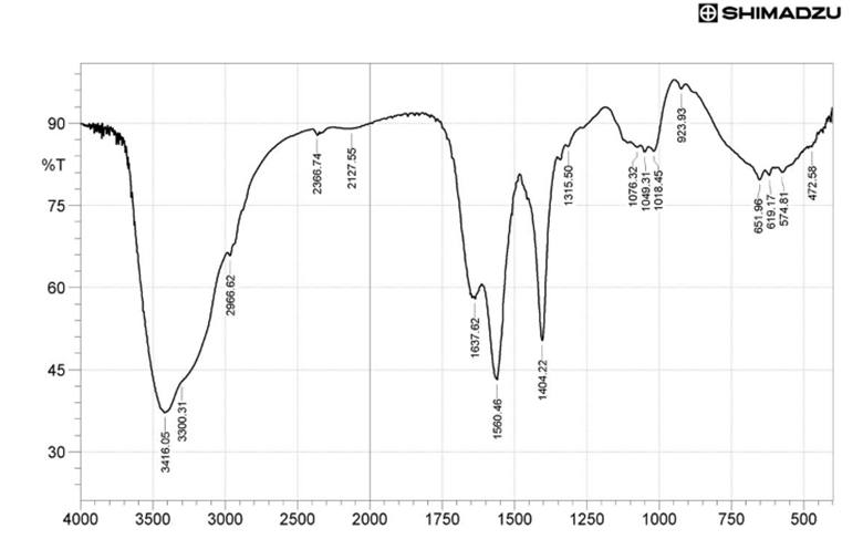
The FTIR spectrum of aqueous leaf extract of Telfairia occidentalis is presented in Figure 1 & Table 2 depicted the data on the peak values and the probable functional groups (obtained by FTIR analysis) present in the aqueous leaf extract of Telfairia occidentalis. The region of IR radiation helps to identify the functional groups of the active components present in extract based on the peaks values of the FTIR spectrum. When the extract was passed into the FTIR, the functional groups of the components were separated based on its peaks ratio. The results of FTIR analysis confirmed the presence of aromatic CH bend, C-O stretch, phenol, aromatic ring stretch, alkenyl stretch, hydroxyl group (alcohol). The absorbance bands analyses in the process are observed in the region between 400–4000 cm-1 are 651.96, 923.83, 1049.31, 1404.42, 1560.46, 1637.62, 2127.55, 2966.62, 3300.31 and 3416.05 cm-1.
| S/N | Peaks wavelength (cm-1) | Functional groups |
|---|---|---|
| 1 | 651.96 | Aromatic CH out-of-plane bend |
| 2 | 923.83 | OH bend |
| 3 | 1049.31 | C-O stretch |
| 4 | 1404.42 | Phenol or tertiary alcohol, OH bend |
| 5 | 1560.46 | Aromatic ring stretch |
| 6 | 1637.62 | Alkenyl C=C stretch |
| 7 | 2127.55 | C –H Stetch |
| 8 | 2966.62 | Methyl C-H asym./ sym. Stretch |
| 9 | 3300.31 | Aromatic C-H stretch, Normal OH stretch |
| 10 | 3416.05 | Hydroxyl group H-bonded OH stretch |
Table 2: FTIR spectral peak valves and functional groups obtained from aqueous leaf extract of Telfairia occidentalis.
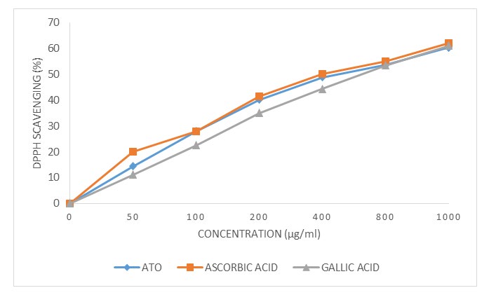
DPPH free radical scavenging activity
The in vitro antioxidant activity of the aqueous leaf extract of Telfairia occidentalis was measured in comparison to the standard antioxidants (Ascorbic and Gallic acids). However, the percentage of DPPH radical scavenging activity occurred in a dose-dependent manner. The aqueous leaf extract of Telfairia occidentalis displayed higher scavenging activity (60.33%) than the standard antioxidant (ascorbic acid) (62.03%) at the concentration of 1000 μg/mL (Figure 2), suggesting that aqueous leaf extract of Telfairia occidentalis has the capacity to reduce oxidative stress caused by free radicals.
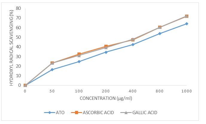
Hydroxyl radical scavenging activity
The hydroxyl radical is the major reactive oxygen species responsible for lipid oxidation and potentially severe biological damage. This assay shows how aqueous leaf extract of Telfairia occidentalis and the standard antioxidants (Ascorbic and Gallic acids) inhibit hydroxyl radical-mediated deoxyribose degradation generated in a Fenton reaction. The hydroxyl radical scavenging capacity of the aqueous leaf extract of Telfairia occidentalis and the standard antioxidants (Ascorbic and Gallic acids) were 63.99%, 71.78% and 72.14% respectively at the concentration of 1000 μg/mL (Figure 3). The results show that aqueous leaf extract of Telfairia occidentalis possesses similar capacity of hydroxyl radical scavenging to the antioxidant standards, suggesting aqueous leaf extract of Telfairia occidentalis could provide a major source of antioxidant. Moreover, the ability of aqueous leaf extract of Telfairia occidentalis to quench hydroxyl radicals might be of direct relevance to the prevention of lipid peroxidation.
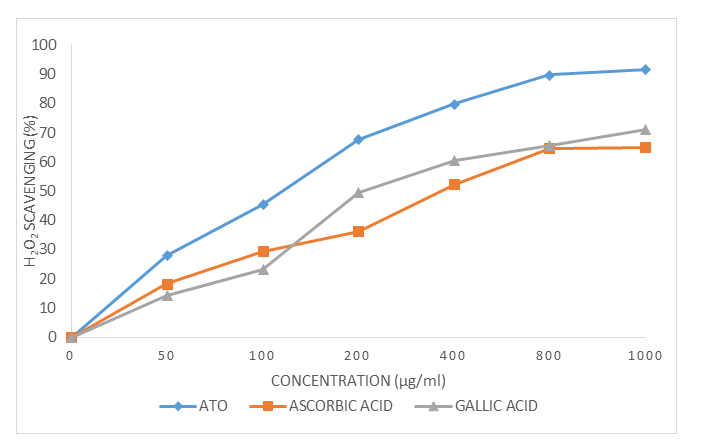
Hydrogen Peroxide Scavenging Activity
The results of the in vitro hydrogen peroxide scavenging activity of aqueous leaf extract of Telfairia occidentalis and the standard antioxidants (Ascorbic and Gallic acids) was shown. The percentage hydrogen peroxide scavenging activities of the extract, ascorbic acid and Gallic acid were
91.53%, 64.95% and 71.21% at the concentration of 1000 μg/mL (Figure 4). Although, the inhibition hydrogen peroxide occurred in a dose-dependent manner, aqueous leaf extract of Telfairia occidentalis has the highest activity, suggesting that the extract has the capacity to reduce oxidative stress caused by hydrogen peroxide.
Discussion
There is an increase in the rate of searching for plants derived drugs and dietary supplements in recent years. Scientists in the field of natural sciences are exploring the nature for natural products and phytochemicals that have therapeutic potentials which could help in the treatment of various disorders/diseases. All plants produce chemical compounds as part of their normal metabolic activities. These phytochemicals are divided into: primary metabolites such as sugars and fats, which are found in all plants; and secondary metabolites compounds which are found in a smaller range of plants, serving a more specific function [17]. It is these secondary metabolites and pigments that can have therapeutic actions in humans and which can be refined to produce drugs [17].
The qualitative phytochemical evaluation of aqueous leaf extract of Telfairia occidentalis showed that the extract is rich in phenol, coumarins, saponin, tannin, anthraquinone, betacyanin, glycosides, oils and resin, quinones, flavonoids, alkaloids, sterols, vitamin C, terpenoids, sterols and phytosterols. These bioactive constituents are known to possess medicinal activity as well as physiological activity [18]. Tannins are polyphenols that are obtained from various parts of different plants belonging to multiple species [19]. They are usually found in larger quantity in the barks of trees where they act as blockage to bacteria and fungi and protect the cell. Tannins are well known for their anti- oxidant and anti-microbial properties, as well as for soothing relief, skin regeneration, anti-inflammatory and diuretics [20]. Flavonoid belongs to the family of polyphenols. They are water insoluble and are found in most plant materials. Flavonoids are well known for their anti-oxidants, anti- carcinogenic, anti-microbial and anti-tumor properties [21]. Epidemiological studies have demonstrated that heart diseases are inversely related to flavonoid intake [22]. Studies have shown that flavonoids prevent the oxidation of low density lipoprotein thereby reducing the risk for the development of atherosclerosis.
FTIR analysis was carried out on aqueous leaf extract of Telfairia occidentalis to identify the functional groups present. The result of the analysis in the spectra in Figure 1
& Table 2 below showed the extract contains phytochemicals with different functional groups like aromatic CH bend, C-O stretch, phenol, aromatic ring stretch, alkenyl stretch, hydroxyl group which shows peaks of 651.96, 923.83, 1049.31, 1404.42, 1560.46, 1637.62, 2127.55, 2966.62, 3300.31 and 3416.05 cm-1. This result further corroborates the presence of phytochemicals in aqueous leaf extract of Telfairia occidentalis as listed in Table 1.
The biological damage caused by oxidative stress includes DNA oxidation, lipid oxidation, protein oxidation, and glycoxidation [23] which have been implicated in the pathogenesis of many diseases such as cancer, neurodegenerative disorders, cardiovascular diseases, diabetes etc. [24]. Oxidative stress is generally characterized by an imbalanced production of ROS and reactive nitrogen species (RNS). Oxidative stress is initiated by free radicals such as the hydrogen peroxide (H2O2), superoxide anion radical (O2 •−), hydroxyl radical (•OH), alkoxyl radicals (RO•), singlet oxygen (1O2), and peroxyl radicals (ROO•), which have a tendency to become stable through electron pairing with biological macromolecules like lipids, proteins, and DNA in healthy human cells. This tendency has led to the assertion that oxidative stress contributes to the pathogenesis of numerous human diseases [25, 26].
Antioxidant defense systems act to remove ROS and prevent cellular damage by quenching free radicals, thereby protecting against diseases [27]. Such defense systems play a vital role in protecting living organisms from the damaging effects of free radical attacks; however, extensive biological damage can occur when the rate of free radical generation exceeds the capacity of this defensive system, leading to elevated ROS levels [28].
Therefore, we quantified the scavenging potential of aqueous leaf extract of Telfairia occidentalis using the DPPH bleaching assay for antioxidant activity [29]. In this assay, the extent of color change is proportional to the potential and concentration of antioxidant activity, conferred by the hydrogen donating ability [30]. In our study, aqueous leaf extract of Telfairia occidentalis showed high scavenging percentage of DPPH, reflecting its potent antioxidant activity (Figure 2). The hydroxyl radical is an extremely damaging ROS formed by successive monovalent reduction of dioxygen (O2), capable of initiating lipid peroxidation which results in severe cell damage in vivo [31]. The short-lived hydroxyl radical is particularly damaging to the polyunsaturated fatty acid of cell membrane phospholipids with harmful effect to the cell [32].
Hydroxyl radicals are generated in the biological system by the Fenton reaction with subsequent degradation of deoxyribose to TBARS which generates a pink chromogen on heating at low pH with TBA [33]. In this study, aqueous leaf extract of Telfairia occidentalis showed promising hydroxyl radical scavenging activity and was capable of protecting deoxyribose in a dose-dependent manner. There is evidence that the hydroxyl radical activity of the extract is directly proportional to its antioxidant activity [34] (Figure 3). In biological systems, aberrant production of free radicals leads to extensive tissue and biomolecules damage, which in turn cause a multitude of human diseases [35, 36].
Hydrogen peroxide occurs naturally at low concentration levels in the air, water, human body, plants, microorganisms and food [37]. H2O2 is rapidly decomposed into oxygen and water and this may produce hydroxyl radicals (•OH) that can initiate lipid peroxidation and cause DNA damage [38]. Aqueous leaf extract of Telfairia occidentalis efficiently scavenged hydrogen peroxide which may be attributed to the presence of phenolic groups that could donate electrons to hydrogen peroxide, thereby neutralizing it into water.
Conclusion
The aqueous leaf extract of Telfairia occidentalis which is used in traditional herbal medicine to treat various ailments was screening for phytochemical analysis and antioxidant activity in this study. The results shown that the aqueous leaf extract of Telfairia occidentalis constist some phytochemicals which are of great health benefits such as phenol, quinones, flavonoids, alkaloids, etc. Also, the in vitro free radical scavenging activities results showed that the extract effectively inhibits free radicals’ generation and reactive oxygen species activities. This stusy suggested that aqueous leaf extract of Telfairia occidentalis may have theurapeutic effects against oxidative stress mediated diseases.
References
-
Edeoga HO, Okwu DE, Mbaebie BO (2005) Phytochemical constituents of some Nigerian medicinal plants. African Journal of Biotechnology 4(7): 685-688.
-
World Health Organization (WHO) (1977) Resolution- promotion and development of training and research in traditional medicine. WHO Document No 30: 49.
-
Kayode AAA, kayode OT (2011) Some medicinal valves of Telfairia occidentalis: A review. American Journal of Biochemistry and Molecular biology 1(1): 30-38.
-
Oladele JO, Oyewole OI, Bello OK, Oladele OT (2017) Cadmium chloride induced electrolyte imbalance, pancytopenia, oxidative stress and renal damage: ameliorating effects of aqueous extract of _Telfairia_ _occidentalis_. Int J Pharmacognosy 4(7): 232-237.
-
Oladele JO, Oyewole OI, Bello OK, Oladele OT (2017) Modulatory properties of _Telfairia occidentalis_ leaf extract on pancytopenia, electrolyte imbalance and renal oxidative damage in rats. J Biosci Biotechnol Discov 2(3): 74-78.
-
Oladele JO, Oyewole OI, Bello OK, Oladele OT (2017) Hepatoprotective Effect of Aqueous Extract of _Telfairia_ _occidentalis_ on Cadmium Chloride-Induced Oxidative Stress and Hepatotoxicity in Rats. Journal of Drug Design and Medicinal Chemistry 3(3): 32-36.
-
Alada ARA (2000) The haematological effects of Telfairia occidentalis diets preparation. Afr J Biomed 3: 185-186.
-
Dina OA, Adedapo AA, Oyinloye OP, Saba AB (2006) Effects of Telfairia occidentalis extract on experimentally induced anaemia in domestic. Afr J Biomed Res 3(3): 181-183.
-
Eseyin OA, Igboasoiyi AC, Oforah E (2005) Studies of the effects of alcohol extract of Telfairia occidentalis on alloxan induced diabetic rats. Global J Pure Appl Sci 11: 85-87.
-
Oboh G, Nwanna EE, Elusiyan CA (2006) Antioxidant and Antimicrobial properties of Telfairia occidentalis (fluted pumpkin) leaf extracts. J Pharmacol Toxicol 1:167-75.
-
Harborne JB (1998) Phytochemical methods a guide to modern techniques of plant analysis. SSBM, Berlin.
-
Trease G, Evans WC (1989) Trease and Evans’ pharmacognosy: a physician’s Guide to Herbal Medicine, 13th edn. Bailliere Tindall, London, pp: 912.
-
Archana P, Samatha T, Mahitha B, Chamundeswari NR (2012) Preliminary phytochemical screening from leaf and seed extracts of _Senna alata_ L. Roxb-an Ethno medicinal plant. Int J Pharm Biol Res 3(3): 82-89.
-
Ilhami GI, Haci AA, Mehmet C (2005) Determination of _in vitro_ antioxidant and radical scavenging activities of Propofol. Chem Pharmacol Bull 53(3): 281-285.
-
Klein SM, Cohen G, Cederbaum AI (1981) Production of formaldehyde during metabolism of dimethyl sulphoxide by hydroxyl radical generating system. Biochem 20(21): 6006-6012.
-
Mensor LL, Menezes FS, Leitao GG, Reis AS, Santos TCD, et al. (2001) Screening of Brazilian plant extracts for antioxidant activity by the use of DPPH free radical method. Phytotherapy research 15 (2): 127-130.
-
Meskin, Mark S (2002) Phytochemicals in Nutrition and Health. CRC Press, pp: 123.
-
Sofowara A (1993) Medicinal plants and Traditional medicine in Africa. Spectrum Books Ltd, Ibadan, Nigeria, pp: 289.
-
Waterman PG, Mole S (1994) Analysis of phenolic plant metabolites. Oxford: Blackwell Scientific Publications.
-
Okwu DE, Okwu ME (2004) Chemical composition of Spondias mombin Linn. Plant Parts. J Sustain Agric Environ 6(2): 140-147.
-
Le Marchand L (2002) Cancer preventive effects of flavonoids-A review. Biomed Pharmacother 56: 296-301.
-
Manikandan L, Senthilkumar GP, Rajesh LT, Suresh R (2006) Cancer chemopreventive agents from medicinal plants. In: Trivedi, P.C. Medicinal Plants: Ethnobotanical approach. Agrobios, India.
-
Halliwell B, Gutteridge JM (2015) Free radicals in biology and medicine. USA: Oxford University Press.
-
Nunomura A, Castellani RJ, Zhu X, Moreira PI, Perry G, (2006) Involvement of oxidative stress in Alzheimer disease. J Neuropathol Exp Neurol 65(7): 631-41.
-
Oladele JO, Oyeleke OM, Oladele OT (2020) Nitrobenzene- induced hormonal disruption, alteration of steroidogenic pathway, and oxidative damage in rat: protective effects of _Vernonia amygdalina_. Clin Phytosci 6: 15.
-
Oladele JO, Oyewole OI, Bello OK, Oladele OT (2017) Hepatoprotective Effect of Aqueous Extract of _Telfairia_ _occidentalis_ on Cadmium Chloride-Induced Oxidative Stress and Hepatotoxicity in Rats. Journal of Drug Design and Medicinal Chemistry 3(3): 32-36.
-
Hossain MS, Reza AA, Rahaman MM, Nasrin MS, Rahat MRU, et al. (2018) Evaluation of morning glory (Jacquemontia tamnifolia (L.) Griseb) leaves for antioxidant, antinociceptive, anticoagulant and cytotoxic activities. J Basic Clin Physiol Pharmacol 28(3): 1-9.
-
Braca A, Sortino C, Politi M, Morelli I, Mendez J (2002) Antioxidant activity of flavonoids from Licania licaniaeflora. J Ethnopharmacol 79(3): 379-381.
-
Hasan SR, Hossain MM, Akter R, Jamila M, Mazumder MEH, et al. (2003) DPPH free radical scavenging activity of some Bangladeshi medicinal plants. J Med Plants Res 3(11): 875-879.
-
Nunes XP, Silva FS, JRGDS A, Barbosa Filho JM, de Lima JT, et al. (2012) Biological oxidations and antioxidant activity of natural products: Phytochemicals as nutraceuticals- Global Approaches to Their Role in Nutrition and Health. London: In Tech.
-
Von Gadow A, Joubert E, Hansmann C (1997) Comparison of the antioxidant activity of rooibos tea (Aspalathus linearis) with green, oolong and black tea. Food Chem 60(1): 73-77.
-
Duan X, Wu G, Jiang Y (2007) Evaluation of the antioxidant properties of litchi fruit phenolics in relation to pericarp browning prevention. Molecules 12(4): 759-771.
-
Halliwell B, Gutteridge JM (1981) Formation of a thiobarbituric-acid-reactive substance from deoxyribose in the presence of iron salts: the role of superoxide and hydroxyl radicals. FEBS Lett 128(2): 347-352.
-
Halliwell B, Gutteridge JM, Aruoma OI (1987) The deoxyribose method: a simple “test-tube” assay for determination of rate constants for reactions of hydroxyl radicals. Anal Biochem 165(1): 215-219.
-
Gülçin İ, Berashvili D, Gepdiremen A (2005) Antiradical and antioxidant activity of total anthocyanins from Perilla pankinensis decne. J Ethnopharmacol 101(1): 287-293.
-
Farombi EO, Awogbindin IO, Farombi TH, Oladele JO, Izomoh ER, et al. (2019) Neuroprotective role of kolaviron in striatal redo-inflammation associated with rotenone model of Parkinson’s disease. Neurotoxicology 73: 132-141.
-
Gulcin I, Berashvili D, Gepdiremen A (2005) Antiradical and antioxidant activity of total anthocyanins from Perilla pankinensis decne. J Ethnopharmacol 101: 287- 293.
-
Sahreen S, Khan MR, Khan RA (2011) Phenolic compounds and antioxidant activities of Rumex hastatus D. Don. Leaves. J Med Plants Res 5: 2755-2765.
- Management of Ear Keloid with Ksharsutra: A Case Study
- Yoga and Global Sustainability: A Holistic Path to One Earth, One Health
- Autoimmune Diseases in Ayurveda: A Narrative Review with Classical and Modern Perspectives
- Management of Cluster Headache Associated with Pituitary Apophysitis by CERT (Chakrasiddh Energy Release Technique): A Case Report on Energy Rebalancing
- Zygophyllum Geslini Coss : Biochemicals and Antioxidant Activity
- Observations of a Beginner Vaidya