Wakouba an Antidiabetic Plant Extract with Protective Effect against Cardiovascular and Nephropathies Complications in Streptozotocin-Induced Diabetic Rats
<p style="text-align: justify;">Background: Diabetes is a disease characterized by hyperglycemia that is often the cause of complications in people with diabetes. The modern antidiabetic drugs generally used do not prevent these complications in these diabetic subjects. Wakouba regenerates β cells of Langerhans, restores insulin production and normalizes blood glucose. Wakouba, could also be a panacea to fight against diabetes and its complications.</p> <p style="text-align: justify;">Aims: The objective of this study is to evaluate the protective role of Wakouba in streptozotocin-diabetic rats (STZ) by studying its effect on catalase activity and nitric oxide (NO) levels in their body during induced diabetes.</p> <p style="text-align: justify;">Materials and Methods: The rats were made diabetic with Streptozotocin 55 mg / kg bw and treated with Wakouba at 1000 and 2500 mg / kg bw. NO rate was evaluated in the rat aorta by the Frouin method. The enzymatic activity of catalase was evaluated in the homogenates of the kidney and the heart by the spectrophotometric method of Elia.</p> <p style="text-align: justify;">Results: During diabetes, the level of NO decreases significantly from 0.51 ± 0.02mM to 0.35 ± 0.01mM in the diabetic rat lot. Treatment of diabetic rats with Wakouba significantly increased (p <0.05) this NO rate until normalization. This NO value varies from 0.35 ± 0.01 mM to 0.45 ± 0.02 mM and then to 0.50 ± 0.03 mM. Treatment with Glibenclamide (Daonil) does not normalize this level of NO which varies from 0.35 ± 0.01mM to 0.37 ± 0.102mM. Regarding the activity of catalase, during diabetes, this activity increases significantly (p <0.05). It evolves from 22.23 ± 0.01mM to 32.13 ± 0.03mM in the kidney and from 13.29 ± 0.02mM to 27.12 ± 0.01mM in the heart. But treatment of diabetic rats with Wakouba at 1000 and 2500mg / kg bw brings these values of catalase back close to the normal. These values decreased significantly (p <0.05) from 32.13 ± 0.03mM to 24.14 ± 0.02mM in the kidney and from 27.12 ± 0.01mM to 17.26 ± 0.03mM in the heart. However, with glibenclamide treatment we noted significant decreases in catalase activity from 32.13 ± 0.03 mM to 27.34 ± 0.04 mM in the kidney and from 27.12 ± 0.01 mM to 25.12 mM in the heart. Wakouba treatment has a better effect than Glibenclamide treatment; because the statistical analysis gives significantly more difference, in terms of catalase activity with Wakouba, than the values obtained with Glibenclamide treatment.</p> <p style="text-align: justify;">Conclusion: In streptozotocin-induced diabetic rats, Wakouba would restore NO-producing cells and restore th their i ntegrity to re-synthesize NO, which will again play a full physiological role in maintaining the nervous s system. , cardiovascular, genitourinary, digestive and immune. Wakouba would also be able to fight against insulin resistance in diabetic rats by restoring the production of NO. Wakouba would therefore act as protective and curative substances that neutralize the harmful activities of certain exogenous and endogenous agents such as ROS by sequestration, thanks to its affinity for the latter, thus reducing their toxic effect in the body.</p>
Introduction
Natural products play an important role in the discovery of new drugs. Nowadays, nearly 50% of the therapeutic agents used, come from natural sources (animals, plants, fungi, algae, etc.) and less than 10% of the plant species have been studied for their biological activities [1, 2]. According to the World Health Organization (WHO), unlike modern care, traditional medicine is accessible, available and popular, as about 80% of africans use it for their health needs. And traditional medicine is to be promoted to fight against diseases [3]. The acute complications of diabetes mellitus lead frequently to admission to emergency department and intensive care unit. The severity of these complications makes the knowledge of their pathophysiology essential to the proper conduct of their treatment. Clinical signs may be related to either metabolic abnormalities or degenerative complications [4]. In diabetic patients, the state of chronic hyperglycemia is responsible for specific complications affecting micro vessels, retinopathy and nephropathy, as well as the nerves, it is neuropathy.
It also participates in the development and complications of arterial lesions that are grouped under the term macroangiopathy. Diabetes mellitus can lead to complications that are currently serious because they are difficult to prevent and treat [4]. Indeed, diabetes can damage: the heart: 50% of diabetics die of a cardiovascular disease (mainly heart disease and stroke) [5, 6]. And people with diabetes are two to four times more prone to cardiovascular risk [7]. Cardiovascular strokes (CVA) are twice as common in people with diabetes and hypertension than in hypertensives only [7]. Diabetic retinopathy is an important cause of blindness and occurs as a result of damage to the small blood vessels of the retina that builds up over time. After 15 years of diabetes, nearly 2% of subjects become blind and 10% have serious visual impairment. About 10 to 20% of people with diabetes die of kidney disease. And 50% are affected by diabetic neuropathy that follows nerve damage. Nephropathy only occurs in the 30 to 40% of diabetics, because of a predisposition, may be genetic; it is the most serious manifestation of microangiopathy, because it leads to kidney disease. This work aims to evaluate some protective effects of Wakouba, an antidiabetics extract from a plant which restores blood and insulin level by restoring the integrity of the Beta cells of Langerhans, in the fight against diabetes complications in diabetic induced rat [5].
Material and Methods
Plant
The slings of the oil palm or Elaeis guineensis Jack were used. These slings were harvested in Sassandra (south- west) of Côte d'Ivoire between March 2017. They were washed with distilled water, cut into small pieces and dried in the open at room temperature 26° to 30°C.). They were subsequently ashed with a muffle furnace at a temperature of 400°C. These ashes were used in the preparation of Wakouba salt.
Preparation of the Extract
The Wakouba salt was prepared according to the method described by Guédé-Guina, et al. [8]. According to this method, 100 g of Elaeis guineensis Jacq ash was collected and dissolved in 1L of distilled water. The aqueous mixture was homogenized for 2 hours at laboratory temperature (25-30°C.) using an IKA MAG magnetic stirrer (USA). The homogenate obtained was filtered twice on hydrophilic cotton and once on 3 mm whatman paper. The collected filtrate was evaporated in an oven at 60°C. The evapora in gray crystals obtained was codified Wakouba which was used in this study as salt extracted from the oil palm.
Induction of Diabetes
The mice of the species Rattus norvegicus of strain Wistar, whose average weight varies between 200 and 220g, were used for the antidiabetic study. These rats, provided by the Pasteur Institute of Côte d'Ivoire (IPCI), were acclimatized for 3 weeks in order to harmonize their physiological state. Eighteen mice were divided into two batches. A batch of 3 mice as control batch, received distilled water and the other test batch of 15 mice received streptozotocin. Permanent hyperglycemia was induced in animals by subcutaneous daily administration of a single dose of 55 mg / kg bw in solution of 0.1 M citrate buffer pH 4.5. Hyperglycaemia was detected after 72 hours. And after 7 days of induction, mice with a blood glucose level greater or equal to 1.75 mg / L are considered diabetic. These animals now constitute the Diabetic batch.
Evaluation of the Activity of Catalase
Catalase was assessed in the homogenates of various organs such as the heart and kidney by the spectrophotometric method used by Elia, et al. [9]. Indeed, catalases are responsible for the degradation of H2O2 in H2O and O2. The assessment method therefore consists in measuring the decrease of the absorbance linked to the disappearance of the hydrogen peroxide substrate of the enzyme. This decrease in hydrogen peroxide, proportional to the activity of catalase, is determined as follows. In a quartz measuring vessel, containing a substrate solution composed of 1 mL of phosphate buffer (0.1 M KH2PO4, pH 7.4), 0.950 mL of H2O2 (0.019 M), 0.025 mL of the source was added enzymatic (organ homogenate). The reaction was followed by recording the absorbance at 560 nm every minute for 2 min.
Evaluation of NO Activity
The NO assessment was carried out by the method of Frouin et al. which is based on the determination of the total nitrite in the sample to be tested [10]. To do this, 2 granules of cadmium were added in 6 test tubes each containing 100 μL of aortic homogenate. The mixture was incubated at 37°C in a water bath for 90 minutes. Then, 100 μL of the Griess reagent and 100 μL of distilled water were added to the tubes. After 10 min incubation at room temperature (30°C), followed by centrifugation at 10,000 rpm for 10 min, the supernatant was recovered and the optical density was determined spectrophotometer (Genesys Thermo Spectronic). At 570 nm.
Statistics Analysis
- Results
- Induction of Diabetes by Streptozotocin in Rats
- Batches
- Désignation
- Administrated doses mg/kg bw
- 1
- Healthy rats « TS »
- 2
- Non treated Diabetic rats « DNT »
- 3
- Diabetics rats treated by Wakouba
- I (WAK1)
- 1000
- 4
- Diabetics rats treated by Wakouba
- II (WAK2)
- 2500
- 5
- Diabetics rats treated by Daonil I (DAO1)
- 10
- 6
- Diabetics rats treated by (Daonil II
- DAO II)
- 20
Table 1: Different doses of Wakouba and Daonil to rats.

0.47 ± 0.07 UI/mL at day14 for untreated diabetic rats. That shows respectively a significant decrease of 34.58% and 86, 40%. At the same time blood glucose level value raised up to 3.64 ± 0.10 g / L, ie a significant increase of 322.87% compared to healthy mice.
Evaluation of the variation of NO Levels in the Aorta of Diabetic Rats Induced and Treated with different Doses of Wakouba and Glibenclamide (Daonil)
Table 1 shows the evolution of NO levels in diabetic, untreated diabetic rats and diabetic rats treated with Wakouba and Glibenclamide (Daonil).
| T | DIAB | DIAB NT | WAK1 | WAK2 | DAO1 | DAO2 | ||||||||||||
|---|---|---|---|---|---|---|---|---|---|---|---|---|---|---|---|---|---|---|
| 0,51±0,02 | 0,35±0,01 | 0,22±0,01 | 0,45±0,02 | 0,5±0,03 | 0,31±0,02 | 0,37±0,02 |
Table 2: Evolution of NO value during the treatment of diabetic rats with Wakouba (a natural vegetable substance) and Daonil (a h
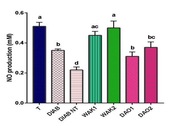
Each bar represent the mean ± SEM, n=3. Letters (a, b, c, d) represent statistical significance. All the groups are compared together. Means with different letters are significantly different (p < 0.05). WAK1: group treated with 'Wakouba' at dose of 1000 mg/kg.pc; WAK2, group treated with 'Wakouba' at dose of 2500 mg/kg.pc; DAO10, batch treated with Daonil at dose of 10 mg/kg.pc; DAO20, batch treated with Daonil at dose of 20 mg/kg.pc Figure 2 shows the variation of the NO level in the samples of diabetic rats, during the treatment of these rats with Wakouba and Glibenclamide (DAONIL). In normal control (T) rats that were not treated with streptozotocin, the value of NO measured was 0.51 ± 0.02 mM. However, after the streptozotocin treatment, we observed in diabetic rats, a decrease in NO level from 0.51 ± 0.02mM to 0.35 ± 0.01mM in the control patient; a very significant decrease of 31.37%. This NO value decreases considerably by more than half and goes from 0.51 ± 0.02mM to 0.22 ± 0.01mM; either a more significant decrease of 56.86%; when diabetic rats are not treated.
However, with Wakouba treatment, the NO level increased from 0.35 ± 0.01mM to 0.45 ± 0.02mM, ie, a significant increase of 28.57% for an administered dose of Wakouba 1000 mg // kg / bw. This level of NO is normalized and goes from 0.35 ± 0.01mM to 0.50 ± 0.03mM, ie a growth of 42.86% for one-dose treatment of Wakouba administered at 2500 mg / kg / day. pc. In fact, we do not statistically record a significant difference between the NO level of healthy control rats (0.51 ± 0.02 mM) and that of diabetic rats treated with Wakouba (0.5 ± 0.03 mM). Which means that Wakouba has normalized the NO rate and, it should be noted that, for the treatment of diabetic rats at different doses of Glibenclamide (Daonil), changes in the level of NO are not significant. And these NO values in the aorta varies from 0.35 ± 0.01mM to 0.31 ± 0.02mM and from 0.35 ± 0.02mM to 0.37 ± 0.02mM, respectively, with Daonil. Statistically, we recorded a significant difference between the NO level of Glibenclamide-treated rats (0.37 ± 0.02mM) and that of healthy control rats (0.5 ± 0.03mM).
Evaluation of the Activity of Catalase in the Different Organs (Kidney and Heart) of Diabetic Rats Induced and Treated with Wakouba and Glibenclamide (Daonil)
| T | DIAB | DIAB NT | WAK1 | WAK2 | DAO1 | DAO2 | |||||||||||||||||||
|---|---|---|---|---|---|---|---|---|---|---|---|---|---|---|---|---|---|---|---|---|---|---|---|---|---|
| Kidney | 22,23±0,01 | 32,13±0,03 | 53,09±0,02 | 28,53±0,01 | 24,14±0,02 | 30,45±0,02 | 27,34±0,02 | ||||||||||||||||||
| Catalase Activity | |||||||||||||||||||||||||
| Heart | 13,29±0,02 | 27,12±0,01 | 42,14±0,02 | 24,04±0,02 | 17,26±0,03 | 27,03±0,47 | 25,12±0,02 | ||||||||||||||||||
Table 3: Evolution of catalase activity in kidney and heart during treatment of diabetic rats with Wakouba, and Glibenclamide (Da
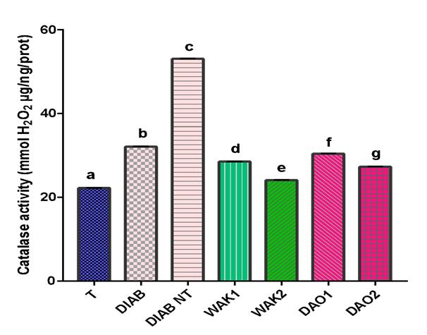
Each bar represent the mean ± SEM, n=3. Letters (a, b, c, d, e, f, g) represent statistical significance. All the groups are compared. Means with different letters are significantly different (p < 0.05).
WAK1: group treated with 'Wakouba' at dose of 1000 mg/kg.pc; WAK2, group treated with 'Wakouba' at dose of 2500 mg/kg.pc; DAO10, batch treated with Daonil at dose of 10 mg/kg.pc; DAO20, batch treated with Daonil at dose of 20 mg/kg.pc.
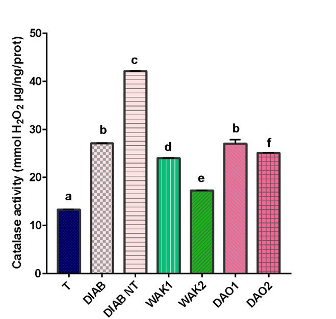
increases of 138.82% and 217.08% respectively. Whatever the organ, we note that catalase activity increases significantly in induced diabetic rats compared to healthy control rats (T). The treatment of diabetic rats with Wakouba at a dose of 1000 mg / kg / pc caused a significant decrease in the catalase activity from 32,13 ± 0.03 to 28.53 ± 0.01 mM in the kidney and from 27.12 ± 0.01mM to 24.04 ± 0.02mM in the the heart; ie significant decreases respectively of 11.20% and 11.36%. For doses of Wakouba administered at 2500 mg / kg / bw, we note more significant decrease of catalase activity. This activity increased from 32.13 ± 0.03mM to 24.14 ± 0.02mM in the kidney; ie a significant decrease of 24.87%. This decrease of catalase activity is also significantly recorded in the heart and goes from 27.12 ± 0.01mM to 17.26 ± 0.03mM; ie a decrease of 36.36% of its activity. This same decrease of catalase activity is also significantly observed with Daonil treatment. However, we note a significant difference of catalase activity between treatment with Wakouba and treatment with Glibenclamide (Daonil). Decreases of catalase activity in diabetic rats treated with Glibenclamide (Daonil) decreased from 32.13 ± 0.03mM to 30.45 ± 0.02mM in the kidney for an administered dose of 10mg / kg / bw and from 27.12 ± 0.01mM to 27.03 ±
0.47mM in the heart; ie repective decreases of 5.23% in the kidney and 0.44% in the heart. With higher doses of Glibenclamide (Daonil at 20mg / kg / bw) we noted significant decreases in catalase activity from 32.13 ± 0.03mM to 27.34 ± 0.02mM in the kidney and from 27 , 12 ± 0.01 mM to 25.12 ± 0.02 mM in the heart; either respective decreases of catalase activity of 14.91% in the kidney and 7.37% in the heart.
Discussion
Variation of NO levels in the Aorta of Diabetic Rats Treated with Different Doses of Wakouba and Glybinclamide (Daonil)
In the normal control rats (T), the rate of NO measured is 0.51 ± 0.02 mM. On the other hand, the induction of diabetes in these rats leaded to a significant decrease of the NO value which passed from 0.51 ± 0.02mM to 0.35 ± 0.01mM in the rat Diabetic control; then from 0.35 ± 0.01mM to 0.22 ± 0.01mM in untreated diabetic rats, ie decreases of 31.37% and 56.86%, respectively (Table 2). Nitric oxide (NO) is a gas whose synthesis has been described in mammals in 1985 [11]. Apart from macrophages, several types of cells have been recognized as capable of secreting NO from NO synthase II: endothelial cells, smooth muscle cells, fibroblasts, epithelial (renal and pulmonary) cells, cardiac myocyte, chondrocytes and hepatocytes [12]. And endogenous synthesis of NO has also been shown in the endothelial cell from different preparations (endothelium isolated, endothelial cells in culture) [13, 14]. It can therefore be deduced that this variation of NO would be related to its quantity produced in the various organs of these diabetic rats. Therefore, this decrease in NO recorded in diabetic rats should be due due to a decrease of the production of NO in the cell during diabetes. In cells, the NO synthesized plays a role in the physiology of many systems: nervous, cardiovascular, genitourinary, digestive, immune. This synthesized NO is also involved in vasodilation and maintenance of vascular integrity in case of aggression [15, 16]. According to some authors, radicals released by neutrophils alter endothelial cells and contribute to increase vascular permeability in endotoxin, ischemia, ethanol or nonsteroidal anti-inflammatory lesions [17]. The overproduction of free radicals leads, during diabetes, to imbalances called oxidative stress, the consequence of which is often the appearance of irreversible damage to the cells.
This reduction of NO production in diabetic rats, therefore, would be the cause of cell dysfunction during diabetes; because most studies in humans show an important role of NO availability decrease in endothelial dysfunction related to hypertension [18]. Therefore Diabetes induced with streptozotocin would have created a dysfunction in the cells involved in the production of NO. As a result, these would no longer be able to synthesize NO normally, hence this decline of NO levels found in these diabetic rats. And this decrease of NO leads to an attack of endothelial cells that can no longer maintain their vascular integrity. This vascular endothelial dysfunction is pharmacologically characterized by the absence of an NO-dependent vasodilator response to acetylcholine [19, 20]. The reduced NO would no longer be sufficient to play effectively, in these induced diabetic rats, its role in the physiology of the nervous, cardiovascular, genitourinary, digestive and immune systems [21]. However, it is important to emphasize that it has been clearly established that the production of NO in humans is considerable, and not negligible, in the immune defense mechanisms against certain types of protozoa, bacteria, fungi and viruses [22]. It has been shown that the induction of iNOS, nitric oxide synthase which produces NO is involved in the mechanism of inflammation, during an aggression of the body by an external agent, and the development of several pathologies including asthma, arthritis, multiple sclerosis, metabolic and neurodegenerative diseases, and cancer [23, 24]. However, with the treatment of diabetic rats by Wakouba, the NO level increases significantly from 0.35 ± 0.01 mM to 0.45 ± 0.02 mM, ie a growth of 28.57% for a dose administered of Wakouba at 1000 mg // kg / pc. This level of NO is normalized and goes from 0.35 ± 0.01 mM to 0.50 ± 0.03 mM, ie a growth of 42.86% for treatment at a dose administered of Wakouba at 2500 mg / kg / bw. . It could then be said that Wakouba administered to diabetic rats at doses of 1000 mg / kg / bw and 2500 mg / kg / bw would contain substances to restore damaged NO-producing cells in diabetic rats. Therefore Wakouba would allow these cells to regain their integrity to synthesize the NO that will play again and fully its physiological role in the nervous, cardiovascular, genitourinary, and digestive and immune systems. It could then be affirmed that Wakouba would participate, in streptozotocin-induced diabetic rats, of the protection and restoration of the nervous, cardiovascular, genitourinary, and digestive and immune systems. Wakouba would protect the nervous, cardiovascular, genitourinary, digestive and immune systems, and also protect the liver, kidney, vessels, genital tract and heart in these diabetic rats. Unlike Wakouba, administration of Glibenclamide (Daonil) to diabetic rats do not significantly increase NO production. Glibenclamide (Daonil) would therefore not allow NO-producing cells to restore and maintain their physiological integrity. In addition, according to Professor Furchgott, the role of the NO - producing enzyme, iNOS, in the relationship between obesity and diabetes has also been demonstrated. The abnormal rate of nitric oxide produced by the action of this enzyme hinder the action of insulin, preventing glucose from entering the muscles; what scientists have called the insulin resistance syndrome that sets the table for type II diabetes [21]. So if Wakouba helps to restore the normal rate of NO, Wakouba would also be able to fight against insulin resistance in diabetics by restoring the production of NO in that case. According to Mahfouz, insulin resistance is a consequence of a decrease of the ability of peripheral target tissues to respond correctly to circulating normal insulin levels [25]. According to some authors, insulin resistance would intervene: 1- With a decrease of the number of insulin receptors that have been observed in adipocytes isolated from obese subjects [26]. According to this theory Wakouba could act directly or indirectly by a mechanism that would increase the number of insulin receptors in adipocytes of obese subjects. 2- Or with an increase of the expression of the PI3-kinase p85α regulatory subunit, which explains some cases of insulin resistance, including that related to obesity [27, 28, 29]. Indeed, the increase of p85α expression would change the stoichiometry between free p85 and the p85- p110 complex and prevent the p85-p110 complex from binding with IRS1 and thus propagating the insulin signal [30]. Then Wakouba, in fighting against insulin resistance, could also act against an increase of the expression of the regulatory subunit of PI3-kinase p85α. Wakouba would thus restore the alterations of the post-receptor insulin pathway to fight against the appearance of resistance to muscle insulin.
Catalase Activity in the Kidney and in the Heart during Treatment of Diabetic Rats with Wakouba, and Glibenclamide (Daonil)
Table 3 shows that in non-diabetic healthy rats, the catalase activity measured in the kidney is 22.23 ± 0.01 mM and 13.29 ± 0.02 mM in the heart. But after the induction of diabetes in these rats with streptozotocin, the activity of catalase increased significantly from
44.53% and goes from 22.23 ± 0.01mM to 32.13 ± 0.03mM in the kidney. And in the heart, this same increase doubled in catalase activity of 104.06% from 13.29 ± 0.02mM to 27.12 ± 0.01mM in diabetic rats. These values grew up respectively to 138.82% and 217.08%. Studies show that catalase is an enzyme that plays an important role in protecting cells against the toxic effects of hydrogen peroxide [31]. This enzyme is widely distributed in our body, with a high concentration in the liver, kidneys and erythrocytes and its expression is regulated at different levels; i.e. the transcriptional, post- translational and translational level [32]. The increase of catalase activity may be due to the high production of Reactive Oxygen Species (ROs) [33, 34, 35]. It can therefore be deduced that diabetes induced in rats would have increased quantity of ROS. Diabetes would have caused an increase of ROS in the body of diabetic rats [36]. Among the factors involved in the dysfunction of the vascular endothelium, oxidative stress plays a preponderant role. ROS are involved in the occurrence of endothelial complications. And so the body, naturally reacts to this aggression by activating more and more catalase to eliminate these ROS. On the other hand, after the treatment given to diabetic rats at the Wakouba dose of 1000mg / kg / bw, we observed significant decreases of the activity of catalase respectively from 32.13 ± 0.03mM to 28.53 ± 0.01 mM in the kidney and 27.12 ± 0.1 mM at 24.04 ± 0.02 mM in the heart; i.e., respective decreases of 11.20% and 11.36%. For doses of Wakouba administered at 2500 mg / kg / bw, we observed more significant decrease of catalase activity from 32.13 ± 0.03 mM to 24.14 ± 0.02 mM in the kidney; ie recorded decrease of 24.87%. This decrease of activity is significantly recorded in the heart and goes from 27.12 ± 0.01mM to 17.26 ± 0.03mM; i.e., a decrease of 36.36% of its activity. Treatment with Wakouba has a much better effect on catalase activity than glibenclamide (Daonil). It can be deduced that administration of Wakouba would reduce the amount of ROS in the organs of diabetic rats, resulting in decreased activity of catalase in these organs. Wakouba would therefore act as protective substances such as albumin, which neutralizes the harmful activities of certain exogenous and endogenous agents by sequestration, thanks to its affinity for the latter, thus reducing their toxic effect in the body [37].
Conclusion
Our study reveals that Wakouba would restore NO production in diabetic rats during treatment. By this restorative activity of Wakouba on the production of NO, Wakouba thus plays a protective role in the nervous, cardiovascular, genito-urinary, digestive and immune systems. Wakouba would therefore have a protective role on the liver, the kidney, the vessels, the genital tract and the heart of these diabetic rats. Wakouba would also be able to fight against insulin resistance of diabetics’ rats. Indeed restoring the production of NO, Wakouba: - Would increases the number of insulin receptors in adipocytes of obese subjects. - Would act against an increase of the expression of the regulatory subunit PI3-kinase p85α. Wakouba would therefore restore the alterations of the post-receptor insulin pathway to fight against the appearance of resistance to muscle insulin. Wakouba would also fight insulin resistance by restoring NO production, since NO would facilitate the binding of insulin to its cellular receptor and it would contribute to insulin-induced vasodilation [38, 39]. Moreover, according to Sydow et al., increased inhibition of NOS may contribute to the development of insulin resistance, in addition to accelerating the development of endothelial dysfunction and hypertension [40]. Besides, insulin- resistant individual’s exhibit impaired insulin vasodilation in skeletal muscle and decreased glucose uptake by insulin. This study also revealed that the administration of Wakouba in diabetic rats induced would reduce the quantity of ROS in their bodies, which would explain the decline of catalase activity in the organs of these diabetic rats treated with Wakouba. Wakouba is therefore a protective substance that neutralizes the harmful activities of certain exogenous and endogenous agents by sequestration, thanks to its affinity for the latter, reducing then their toxic effect in the body. It is now accepted that high concentrations of glucose in extra and intracellular media induce oxidative stress, defined as an imbalance in the balance between pro-oxidants and antioxidants [41]. According to authors, the cellular over expression of antioxidant enzymes, such as catalase, is a response to oxidative stress following high glucose levels [42]. These studies on Wakouba should be deeper by testing its effects. We need to test the effects of wakouba on the blood level of total cholesterol, Low Density Cholesterol (LDC), High Density Cholesterol (HDC) and triglyceride on rats suffering from diabetic complications.
Ethical Approval
As per international standard or university standard written ethical permission has been collected and preserved by the authors.
References
-
Balunas M, Kinghorn J (2005) Drug discovery from medicinal plants. Life science 78(5): 431-441.
-
Lioret J (2010) Human health benefits supplied by Mediterranean marine biodiversity. Marine Pollution bulletin 60(10): 1640-1646.
-
Organisation mondiale de la Santé (2000) Promoting the Role of Traditional Medicine in Health Systems: a Strategy for the African Region 2001–2010. Harare, Organisation mondiale de la Santé.
-
Guillausseau PJ (1995) L diabète non insulino – dépendant. Collection stratégies thé rapeutiques, édition Espaces 34, Montpellier.
-
Maunand B (2002) Diabéto, l’infermière en diabétologie. Deuxième édition, Lamarre , Paris .
-
Vanderwood KK, Hall TO, Harwell TS, Butcher MK, Helgerson SD (2010) Implementing a state-based cardiovascular disease and diabetes prevention program. Diabetes Care 33(12): 2543-2545.
-
(2001) Diabete Et Maladies Cardiovasculaires. Il est temps d’agir. Ann Federation Internationale du Diabete, pp: 2.
-
Guédé-Guina F, Vangah-Manda M, Harouna D, Bahi C (1993) “Potencies of MISCA, a plant source concentrate against fungi”. Mycol Med 5: 225-229.
-
Elia AC, Galarini R, Taticchi MI, Dorr AJ, Mantilacci L (2003) Antioxidant responses and bioaccumulation in Ictalurus melas under mercury exposure. Ecotoxicology and Environmental Safety 55(2): 162- 167.
-
Frouin et al. (1976)
-
Marletta MA, Yoon PS, Iyengar R, Leaf CD, Wishnok JS (1988) Macrophage oxidation of L-arginine to nitrite and nitrate; nitric oxide is an intermediate. Biochemistry 27(24): 8706-8711.
-
Moncada S, Palmer RM, Higgs EA (1991) Nitric oxide: physiology, pathophysiology and pharmacology. Pharmacol Rev 43(2): 109-142.
-
Kelm M, Feelisch M, Spahr R, Piper HM, Noack E, et al. (1988) Quantitative and kinetic characterization of nitric oxide and EDRF released from cultured endothelial cells. Biochem Biophys Res Commun 154(1): 236-244.
-
Collin S, Levy B (2008) Physiopathologie de l’hyporéactivité vasculaire au cours des états de choc. Réan 17(2): 101-110.
-
Holzer P, Livingston EH, Guth PH (1994) Neural, metabolic, physical, and endothelial factors in the regulation of the gastric circulation. In: Johnson LR, ed. Physiology of the gastrointestinal tract. 3rd (Edn.), New York: Raven, pp: 1311-1329.
-
Brain SD, Williams TJ, Tippins JR, Moris HR, Macintyre I (1985) Calcitonin gene-related peptide is a potent vasodilatator. Nature 313(5997): 54-56.
-
Smith SM, Holm-Rutili L, Perry MA, Grisham MN, Arfors KE, et al. (1987) Role of neutrophils in hemorrhagic shock-induced gastric mucosal injury in the rat. Gastroenterology 93(3): 466-471.
-
Alain, Justine Wawa, Anatole Koutou, Jean David N'guessan, Joseph Allico Djaman (2016) Anti-radical mechanism of the antihypertensive effect of Wakouba an extract from palm oil. International Journal of Innovation and Scientific Research 24(1): 95-106.
-
Celermajer DS, Adams MR, Clarkson P, Robinson J, McCredie R, et al. (1996) Passive smoking and impaired endothelium-dependent arterial dilatation in healthy young adults. N Engl J Med 334(3): 150- 154.
-
Celermajer DS, Sorensen KE, Georgakopoulos D, Bull C, Thomas O, et al. (1993) Cigarette smoking is associated with dose-related and potentially reversible impairment of endothelium-dependent dilation in healthy young adults. Circulation 88(5): 2149-2155.
-
R F Furchgott (1993) The discovery of endothelium- dependent relaxation. Circulation 87(suppl. V): V3-8.
-
Bogdan C (2001) Nitric oxide and the immune response. Nat Immunol 2(10): 907-916.
-
Kroncke KD, Fehsel K, Kolb-Bachofen V (1998) Inducible nitric oxide synthase in human diseases. Clin Exp Immunol 113(2): 147-156.
-
Kroncke KD, Fehsel K, Kolb-Bachofen V (1997) Nitric oxide: cytotoxicity versus cytoprotection--how, why, when, and where? Nitric Oxide 1(2): 107-120.
-
Rana Mahfouz (2015) Insulinorésistance musculaire induite par les céramides : étude des mécanismes d'action et de l'implication du transporteur CERT.
-
Molina JM, Ciaraldi TP, Brady D, Olefsky JM (1989) Decreased activation rate of insulinstimulated glucose transport in adipocytes from obese subjects. Diabetes 38: 991-995.
-
Barbour LA, Shao J, Qiao L, Leitner W, Anderson M, et al. (2004) Human placental growth hormone increases expression of the p85 regulatory unit of phosphatidylinositol 3- kinase and triggers severe insulin resistance in skeletal muscle. Endocrinology 145(3): 1144-1150.
-
Barbour LA, Mizanoor RS, Gurevich I, Leitner JW, Fischer SJ, et al. (2005) Increased P85alpha is a potent negative regulator of skeletal muscle insulin signaling and induces in vivo insulin resistance associated with growth hormone excess. J Biol Chem 280(45): 37489-37494.
-
Bandyopadhyay GK, Yu JG, Ofrecio J, Olefsky JM (2005) Increased p85/55/50 expression and decreased phosphotidylinositol 3-kinase activity in insulin-resistant human skeletal muscle. Diabetes 54(8): 2351-2359.
-
Draznin B (2006) Molecular mechanisms of insulin resistance: serine phosphorylation of insulin receptor substrate-1 and increased expression of p85alpha: the two sides of a coin. Diabetes 55(8): 2392-2397.
-
Hwang TS, Choi HK, Han HS (2007) Differential expression of manganese superoxide dismutase, copper/zinc superoxide dismutase, and catalase in gastric adenocarcinoma and normal gastric mucosa. Eur J Surg Oncol 33(4): 474-479.
-
Nishikawa M, Hashida M, Takakura Y (2009) Catalase delivery for inhibiting ROS mediated tissue injury and tumor metastasis. Advanced Drug Delivery Reviews 61(4): 319-326.
-
Ranieri A, Castagna A, Scebba F, Careri M, Zagnoni I, et al. (2005) Oxidative stress and PC Characterisation in bread wheat exposed to Cd excess. Plant Physiology and Biochemistry 43(1): 45-54.
-
Iannone MF, Rosales EP, Goppa MD, Benavides MP (2010) Reactive Oxyd=gren species formation and cell death in Catalase deficient tobacco leaf disks exposed to Cadmium Protoplasma 245: 15-27.
-
Jin X, Yang X, Mahmood Q, Islam E, Liu D, et al. (2008) Response of antioxidant Enzyme, ascorbate and glutathione metabolism Towards Cd in hyperaccumulator and non hyperaccumulator ecotypes of Sedum Alfredii H. Environmental Toxicology and Chemistry 23(4): 517-529.
-
Jennifer Baraka-Vidot (2014) Stress oxydant et pathologie diabétique { l’île de La Réunion – Identification et caractérisation des propriétés structurales et fonctionnelles de l’albumine glyquée. Médecine humaine et pathologie. Université de la Réunion,
-
Nihar Ranjan Singh PR, Rondeau P, Emmanuel B (2010) Identification of Up-Regulated Low Molecular Weight Proteins in Human Adipocytes Treated with Glycoxidized Albumin. Open Obes J 2:110-115.
-
Scherrer U, Randin D, Vollenweider P, Vollenweider L, Nicod P (1994) Nitric oxide release accounts for insulin's vascular effects in humans. J Clin Invest 94 (6): 2511-2515.
-
Ragoobirsingh D, McGrowder D, Dasgupta T, Brown P (2004) The effect of nitric oxide on glucose metabolism. Mol Cell Biochem 263(1-2): 29-34.
-
Sydow K, Mondon CE, Cooke JP (2005) Insulin resistance: potential role of the endogenous nitric oxide synthase inhibitor ADMA. Vasc Med 10(S1): S35-43.
-
Giugliano D, Ceriello A, Paolisso G (1996) Oxidative stress and diabetic vascular complications. Diabetes Care 19(3): 257-267.
-
Ceriello A, Dello Russo P, Amstad P, Cerutti P (1996) High glucose induces antioxidant enzymes in human endothelial cells in culture. Evidence linking hyperglycemia and oxidative stress. Diabetes 45(4): 471-477.
- Psychogenic Erectile Dysfunction in Late Adulthood: A Case Report on Clinical Intervention and Intimacy Restoration
- Clinical Trials on COVID-19 in 2025: A New Chapter in Global Health Research
- Innovations and Challenges in Contemporary Medical Clinical Trials: An Editorial Perspective
- Innovations and Challenges in Contemporary Medical Clinical Trials: A Critical Perspective
- Reimagining Clinical Trials: The Power of Continuous Feedback from Medical Reports
- Factors Influencing Brain Drain: Perspectives from a Medical School in Turkey