Theory and Modelling of Ion Track-Based Biosensors for CBRN- Agents Detection in Medical Applications
Pressing challenges of recent decades, associated with agents that are aggressive towards humans - substances and radiation of chemical, biological, radiological, and nuclear (CBRN) agents - require scientific and technological responses. These responses lie in the areas of agent detection and protection from them. The mentioned bio destructive agents can be divided into 2 groups: 1) chemical and biochemical, and 2) radiative (leading to chemical destruction of biomass). In this study, we consider models of universal track nanosensors that are capable of producing a correlated electrical response to the flow of aggressive and non-aggressive active agents.
Introduction: CBRN-Agents Nature
Contemporary extremist groups have a wide variety of potential agents and delivery means to choose from chemical, biological, radiological, or nuclear (CBRN) attacks. Identification, protection and decontamination are the main scientific and technological responses for the modern challenges of CBRN events [1]. Decontamination, in general, is defined as the removal of hazardous materials from the areas where they are not wanted. Decontamination is utilized to reduce the dose that humans or biological organisms may receive from a component or surface, to reduce the potential for airborne CBRN-agents, or to reduce the disposal cost associated with components or the materials [1]. An expanded abbreviation can be used for destructive agents, particularly, the additional symbol ‘E’ in the acronym CBRNE means ‘Explosive’ for Chemical, Biological, Radiological, Nuclear, and Explosive materials (so called ‘SEE-burn’) which are considered very dangerous for people, animals, and the environment. Usually CBRNE materials are used for weapons of mass destruction. However, the focus of our attention is on CBRN-agents, which can be detected, controlled, and managed. Typical CBRN-agents are presented in Table 1.
| Agent groups and types | Identification features | ||||
|---|---|---|---|---|---|
| Chemical | |||||
| Cyanides | |||||
| Sodium or potassium cyanides | Sodium or potassium cyanides are white-to-pale yellow salts that can be easily used to poison food or drinks. | ||||
| Hydrogen cyanide and cyanogen chloride | Hydrogen cyanide (HCN) and cyanogen chloride (ClCN) are colorless-to-pale yellow liquids that will turn into a gas near room temperature. HCN has a characteristic odor of bitter almonds, and ClCN has an acrid choking odor and causes burning pain in the victim’s eyes. | ||||
| Mustard Agent | Mustard is a blister agent that poses a contact and vapor hazard. Its color ranges from clear to dark brown depending on purity, and it has a characteristic garliclikeodor. Mustard is a viscous liquid at room temperature. | ||||
| Nerve Agents | Sarin, tabun, and VX are highly toxic military agents that disrupt a victim’s nervous system by blocking the transmission of nerve signals. | ||||
| Toxic Industrial Chemicals | Chlorine and phosgene are industrial chemicals that are transported in multiton shipments by road and rail. The effects of chlorine and phosgene are similar to those of mustard agent. Organophosphate pesticides such as parathion are in the same chemical class as nerve agents. | ||||
| Biochemical | |||||
| Anthrax | Bacillus anthracis, the bacterium that causes anthrax, is capable of causing mass casualties. Symptoms usually appear within one to six days after exposure and include fever, malaise, fatigue, and shortness of breath. | ||||
| Botulinum toxin | Botulinum toxin is produced by the bacterium Clostridium botulinum, which occurs naturally in the soil. Crude but viable methods to produce small quantities of this lethal toxin have been found in terrorist training manuals. | ||||
| Ricin | Ricin is a plant toxin that is 30 times more potent than the nerve agent VX by weight and is readily obtainable by extraction from common castor beans. | ||||
| Viruses Biological warfare (BW) | Germ warfare is the use of biological toxins or infectious agentssuch as bacteria, viruses, and fungi with the intent to kill or incapacitate humans, animals or plants as an act of war. Biological weapons (‘bio-agents’) are living organisms or replicating entities (viruses, which are not universally considered "alive") that reproduce or replicate within their host victims. Entomological (insect) warfare is also considered a type of biological weapon. This type of warfare is distinct from nuclear warfare and chemical warfare, which together with biological warfare make up NBC, the military acronym for nuclear, biological, and chemical warfare using weapons of mass destruction (WMDs). |
Table 1: Typical Radiological and Nuclear CBRN-agents. Different types of ionizing radiation have a different destructive effect
radioactive materials is commonly available and could be used in any RDD, including 137Cs, 90Sr, 60Co. Moreover, these agents can also be constituent parts of the devices and tools used in hospitals, universities, factories, construction companies, and laboratories, being possible sources for such radioactive materials. A passive RDD is a system in which unshielded radioactive material is dispersed or placed manually at the target. An explosive variant of RDD (a dirty bomb) is any system that uses the explosive force of detonation to disperse radioactive material. An atmospheric RDD is any system in which radioactive material is converted into a form that is easily transported by air currents. Another type of aggressive action is the radiation, which outruns any direct contact with an aggressive material. Subsequent destructive biochemical reactions are additional destructive post-factors that can be identified biochemically. Radiations induces the radiation sickness, resulting from the effects of various types of ionizing radiation and characterized by symptoms, depending on the type of damage, its dose, the location of the radiation source, the distribution of the dose in time and the body of a living being (for example, a person). The most important types of ionizing radiation: X-rays, γ-rays, β+,-- particles, α-particles, n-neutrons, p-protons (Table 2).
| Agent groups and types | Identification features | |||
|---|---|---|---|---|
| Radiological and Nuclear Devices | An RDD (Radiological Dispersion Device) is a conventional bomb not a yield producing nuclear device. RDDs are designed to disperse radioactive material to cause destruction, contamination, and injury from the radiation produced by the material (137Cs, 90Sr, 60Co). | |||
| Improvised Nuclear Device (IND) | An IND is intended to cause a yield-producing nuclear explosion. An IND could consist of diverted nuclear weapon components, a modified nuclear weapon, or indigenous-designed device. | |||
| Radiological | ||||
| 131I | -, - decays, half-life — appr. 8 days. -decay mutations and cell death, as well as - surrounding tissues to a depth of several millimeters. It concentrates mainly in the thyroid gland. | |||
| 90Sr | half-life — appr 28,8 years. 90Sr falls into the environment mainly with emissions from nuclear power plants and nuclear explosions. It is extremely dangerous. It is deposited, mainly, in bone tissues. | |||
| 137Cs | half-life - 30 years. One of the main components of radioactive contamination of the biosphere. The release of 137Cs into the environment occurs mainly as a result of accidents at nuclear power plants and nuclear weapons tests. | |||
| 60Co | half-lifeappr. 5,3 years | |||
| 241Am | half-lifeappr. 433 years | |||
| note | The contribution of these radioactive components at Chernobyl event 1986, April was (appr.): 131I - 1,8·1018 Bq, 137Cs . - 8,5·1016Bq, 90Sr — 1·1016 Bq. The total activity of substances released into the environment was, according to various estimates, up to 14·1018 Bq. | |||
| Nuclear atomic warfare, thermonuclear warfare | ||||
| α-particle β- particle γ-rays nneutrons | Basic radiations: 241Am237Np + 4α ,90Sr90Y +β , γ-rays, X-rays - Nuclear atomic warfare, 95 93 2 38 39 X-rays - thermonuclear warfare hydrogen bomb n-neutrons - Neutron bomb pptotons - the sources of intense proton radiation are accelerators of charged particles (till 10 GeV). Even greater energy are found in outer space. Proton radiation is the main component of galactic and solar cosmic radiation. |
Table 2: Typical Radiological and Nuclear CBRN-agents. Different types of ionizing radiation have a different destructive effect
Therefore, to describe the effect of radiation on living organisms, the concept relative biological effectiveness of RBE of radiation is introduced [2].
RBE of ionizing radiation is a dimensionless coefficient characterizing the efficiency biological effect of various ionizing radiations, which is defined as the ratio of the dose of some of the reference radiation D0 to the dose of this radiation Dx: RBE = D0/Dx [ 2 ]. As a quality of standard take X-rays with a certain spectrum, D0 and Dx. RBE is measured by Quality factor (Q). Coefficient (Q) to account for the biological effectiveness of different types of ionizing radiation in determining the equivalent dose. To obtain an equivalent dose, the absorbed dose of the radiation in question must be multiplied by the quality factor. For X-ray, beta and gamma radiation, the coefficient Q = 1, proton and neutron radiation (fast neutrons) Q = 10, alpha radiation Q = 20. For metrology and control of aggressive and biologically active agents it is essential to observe the evolution of the decontamination level. The effectiveness of the decontamination can be expressed as decontamination factor (DF). It is the ratio of the contamination level of a material before decontamination to the contamination level of a material after decontamination. DF=LB/LA, where LB is the contamination Level of a material or Component Before the decontamination application, LA is the contamination Level measured immediately After decontamination application. A decontamination process that removes material will result in a DF greater than 1. The percentage of contamination removed from the surface can be given by the Percent contamination removed = (1-1/DF) × 100; If DF = 10, the percent contamination removed = 90% If DF= 100, the percent contamination removed = 99% [1].
Bionanosensors: Polymer Nanoporous Model Structures
Since the 1960s, it has been known that energetic (with tens of MeV or more) heavy (with atomic masses being usually larger than that of Ar) ion irradiation (‘swift heavy ions’, SHI) introduces very narrow (~ some nm) but long (typically 10-100 μm) parallel trails of damage in irradiated polymer foils, the so-called latent ion tracks. The damage shows up primarily by the formation of radiochemical reaction products. Whereas the smaller ones readily escape from the irradiated zone, thus leaving behind themselves nanoscopic voids, the larger ones tend to aggregate towards carbonaceous clusters. Thus, emerging structural disorder along the tracks modifies their electronic behaviour. The newly created intrinsic free volume enables electrolytes to penetrate into the polymer, thus forming parallel liquid nanowires. In case of tracks penetration through all the foil, the conducting connections emerge between the front and back sides of the foil. The ion track technology is particularly intended to biosensing applications. In this case, the ion tracks are functionalized directly by attaching organic or bioactive compounds (such as enzymes) to their walls. Consider the scheme of the glucose detection in the blood (Figure 1).
![Figure 1: General scheme describing the detection scheme and modified polymer. Principle arrangement of experimental setup to study voltage-current dependences in ion track-containing foils embedded in electrolytes The description of the sensing reaction of glucose with the enzyme GOx looks as follows: a) the overall net reaction is: Glucose (C6H12O6) + O2 (due to enzyme-induced oxidation) gluconic acid (C6H12O7) + O; b) this remaining O attaches to some H2O to form peroxide H2O2 ; c) the product: gluconic acid dissociates around pH=7. Thus, the conductivity of the liquid changes (essentially if the product is enriched in the track’s confinement); this is what is measured by the sensor [3,4]. In particular, a complicated biochemical kinetics of basic reaction of glucose detection depends on track qualities (e.g., track creation mechanism, foil material properties), enzyme (GOx) distribution on the track surface, geometry of the etched track etc. All these factors are the subject for the nearest special research. Moreover, the detailed kinetics of reaction is the object of 3D-modelling to design the optimal geometry of nanosensor active space. This allows creating optimal nanosensors with the increased efficiency. Experimental and theoretical calibration dependences demonstrate similar trends. The proposed device can serve to detect physiologically relevant glucose concentrations.](/fulltextimages/1716/fig_1.jpeg)
Figure 1: General scheme describing the detection scheme and modified polymer. Principle arrangement of experimental setup to study voltage-current dependences in ion track-containing foils embedded in electrolytes The description of the sensing reaction of glucose with the enzyme GOx looks as follows: a) the overall net reaction is: Glucose (C6H12O6) + O2 (due to enzyme-induced oxidation) gluconic acid (C6H12O7) + O; b) this remaining O attaches to some H2O to form peroxide H2O2 ; c) the product: gluconic acid dissociates around pH=7. Thus, the conductivity of the liquid changes (essentially if the product is enriched in the track’s confinement); this is what is measured by the sensor [3, 4]. In particular, a complicated biochemical kinetics of basic reaction of glucose detection depends on track qualities (e.g., track creation mechanism, foil material properties), enzyme (GOx) distribution on the track surface, geometry of the etched track etc. All these factors are the subject for the nearest special research. Moreover, the detailed kinetics of reaction is the object of 3D-modelling to design the optimal geometry of nanosensor active space. This allows creating optimal nanosensors with the increased efficiency. Experimental and theoretical calibration dependences demonstrate similar trends. The proposed device can serve to detect physiologically relevant glucose concentrations.
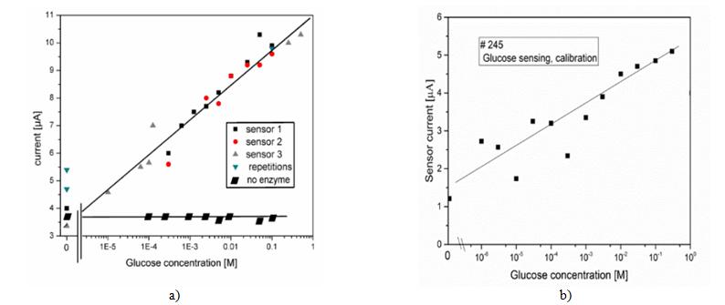
The catalytic sensor can be made re-usable due to the formation of diffusible products from the oxidative biomolecular recognition event. Moreover, we can develop a multi-agent packet nanosensor, suitable for application as a human breathing analyzer in relation to cancer detection, hepatitis, and so on [4, 5, 6, 7, 8]. The main detection problems of various agents in the proposed schemes of nanotrack devices are connected with the exploration and creation of effective chemical reactions capable of producing stable positive and negative ion fluxes within the working zone (as an electrolyte, Figure 1). These ion fluxes should be strongly correlated with the tested agent concentrations [3, 4, 5, 6, 7, 8]. The detection of glucose concentration in the human blood on the polymer nanotrack based technology has well-tested and stable experimental results [6, 9]. The recent advancements in the field of nanosensor design allow monitoring and tracking biomolecules in such areas as the environment, food quality and healthcare. The presently developed ion track-based nanosensors (Figure 2) provide high sensitivity, reliable calibration (Figure 3), small power and low cost.
![Figure 2: Favourable platform for measuring enrichment of chemical reaction products via electrical current transmitted through the nanopores: a) nanosensor system prototype [8]; b) principal electrical scheme of the sensor current [8].](/fulltextimages/1716/fig_2.jpeg)
+ → + GOx C H O O C H O O
6 12 6 2 6 12 7
1 , 2 + → → +
catalaza O H O H O H O O H O
2 2 2 2 2 2 2
− + → + C H O C H O H
6 12 7 6 12 7
1 : 2 2 2 : 2 2
+ − → + + Anode H O O H e
2 2 + − + →
Cathode H e H
2 + → + :
Ultimate reaction C H O H O C H O H
6 12 6 2 6 12 7 2
![Figure 4: General model of glucose detection process on the ion track-containing foils embedded in electrolyteand basic set of biochemical reaction [3,5,8]. Simulation schemes are directed on concentrations of H+ which are equal to glucose concentrations. In cases of stable of glucose inputs we obtain can write typical simulation equations which lead to the saturated values of H+ ions concentrations (Figure 5):](/fulltextimages/1716/fig_4.png)
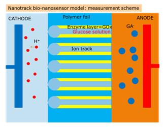
Figure 4: General model of glucose detection process on the ion track-containing foils embedded in electrolyteand basic set of biochemical reaction [3, 5, 8]. Simulation schemes are directed on concentrations of H+ which are equal to glucose concentrations. In cases of stable of glucose inputs we obtain can write typical simulation equations which lead to the saturated values of H+ ions concentrations (Figure 5):
=− = − =− + =
d C H O w dt d H O w w dt
[ ]
6 12 6 1 [ ]
2 2 1 2 (10.1) d O w w dt
[ ]
2 1 2 d H w dt
+ [ ] 2
1
$$ \mathrm {w h e r e} w _ {1} = k _ {1} \left[ C _ {6} H _ {1 2} O _ {6} \right] \left[ O _ {2} \right] \left[ G O x \right], w _ {2} = k _ {2} \left[ H _ {2} O _ {2} \right] \mathrm {a r e} $$ the reactions rates, 1_k_ and 2_k_ are simulation parameters.
We can point out that theoretical simulation results demonstrate similar linear trend in comparison to experimental data (Figures 3, Figure 5b).
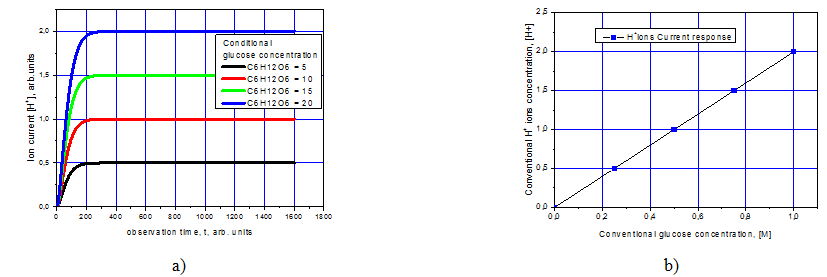
Figure 5: a) Simulation of H+ ion current via observation time in case of saturation; b) Theoretical model of typical calibration dependence based on chemical kinetics results. Simulation of induced H+ ioncurrent via glucose concentration Taking into account the probable process of H+ ions recombination we can modify simulation equations (10.1) as:
$$ \left\{ \begin{array}{l} \frac {d}{d t} \left[ C _ {6} H _ {1 2} O _ {6} \right] = - w \\ \frac {d}{d t} \left[ H _ {2} O _ {2} \right] = w _ {1} - w _ {2} \\ \frac {d}{d t} \left[ O _ {2} \right] = - w _ {1} + w _ {2} \\ \frac {d}{d t} \left[ H ^ {+} \right] = 2 w _ {2} - 2 w _ {1} \end{array} \right. $$ d C H O w dt d H O w w dt [ ]
Novel Bio-Agents Devices: Technological Solutions
The main idea of the modification of the considered bionanosensor model requires finding effective detection reactions of the controlled agents that will provide a stable and correlated electrical response. An equally fundamental problem is the creation of a multi-packet nanosensor tuned to a set of agents. The market presents various technological solutions for the denoted problems. Consider some of them.
The Breath Analyzer Mint
The company Breathometer (USA) began selling a mobile oral health analyser. The device is called Mint. However, this device has very limited applications. Mint is a compact device with built-in sensors to analyse the condition of the mouth. A person needs to keep the mouth closed for about 30 seconds and then put the mouthpiece of the device into the mouth. The gadget sucks in the air, passes it through special analysers and produces a result: from ‘A’ – ‘everything is fine’ up to ‘F’ – ‘it's time to see a doctor’. Mint sensors detect volatile sulphur compounds. They are the cause of bad breath from the mouth when, for example, periodontal disease occurs. The manufacturer notes that the device is designed for a primary diagnosis, not for the treatment. All data appears in the company application, which you need to install on your smartphone. Mint is able to detect very approximately only sulphur components and it is doubtful that this metrological scheme can be expanded for other chemical and biochemical groups of agents.
AI-Powered Breath Detector
Technion−Israel Institute of Technology has developed a smart breath sensor based on artificial intelligence (AI) abilities. The developed sensor detects many different molecules and finds correlations of the tested biomarkers to 17 different diseases. Before modern methods of medical laboratory research became available, doctors often diagnosed certain diseases, sniffing the patient's exhaled air. Scientists for many years worked on the creation of analytical tools that could simulate this ability to detect disease by smell. Recently, the researchers reported in the journal ACS Nano [10] that they identified the presence of a unique breathing spectrum of the ‘breath print’=breath spectrum’ for each disease. Using this information, they created a device analysing samples of exhaled air to classify and diagnose several different types of disease. The researchers developed a grating made up of nanoscale sensors, which represents an array of specially prepared micro-sensors from gold nanoparticles, each of which contains a randomly organized network of carbon nanotubes. Based on the analysis of the results obtained using artificial intelligence methods, scientists were able to use this lattice to classify and diagnose various diseases. They used mass spectrometry to identify those respiratory components that are associated with diseases. The exhaled air contains nitrogen, carbon dioxide and oxygen, as well as small amounts of more than 100 other chemicals. Researchers managed to find out that each disease has a unique volatile "breathprint", based on different concentrations of 13 components. The experiments also showed that the presence of one disease will not interfere with the detection of the other, which was the prerequisite for developing a practical device for detecting and diagnosing various diseases, and non- invasive, inexpensive and portable. The breath analyser diagnoses the disease by the exit. Already, the respiratory analyser is able to diagnose 17 diseases in just one exhalation. In recent years, a number of specific tests have been developed to determine hypertension, tuberculosis, certain forms of cancer and other diseases by breathing the patient. The device contains an array of specially prepared micro-sensors from gold nanoparticles, each of which contains a randomly organized network of carbon nanotubes. The device can be used to collect breath samples of numerous patients with various diseases, and to correlate the information obtained with certain diseases using software based on artificial intelligence, which revealed patterns and deviations in the concentration of various gases. As a result, researchers were able to accurately determine the disease in 90% of cases. As the test studies showed, the average accuracy of diagnosing this system was not less than 86% for 17 diseases, including several cancers, kidney failure, Crohn's disease, multiple sclerosis, two types of Parkinson's disease etc.
Laser Spectroscopic Techniques of Breath Analysis
| Detection of Sarinand Other | Toxic Synthetic |
|---|---|
| Organophosphorus Compounds |
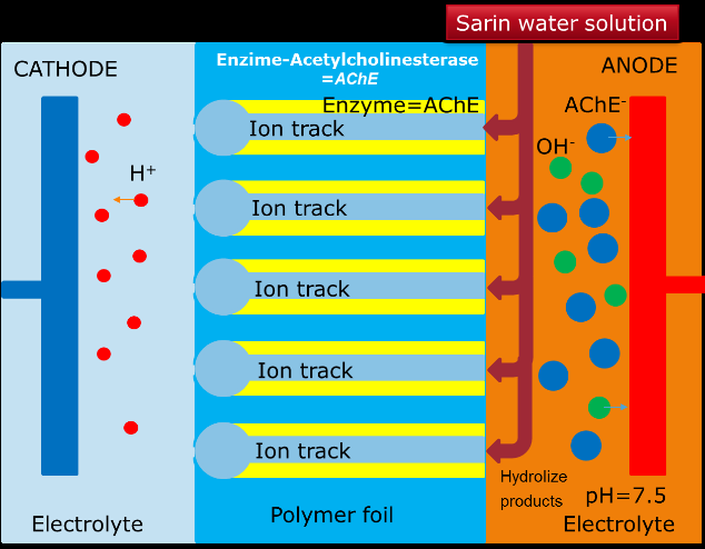
The most dangerous in emergency and terrorist relationships are organophosphorus poisoning substances - sarin, soman, substance VX, related to nerve agents [12]. The extremely high toxicity of synthetic organophosphorus compounds, which exceeds many times the known poisonous substances, the rapid development of poisoning (within a few minutes) require the creation of devices capable of accurately determining the chemical nature of a hazardous substance within a short time interval (5 to 30 s). The solution of this chemical-analytical problem is possible with the use of special high-speed equipment that can work stably at control sites (in the field_). When choosing the most suitable detection method, the following criteria are required: 1) the ability of the method to provide a direct and specific measurement of the analytic signal of the compound being determined; sensitivity; 2) the operating range of concentrations; 3) the detection limit; 4) information; 5) the influence of interfering components and factors; 6) the possibility of automation. There are some basic groups of detection methods: chromatography, mass spectrometry IR spectroscopy and enzymatic tools. _Enzymatic methods: Enzymatic method is more preferable from the point of view of interpretation of direct correlations ‘agent-electric response’. The use of enzymatic reactions is a particular case of kinetic analysis methods based on measuring the rate of the indicator catalytic reaction in the presence of various amounts of detectable substances. Only about 20 enzymes are of practical interest. They have high specificity and provide low detection limits. Among them, cholinesterases, related to hydrolase enzymes, are widely used [13]. Sarin detection: Let us consider some details of the detection of sarin as an example of a very agrressive and dangerous chemical agent. Sarin (C4H10FO2P) is an organophosphorous toxic agent, isopropyl ether of methylphosphonic acid fluoride, a liquid without colour and odour.At the room temperature, sarin is a colourless liquid with a faint smell of flowering apple trees.Miscible with water and organic solvents in all respects. The relatively high pressure of its vapour causes it to evaporate rapidly (about 36 times faster than, e.g., tabun). In the gaseous state, sarin is also colourless and odourless. Sarin molecule is chiral because it contains four different chemical substituents combined by a phosphorus atom into a tetrahedron. Sarin is biologically active and has a great affinity for the enzyme acetylcholinesterase. Note. Acetylcholinesterase (acetylcholine acetylhydrolase), enzyme class hydrolase, catalyzing hydrolyzacetylcholine, as well as other ethers of choline:
→ + - 3 2 2 3 3 2 + - 3 2 2 3 3 [CH COOCH CH N (CH ) ]OH + H O .
CH COOH +[HOCH CH N (CH ) ]OH
Optimal catalytic activity of acetylcholinesterase is observed at pH=7.5-8.5. Sarin inhibits the enzyme acetylcholinesterase by forming a covalent compound with that part of the enzyme where acetylcholine undergoes hydrolysis. The general scheme of enzymatic sarin detection using the developed nanotrack sensor technique is presented in Figure 7.
Conclusion: Multiagent Nanotrack Based Nanosensor Model
The creation of novel biosensors and their further improvements require a careful study of the mechanisms of electrolytes passing through the tracks [14, 15]. Experimental and theoretical calibration dependences demonstrate similar trends. The proposed device can serve to detect physiologically relevant glucose concentrations. The catalytic sensor can be made re-usable due to the formation of diffusible products from the oxidative biomolecular recognition event. Moreover, we can develop a multi-agent packet nanosensor, which can be used as a human breathing analyzer in relation to cancer detection, hepatitis, and many other diseases (Figure 8) [16].
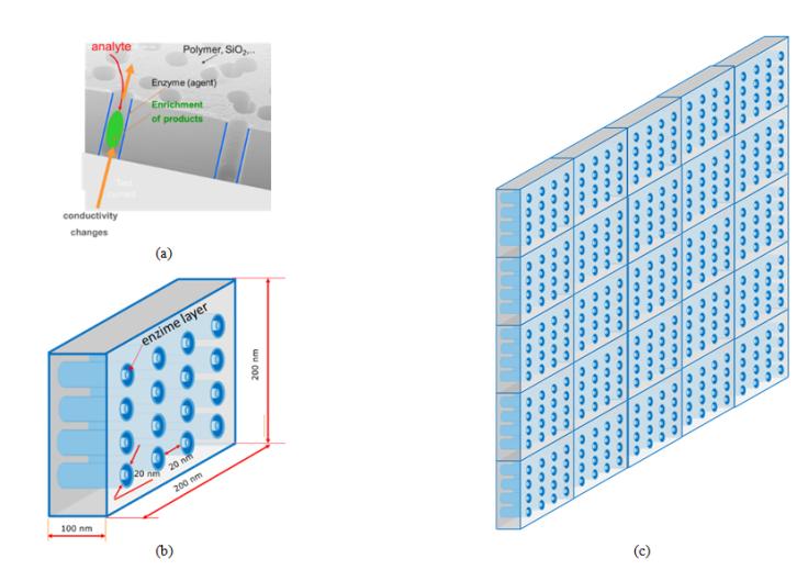
Figure 8: Sensing of chemical reaction products of an analyte with a specific enzyme via the track conductivity: a) nanotracks in the foil; b) model of nsnocell with nanotracks including to corresponding tested bio-agent; c) multi-agent testing nanocell matrix (5x5), where the matrix any cell element independently oriented on particular bio-agent. Every nanocell (Figures 2,4) has an independent electrical metrology scheme. Moreover, sensitive cells for particular agents can be distributed along the sensitive surface of multi-agents flux. All electrical responses corresponding to a particular agent accumulated digitally as calibrated medical data on the special integrated screen. In this way, we can instantly obtain the information about various agent concentrations in real-time. This multi-agent detector can be used as a device for the analysis of the human breath supplying a spectrum of medical information in a non-invasive way. This is very important for the evaluation of post effects of aggressive agents (e.g. CBRN).
Acknowledgement
This research has been partially supported by grant ‘Nanostructures for bacteria detection and study’ (NANOBAC) (01.10.2015-31.12.2017) the Ministry of Education and Science of the Republic of Kazakhstan. Authors are also thankful to Prof LitalAlfonta (Ben-Gurion University, Israel) and Prof Eugene Kotomin (Institute of Solid State Physics, University of Latvia) for support and evaluation of theoretical aspects of research.
References
-
Kumar V, Goel R, Chawla R, Silambarasan M, Sharma R K (2010) Chemical, biological, radiological, and nuclear decontamination: Recent trends and future perspective. J Pharm Bioallied Sci 2(3): 220-238.
-
Sinclair D W (2003) Relative biological effectiveness (RBE), quality factor (Q) and radiation weighting factor (Wr). Ann ICRP 33(4): 1-117.
-
Fink D, Klinkovich I, Bukelman O, Marks RS, Kiv A, et al. (2009) Glucose determination using a re-usable enzyme-modified ion track membrane sensor. Biosensors and Bioelectronics 24(8): 2702-2706.
-
Wang J (2008) Electrochemical Glucose Biosensors. Chem Rev 108(2): 814-825.
-
Fink D, Kiv A, Shunin Y, Mykytenko N, Lobanova- Shunina T, et al. (2015) The nature of oscillations of ion currents in the ion track electronics. Computer Modelling and New Technologies 19(6): 7-13.
-
Fink D, Gerardo Munoz H, Alfonta L, Mandabi Y, Dias JF, et al. (2012) Status and Perspectives of Ion Track Electronics for Advanced Biosensing. In: Shunin Yu and Kiv A (Eds.), Nanodevices and Nanomaterials for Ecological Security (NATO Science for Peace Series B - Physics and Biophysics), Springer Verlag Heidelberg, pp: 269-279.
-
Shunin Yu, Alfonta L, Fink D, Kiv A, Mansharipova A, et al. (2016) Modelling and simulation of electric response of nanocarbon nanocomposites and nanoporous polymer based structures for nanosensor devices. In: Theses of the 14th Int. scientific conference Information Technologies and Management, ISMA University, Riga Latvia.
-
Shunin Yu, Fink D, Kiv A, Alfonta L, Mansharipova A, et al. (2016) Theory and modelling of physical and bio-nanosensor systems. In: Proceedings of the 5th Int Workshop Nanocarbon Photonics and Optoelectronics, Holiday Club Saimaa, Finland.
-
Fink D, Vacik J, Hnatowicz V, Muñoz Hernandez G, et al. (2017) Diffusion kinetics of the Glucose/Glucose Oxidase system in swift heavy ion track-based biosensors. Nucl Instr Meth B 398: 21-26.
-
Nakhleh MK, Amal H, Jeries R (2017) Diagnosis and Classification of 17 Diseases from 1404 Subjects via Pattern Analysis of Exhaled Molecules. ACS Nano 11(1): 112-125.
-
Wang Ch, Sahay P (2009) Breath Analysis Using Laser Spectroscopic Techniques: Breath Biomarkers, Spectral Fingerprints, and Detection Limits. Sensors 9(10): 8230-8262.
-
Types of Chemical Weapons FAS-Federation of American Scientists.
-
Aziz A, Palleschi G (2004) Phoshate, nitrate, and sulfate biosensors. Anal Lett 37(1): 1-19.
-
(2012) Nanodevices and Nanomaterials for Ecological Security. In: Yu Shunin and A Kiv (Eds.), NATO Science for Peace Series B - Physics and Biophysics, Heidelberg Springer Verlag, pp: 363.
-
(2004) Fundamentals of Ion-Irradiated Polymers. In: D Fink (Eds.), Springer Series in Materials Science 63, Berlin Heidelberg.
-
Shunin Yu, Bellucci S, Gruodis A, Lobanova-Shunina (2018) Nonregular Nanosystems. Theory and Applications, Heidelberg Springer Verlag, pp: 406.
- Solution-Processed Chiral Perovskites for Biomedical Applications
- Nanotechnology in Health Chemistry and Medicine: Current Challenges and Future Directions
- Human Exposure to Micro- and Nanoplastics: Pathways, Toxicity, and Intervention Strategies
- Exosome Nanomedicine for Cancer Therapy
- Micro and Nanoplastics–Plastisphere, Biotoxicity, Impact on Human Health, and Mitigation Strategies
- Process Validation of Cefixime Powder for Suspension Dosage Form, 50 mL