Innovative Treatment Strategy for Cancer with Nanotechnology: A Review
By reducing collateral toxicity to nonmalignant cells, nanotechnology has the potential to improve the efficacy and selectivity of chemical, physical, and biological methods for inducing cancer cell death. More and more materials at the nanoscale are being actively and passively targeted to specifically target cancer cells. This review focuses on how different tactics with distinct identifying qualities set nanoparticles apart from earlier anticancer medicines in terms of their capacity to recognize cells. Additionally, it talks about how precise medication delivery via nanoparticles inside the cells has been the subject of numerous successful studies, as well as how nanoparticles can be used to treat cancer specifically while removing the side effects of conventional medicines.
Introduction
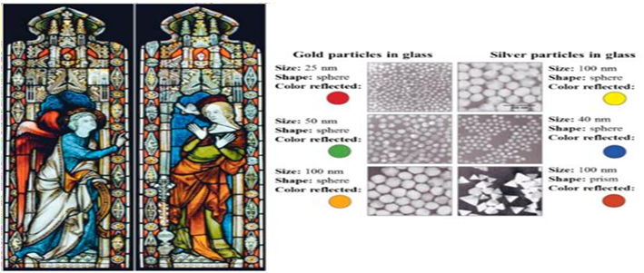
Statistics showing that cancer incidence, prevalence, and mortality continue to be extremely high levels make it clear that an improved technology is required to play a significant role in cancer treatment. By 2030, it is anticipated that there will be 13.1 million cancer-related deaths [1]. We must first comprehend how cancer originates before we can comprehend how lifestyle modifications could lower the risk for the most prevalent tumours [2]. Surgery, chemotherapy, and radiotherapy have all been proven to be successful cancer treatments during the previous century. Treatment is more challenging, though, if the tumor has spread. Even in these situations, existing treatment modalities have the potential to make cancer more of a chronic condition. For particular cancer forms, such as glioblastoma, where a combination of early identification, surgery, chemotherapy, and radiotherapy cannot improve survival beyond 1-2 years, there are still enormous difficulties to be overcome [3]. Unfortunately, nonspecific targeting of anticancer drugs results in a number of side effects, and those agents’ poor drug delivery in most situations prevents them from producing the intended results. The creation of cancer drugs involves a highly complex a process connected to cutting-edge electronic engineering and polymer chemistry. Differentiating between dangerous and healthy bodily cells is the fundamental problem with cancer therapies. Because of this, the primary goal is to develop a medication that can recognize cancer cells and inhibit their development and multiplication. Conventional chemotherapy falls short of selectively targeting malignant cells without interfering with healthy body cells. As a result, they have severe side effects, such as organ damage, which makes it difficult to cure them and eventually leads to low survival rates [4]. Cancer is not a single disease, but rather a variety of disorders that can affect different organs or systems in different ways. Numerous cases of cancer could be prevented; according to some estimates, about 30% of cancer deaths are linked to smoking or other lifestyle choices or dietary habits that might be changed to reduce the risk [1]. By providing superior pharmacokinetic features, prolonged blood circulation time, cellular uptake, and volume of distribution, and half-life, Nano carrier use, on the other hand, results in increased therapeutic index and tumor tissue concentrations of the drugs and can improve the efficacy of currently used regimens. These are key factors for an improved therapeutic window and subsequent clinical success [5]. History of Nanotechnology The inventor of contemporary nanotechnology is physicist Richard Feynman, who won the Nobel Prize in physics in 1965. He proposed the idea of influencing matter at the atomic level in a talk titled “There’s Plenty of Room at the Bottom” during the 1959 American Physical Society meeting held at Caltech. With this innovative concept, new methods of thinking were demonstrated, and Feynman’s theories were later found to be true. He is regarded as the founding father of contemporary nanotechnology because of these factors. Norio Taniguchi, a Japanese scientist, was the first to use the term “nanotechnology” to describe semiconductor operations about 15 years after Feynman’s talk [6]. One of the most intriguing examples of nanotechnology in the ancient world was presented by the Romans in the fourth century AD, who employed nanoparticles and structures. One of the most remarkable works of ancient glass art is the Lycurgus cup, which is part of the British Museum collection. It is the earliest well-known instance of dichroic glass. Dichroic glass refers to two distinct glass kinds that, depending on the illumination, can change colour. To understand the dichroism phenomena, scientists examined the cup in 1990 using a transmission electron microscope (TEM) The presence of nanoparticles with a diameter of 50 to 100 nm is what causes the dichroism (two colours) that has been seen. According to X-ray examination, these nanoparticles are made of an alloy of silver and gold (Ag-Au) The Au nanoparticles absorb light at a wavelength of about 520 nm, which gives them a red tint. The green hue is a result of colloidal dispersions of Ag nanoparticles with a size > 40 nm scattering light, whilst the red-purple colour results from the absorption by the larger particles.

It is acknowledged that one of the earliest artificial nanomaterials is the Lycurgus cup. A similar effect seen in Church windows from the late Middle Ages exhibit a similar phenomenon, displaying dazzling red and yellow colours as a result of the fusion of Au and Ag nanoparticles into the glass. Figure provides an illustration of how these nanoparticles of various sizes affect stained glass windows [7].
Nanotechnology in Cancer Targeting
Selective cancer targeting has undergone a major transformation thanks to nanotechnology. Nanoparticles can be programmed to target certain cells by making numerous alterations to their size, shape, chemical, and physical properties, among other things. They have the option of actively or passively targeting the cancerous cells. Active Targeting: - In the case of active targeting, chemotherapeutic agent-containing nanoparticles are created in a way that they interact with the damaged cells directly. As a result, the nanoparticles surface has been altered to specifically target malignant cells. For molecular recognition, targeted chemicals are typically added to the surface of nanoparticles. Specifically crafted nanoparticles go after malignant cells [1].
Types of Nanoparticles
Lipid-Based Nanoparticles- Liposomes: Due of their biocompatibility and biodegradability, liposomes is among the delivery technologies that have received the most research. These nanoparticles mostly consist of phospholipids, which are arranged in a bilayer form as a result of their amphipathic characteristics. They develop vesicles when there is water present, which increases the solubility and stability of anticancer medications once they are loaded into their structure. Drugs that are either hydrophilic or hydrophobic can be encapsulated by them other substances, such as cholesterol, can be included in their formulations in addition to phospholipids. This increases the permeability of hydrophobic medicines through the bilayer membrane and increases the stability of these nanoparticles in blood [8]. Solid Lipid Nanoparticles: Nanoparticles, Solid Lipid Nanoparticles, Nano suspension, Nano emulsion, and Nano crystals are a few of the significant Drug Delivery Systems created employing the concepts of nanotechnology. Solid lipid nanoparticles are the primary topic of this essay (SLNs). Solid Lipid Nanoparticles, which were first introduced in 1991, are a different and superior carrier system. It has been demonstrated that using solid lipid instead of liquid lipid improves stability of included chemically-sensitive lipophilic components and increases control over the release kinetics of encapsulated chemicals. Numerous physicochemical traits connected to the lipid phase’s physical state are the cause of these possibly advantageous effects [9].
Advantages of Solid Lipid Nanoparticles:
- Possibility of targeted medicine delivery and controlled drug release.
- Improved drug’s stability
- High drug payload
- Stay away from organic solvents
- Increased bioavailability of bioactive substances that have been trapped
Disadvantages of Solid Lipid Nanoparticles:
- Particle expansion
- Unknown gelation tendency
- Sometimes blast release
Solid Lipid Nanoparticle Preparation Techniques:
- Homogenization under high pressure
- Cold homogenization
- Hot homogenization
- High-speed homogenization/ultra sonication
- Ultrasonic probe cleaning;
- Ultrasonic bath cleaning
- The method of solvent evaporation
- The solvent emulsion-diffusion technique
- The use of supercritical fluid
- A approach based on micro emulsions
- The dual-emulsion technique
- The method of precipitation
- Dispersion of film ultrasonic
- Solute Injection Method
Ceramic Nanoparticles
Ceramic nanoparticles (CeNPs) are inorganic metalloid solids composed of oxides, carbides, carbonates, and phosphates formed by high-temperature heating followed by fast cooling. They can exist in amorphous, crystalline, dense, porous, or hollow states. These NPs are chemically immobile and have a high heat endurance. They are employed in chemical processes, photo degradation of dyes, photo catalysis, and imaging applications because of these features [10].
Toxicity of the Ceramic Nanoparticles
The European Society of Biomaterials defines biocompatibility as “the capacity of a material to perform with an adequate host reaction in a specific application.” Regardless of the distinct nature of each type of inorganic nanoparticle (silica, calcium, carbon, etc.), their small size, within the range of cellular organelles and compartments, can have a number of negative effects. The contact with biological entities is substantially enhanced by their distinctive high surface area to volume ratio. The production of reactive oxygen species which can induce undesirable side effects, is one of the most common outcomes of nanoparticle exposure [11].
Carbon-Based Nanoparticles
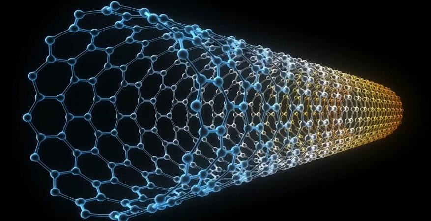
Carbon-based nanomaterials have piqued the interest of researchers in biological fields such as improved imaging, tissue regeneration, and medication or gene delivery. Carbon-based nanomaterials, due to their distinct chemical and physical properties, are promising prospects for a wide range of biomedical applications, including cancer imaging, targeted photo thermal therapy, photo acoustic imaging, drug administration, and tissue engineering. CNTs are moderately flexible carbon compounds with a peculiar structure that allows them to pass through cell membranes and appear to enter the cytoplasm via a “snaking effect.” The snaking effect of carbon nanotubes refers to their capacity to increase membrane penetration and entry into cells via a spiraling or winding motion as well as strong connections with specific proteins [12].
Single Wall Carbon Nanotubes
SWCNTs were used as a diagnostic and therapeutic agent—an integrated “theranostic” Nano platform for both microwave detection and treatment of breast cancer. SWCNTs are promising candidates for tumor-targeting applications due to their size and unique physiochemical characteristics SWCNT-based contrast agents have already demonstrated promise in molecular imaging modalities such as magnetic resonance, positron emission tomography, nuclear, and photo acoustic imaging [13]. Another promising and rising application for CNTs is in diagnostic imaging. Ultrasonography is a common imaging technology due to its low cost per test and inherent safety. The therapeutic use of Ultrasound (US) incorporates sound waves with frequencies ranging from 2 to 12 MHz and spatial resolution ranging from 0.2 to 1 mm. Delogu, et al. proved that functionalized MWCNTs have enhanced qualities as ultrasonic contrast agents [14]. New Carbon nanotubes (CNTs) can be created by heating carbon black and graphite in a controlled flame environment. The main issue with this approach is that the CNTs obtained are uneven in shape, size, mechanical strength, quality, and purity. Techniques such as electric arc discharge, laser ablation, and catalytic hydrocarbon decomposition have been proposed to avoid these issues. Varied varieties of CNTs with different properties can be generated depending on the manner of synthesis [15].
Quantum Dots
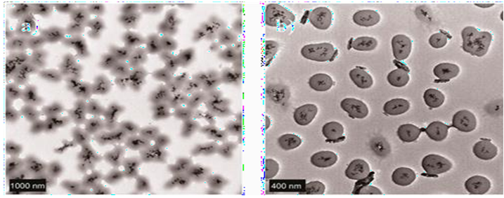
QDs are semiconductor Nano crystals having a quantum- confinement feature that allows them to glow from visible to infrared wavelengths when excited. A single QD typically includes between 100 and 100,000 atoms in its crystal core. QDs are typically 2-10 nm in diameter and range in size from nanometers to micrometers [16]. Graphene Quantum Dots: Graphene quantum dots (GQD), which are fragments of a single-layer two-dimensional graphene, are thought to be the next generation of carbon- based nanomaterials with immense biomedical promise. According to recent research on this family of nanomaterials, GQD are less poisonous and more hydrophobic than graphene and have a sustained bright fluorescence. Their intrinsic fluorescence has sparked interest in anticancer therapy because it may allow for the efficient surveillance of human cells in vitro. Similarly to graphene and GO, the availability of additional active groups on the GQD surface allows for multimodal conjugation, making them suitable carriers for cancer cell treatment and surveillance. Furthermore, due to their distinct structural features, GQD have been demonstrated to improve the chemotherapeutic efficacy of anticancer medications that are poor due to drug resistance. Recent research has shown that GQD can efficiently enhance the nuclear accumulation of medicines such as doxorubicin and cisplatin. These drugs’ DNA cleavage activity and cytotoxicity are significantly increased. These exceptional biological properties highlight GQD’s superiority over modified graphene or GO, as well as many other nanoparticle- based delivery systems [17]. Carbon Quantum Dots: Discovery of CQDs during the separation and purification of single-walled carbon nanotubes (SWCNTs) in 2004 sparked additional research to leverage CQD fluorescence features and produce a new class of practical fluorescent nanomaterials. 6 The term “carbon quantum dots” was coined in 2006 by who presented a synthetic approach to manufacture CQDs with greatly increased fluorescence emissions by surface passivation. The top-down route7-9 and the bottom-up route are used to synthesize CQDs. 3,10-12 CQDs are typically quasi-spherical nanoparticles with amorphous to Nano crystalline cores made up of graphitic or turbostratic carbon (sp2 carbon) or graphene and graphene oxide sheets joined by diamond-like sp3 hybridized carbon insertions [18]. Non-Chemotoxic Induction of Cancer Cell Death Using Magnetic Nanowires: Magnetic fields can be used to modify magnetic nanoparticles (MNPs) remotely. MNPs can be captured, concentrated [9, 10, 11, 12], or employed in cell separation using direct current fields [13, 14, 15]. MNPs can be heated [18] or rotated in alternating fields [19, 20], and in the case of elongated structures, they can transmit forces or torques to anything they come into contact with the study of magnetic nanostructures for biomedical applications has lately increased. The majority of earlier experiments used magnetic nanobeads, although subsequent investigations have used Magnetic nanowires (NWs) have also been reported to have a variety of bio applications. NWs outperform nanobeads in terms of magnetic moments per unit volume and surface area to volume ratios, allowing them to bind to cells more efficiently and produce purer populations during cell separation. Fe and Ni are the two magnetic materials commonly employed as NWs in biological applications. Fe NWs tend to aggregate more than Ni because they have a greater remanence magnetization value, making Ni a better material in that sense [19]. Furthermore, Ni NWs have been demonstrated to be effective in cell separation, manipulation, and purification as well as in delivering payloads of diverse macro-particles, including biological entities. Recent research has looked into their use as therapeutic agents for hyperthermia and cell inflammation induction in human embryonic cell cultures [20]. They have also been demonstrated to function as apoptotic agents for pancreatic cancer cells. Despite its documented genotoxicity and cytotoxicity in Ni-containing dust particles, the high number of research that use Ni NWs in contrast to Fe may suggest that Ni is a superior potential material. Despite the fact that no work has been done directly comparing the cytotoxicity of Fe and Ni NWs under the same experimental conditions (cell line, incubation times, concentrations), a cross-comparison of studies reveals that Fe NWs are significantly less toxic at a given concentration than Ni NWs. However, as previously stated, the higher aggregation of Fe NWs limits the usage of pristine NWs, necessitating passivation to overcome this limitation. In addition to work on biological and biomedical applications, fundamental research on internalization into different cell lines, cytotoxicity reliance on incubation duration and NW concentration, and length-dependent cytotoxicity have been described. Furthermore, elevated amounts of reactive oxygen species, cell viability loss, and cell membrane leakage have been demonstrated to be activated just by incubating cells with Ni NWs. NWs with diameters ranging from 150 to 280 nm were employed in the investigations listed here. Optical microscopy and quantitative real-time polymerase chain reaction (PCR) were employed to measure cell damage. Only Fung, et al. 40 employed a colorimetric approach to assess cell viability before and after the administration of the low-frequency AMF. We used a low-frequency, small- amplitude AMF on cells that had been treated with Ni NWs in this investigation. In comparison to previous studies, the applied magnetic field was much weaker in our experimental setup, and the NWs were about one order of magnitude thinner. The motivation for this experimental setup is to use a non-chemotoxic method of inducing cancer cell death. We investigate NW-cell interactions using transmission electron microscopy (TEM) pictures that highlight the internalization process of magnetic NWs and their position within cancer cells. We also consider AMF- cell interactions when a magnetic field is administered to cells in culture for short periods of time (10 or 30 minutes) [21].
Fullerene
One of the primary issues with these approaches is poor pharmacokinetics and untargeted drug delivery; as a result, in some applications, such as chemotherapy, there are rising adverse effects from the medicine and the effectiveness of therapy may diminish. In addition to medicine administration, biological material such as DNA and numerous tiny molecules must be delivered past the cell membrane in some disorders. Indeed, the delivery of such compounds to the nucleus and other organelles can bring new obstacles due to the cell’s unique barriers and peculiarities. To address these issues, nanotechnology research has provided a variety of strategies to improve drug delivery effectiveness. For example, the pioneer nanoparticle: fullerene, with its unique geometry, size, and surface properties, has a spherical structure with a strong a polar feature. Because of these properties, fullerenes can be employed in lipid- like systems, acting as a reservoir and even traversing cell membranes. Fullerenes, often known as Buckminsterfullerene’s or Bucky balls, are a type of carbon nanomaterial allotrope. Fullerenes have a structure made up of sp2.
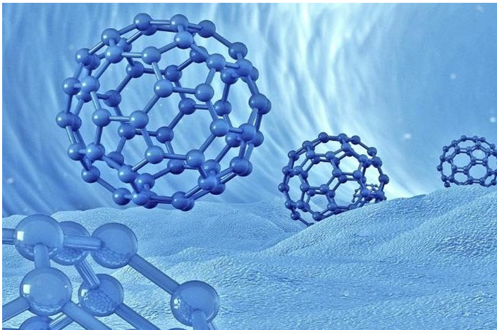
Carbons with distinct chemical and physical properties, as well as a highly symmetrical cage of varying sizes (C60, C76, etc.). C60 is the most prevalent fullerene in the synthesized mixture. It is made up of 60 carbon atoms connected by C5- C5 single bonds (12 pentagons) and C5=C6 double bonds (20 hexagons). Each fullerene has 2n + 20 carbon atoms and ‘n’ hexagons. At 1000°C, C60 and C70 are created, and the concentration increases as the pulse duration increases. Among other things, the dual behavior of C60 among reactive oxygen species (ROS) gives this nanoparticle the potential to act in different ways in different settings. C60 has the ability to create oxygen species when exposed to visible light in some instances, making it a viable option for photodynamic treatment (PDT). In other circumstances, it inhibits ROS production, which might be employed as a neuroprotective drug. Further research is needed to determine the mechanism of this activity. This molecule’s limited solubility in many organic solvents and insolubility in water provide challenges for using it in biological applications. Accordingly, materials’ hydrophilicity in biological systems is more significant than their hydrophobicity, and numerous techniques have been employed to boost their hydrophilicity and water solubility. Two-phase colloidal solution preparation, the creation of fullerene derivatives, fullerene polymers, encapsulation in specialized carriers (cyclodextrins, calixarenes, polyvinylpyrrolidone, micelles, liposomes, etc.), and chemical modification [by adding hydrophilic elements like amino acids, carboxylic acids, polyhydroxyl groups (fullerenes), and amphiphilic polymers], among others [22].
Applications of Fullerene in Pharmaceutics:
- Delivering drugs topically- [22]
- Cancer treatment-
- A photodynamic treatment-
In Vivo Testing of Multifunctional Single Dendrimer Nano Devices
After the in vitro investigations were finished, we started animal tests to look at the distribution and effectiveness of the targeted nanotherapeutic. The data in this subsection describe the therapeutic effect of drug-dendrimer conjugates on tumour cells when administered in vivo [23]. Nanomedicine: Due to the leaky tumour vasculature and inadequate lymphatic drainage, Nano medicine has emerged as a viable alternative technique that permits the selective accumulation of systemically delivered chemotherapeutics in the tumour tissues In the past three decades, a great deal of research has been devoted to creating cancer nanomedicines that can effectively transport chemotherapeutic chemicals to the targeted areas while avoiding side effects and overcoming biological obstacles. on wholesome tissues In order to improve the targeting selectivity of chemotherapeutics, the following two crucial methods have been adopted in the development of cancer nanomedicines: surface- Nano engineering of nanoparticles (NPs) by conjugating corresponding active recognition moieties to their surfaces; and improved accumulation and efficacy of therapeutic payloads by overcoming biological barriers and improving tumour penetration of Nano medicine. Numerous NPs are appealing for use in the creation of cancer nanomedicines because of their high surface area-to-volume ratios, controlled shapes and sizes, and special features. The primary benefits of their use include: enhancing the pharmacokinetic and pharmacodynamics profiles of currently available anticancer compounds; minimizing the toxic and adverse effects of anticancer medications on healthy cells; facilitating more sensitive cancer detection; enhancing therapeutic efficacy and specificity; demonstrating the viability of combining cancer therapies; facilitating cost-effectiveness and improving quality of life; and providing an applicability for various administrative settings. The anticipated clinical translation rate of promising nanomedicines from bench to bedside is often less than 10%, which has caused a significant increase in awareness among researchers and regulators [24].
- Advantages of Nanomedicine: Because it has the ability to enable the preferential delivery of medications to tumours due to the EPR effect and can administer more than one therapeutic agent for combination therapy, Nano medicine for cancer therapy has advantages over conventional medicine. Due to the distinct Nano size (1-100 nm) and high surface-to-volume ratio, nanotechnology has had a significant impact on drug delivery. Due to their larger size relative to the free drug, Nano formulations can prevent medications from being quickly removed through the renal system when intravenously injected into the body. This frequently leads to a prolonged blood circulation period with a higher possibility of accumulating in the target tissue. Nano formulations can reduce adverse effects, making the medications used more suitable for therapeutic use. Additionally, by using passive targeting mediated by the EPR phenomenon, Nano carriers can leak out of fenestrated tumour vascular walls and aggregate in the tumour tissue that frequently lacks efficient lymphatic drainage. As a result, the use of nanotechnology in drug administration can considerably increase therapeutic efficacy while reducing pharmacological side effects and systemic toxicity [24]. Nanobots: The challenging subject of Nano robotics, which deals with minute objects at the molecular level, is growing. Nano robots are classic nano electromechanical devices created to carry out particular tasks precisely at Nano scale scales. Its size gives it an edge over traditional medicine. The pattern of deposition and serum lifetime are influenced by particle size. As a result, the therapeutic effect of nanoscale medicines can be felt sooner and at lower concentrations. By directing carriers to a specified location, it also supplies materials for controlled drug delivery. Applications of nanorobots and nanoscale- structured materials inside the body to diagnose and treat disease are particularly intriguing in the field of biomedical engineering. A wide range of capabilities should be made possible by the swift advancement in nanoscale device engineering. One source of components for nanorobots is the continual development of electronics, sensors, and motors at the molecular scale. The capacity to programme bacteria has been demonstrated to create computation for Nano devices. Nano systems for controlling nanorobots to carry out specific tasks in medicine are being developed. This could lead to advancements in nanotechnology automation. A medical Nano robot’s main component will probably be carbon, most likely in the form of diamond or diamonded/ fullerene Nano composites. Numerous others light elements, including silicon, fluorine, oxygen, nitrogen, hydrogen, and sulphur, will serve specific functions in nanoscale gears and other parts. The various parts of the nanorobot design could include molecular computers, onboard molecular computers, motors, manipulators, and onboard sensors.. In contrast, self-assembly is far less laborious since it makes use of some molecules’ innate propensity to seek out one another. Self-assembling components allow researchers to create the necessary configurations by simply adding billions of them to a beaker and letting their natural affinities connect them together. Manufacturing methods that can construct a molecular structure through computer models of diamond mechanosynthesis are required to create complicated nanorobotic systems [25].
- Nanorobots in Cancer Detection and Treatment: The creation of nanorobots may lead to significant improvements in the detection and treatment of cancer. Due to the fact that conventional therapies like chemotherapy and radiation therapy frequently result in the destruction of more healthy cells than diseased ones, nanorobots could be very beneficial and promising for patient care. From this perspective, it offers cancer patients a no depressive kind of therapy. By examining their surface antigens, the Nanorobots will be able to discriminate between various cell kinds, such as cancerous and healthy cells (they are different for each type of cell). Chemotactic sensors that are tuned to the specific antigens on the target cells are used to achieve this. Another strategy employs cutting-edge methodology to develop distributed collective control for the fight against cancer. They may be configured to recognize various concentrations of beta-catenin and E-cadherin in both the primary and metastatic phases using chemical sensors. Then, and only then, will medical nanorobots eliminate these cells. One may take into account the following control techniques: Random: nanorobots moving passively through the fluid only collide with the target as a result of Brownian motion
- Follow Gradient: Nanorobots measure the gradient and follow it until they reach the destination. They continuously scan concentration intensities for E-cadherin signals. The nanorobot interprets the signal as being a false positive and continues to flow with the fluid if the gradient estimate performed after signal detection does not reveal any additional signal in 50 ms.
- Follow Gradient with Attractant: like above, but as nanorobots reach the destination, they also release a separate chemical signal that helps other robots discover the target more easily. As a result, a larger gradient of E-cadherin signal intensity is used as a chemical parameter identification while directing nanorobots to locate cancerous tumours. To determine the strength of E-cadherin signals, integrated Nano sensors can be used. Consequently, they can be used productively to combat cancer [25].
Active Drug Delivery by Micro-/Nanobots
The development of micro-/nanorobots for active drug delivery has progressed from the test tube to the cellular level and to living animals as a result of the tireless work of researchers over the past few decades. Model pharmaceuticals can currently be loaded onto micro-/ nanorobots using a variety of techniques, such as layer-by- layer (LbL) encapsulation, physical adsorption, electrostatic contact, and more. Typically, disease-related biochemical gradients or magnetic fields are used to direct drug-loaded micro- or nanorobots toward a predetermined location. After reaching the desired location, physiological conditions (such as pH) or outside forces (such as NIR light, ultrasound, and magnetic fields) can cause the release of medications that have been encapsulated. For further details on these active medication delivery systems based on micro-/nanorobots, readers might refer to. This section provides a summary of current advancements in the use of micro- and nanorobots for active medication delivery in vitro and in vivo [26].
Nanorobot Types
Some researchers categorized nanorobots used for therapeutic and drug delivery based on the uses they are used for, which are listed below. Pharmacyte: Pharmacyte is a medical nanorobot with a size of 1-2 mm and the capacity to carry up to 1 mm3 of a specific medicine in tanks. They are managed by mechanical sorting pumps and systems. They are given chemotactic sensors or molecular markers, which ensure complete targeting precision. The onboard power source is glucose and oxygen drawn from local surroundings like blood, intestinal fluid, and cytosol. The nanorobots can be removed or recovered using centrifuge nan apheresis after finishing their tasks. Diagnosis and Imaging: They have microchips with human molecules overlay for diagnosis and imaging. When the molecules identify an illness, the chip is expected to send an electrical signal. Gives an illustration of specialized sensor nanobots that can be injected into the blood beneath the skin, where they can check the contents of the blood and detect any potential ailments. They can also be used to track blood sugar levels. The low cost of production and ease of manipulation are advantages. Respirocyte: Respirocyte is a nanorobot that resembles an artificial red blood cell and is an artificial oxygen carrier. Endogenous serum glucose provides the power. In comparison to RBCs, this artificial cell can deliver 236 times more oxygen to the tissues per unit volume. It can also deliver acidity. Microbivores: It is an oblate spheroidal device with 3.4 m along its major axis and 2.0 m along its minor axis for nanomedical applications. Up to 200 pW can be continuously used by the nanobot. Utilizing this ability, imprisoned microorganisms are digested. In terms of volume/sec digested per unit volume of phagocytic agent, phagocytes are able to phagocytose roughly 80 times more effectively than macrophage agents. Clottocytes: Clottocytes are a special kind of nanorobot that can produce artificial mechanical platelets to produce “immediate” hemostasis. It is common knowledge that platelets are roughly spheroidal, nucleus-free blood cells with a diameter of 2 m. At the site of the bleeding, platelets unite. They become tacky and activated there, forming a tampon that helps seal the blood vessel and stop the bleeding. Additionally, they deliver elements that support coagulation. Chromallocyte: The Chromallocyte would completely swap out individual cells’ chromosomes, undoing the consequences of genetic disorders and other accumulated gene damage, and stop ageing. Repair machines will be able to repair the entire cell by first assessing the situation inside the cell by looking at its contents and activity, and then acting by moving along molecule-by-molecule and structure-by- structure [27].
- Advantages of Nanorobots:
- Use of more bioavailable nanorobot medication delivery technologies
- targeted therapy, such as treating only cancerous cells
- reach a portion of the human anatomy that is difficult to operate on when lying on the operating table
- The benefits of a large interfacial area during mass transfer can be realized as drug molecules are transported by nanorobots and released where necessary
- non-intrusive method
- Nobs for fine-tuning the amount, frequency, and time of release are part of a computer- controlled process
- improved accuracy
- avoiding unintended side effects by rendering the drug inactive when therapy is not required
- Small size: The maximum size of a nanorobot is 3 microns, allowing it to readily pass through the body without obstructing capillary flow [27].
Conclusion
Numerous aspects of cancer therapy have already changed as a result of nanotechnology, which is also drastically changing the course of cancer treatment. It has greatly influenced the ability to overcome the limitations of conventional chemotherapies, identify malignant cells selectively, and administer medications in a targeted manner. While some nanotechnology-based products have already been put on the market, many are still in the development and clinical study stages. These cutting-edge active or passive targeting strategies can drastically lessen the side effects of traditional chemotherapies while also increasing the survival rate. The application of nanotechnology in targeted cancer treatment that prevents potentially fatal side effects has the potential to progress clinical practise towards a life-saving approach, since cancer is one of the deadliest diseases.
References
-
Gmeiner WH, Ghosh S (2004) Nanotechnology for cancer treatment. Nanotechnology Reviews 3(2): 111-122.
-
Barnard RJ (2004) Prevention of Cancer through Lifestyle Changes. Evidence-Based Complementary and Alternative Medicine 1(3): 233-239.
-
Shewach DS, Kuchta RD (2009) Introduction to Cancer Chemotherapeutics. Chemical Reviews 109(7): 2859- 2861.
-
Sutradhar KB, Amin MdL (2014) Nanotechnology in Cancer Drug Delivery and Selective Targeting. ISRN Nanotechnology 2014: 1-12.
-
Aslan B, Ozpolat B, Sood AK, Lopez Berestein G (2013) Nanotechnology in cancer therapy. Journal of Drug Targeting 21(10): 904-913.
-
Hulla J, Sahu S, Hayes A (2015) Nanotechnology: History and future. Human & Experimental Toxicology 34(12): 1318-1321.
-
Bayda S, Adeel M, Tuccinardi T, Cordani M, Rizzolio F (2019) The History of Nanoscience and Nanotechnology: From Chemical–Physical Applications to Nanomedicine. Molecules 25(1): 112.
-
García-Pinel B, Porras Alcalá C, Ortega-Rodríguez A, Sarabia F, Prados J, et al. (2019) Lipid-Based Nanoparticles: Application and Recent Advances in Cancer Treatment. Nanomaterials (Basel) 9(4): 638.
-
Yadav N, Khatak S, Sara S (2013) Solid lipid nanoparticles: a review. International Journal of Applied Pharmaceutics 5(2): 8-18.
-
Bhardwaj P, Singh B, Behera SP (2021) Green approaches for nanoparticle synthesis: emerging trends. Nanomaterials 2021: 167-193.
-
Baeza A (2014) Ceramic Nanoparticles for Cancer Treatment. In: Vallet Regí M (Ed.), Bio‐Ceramics with Clinical Applications, Wiley Online Library.
-
Zhang Y, Petibone D, Xu Y, Mahmood M, Karmakar A, et al. (2014) Toxicity and efficacy of carbon nanotubes and graphene: the utility of carbon-based nanoparticles in Nano medicine. Drug Metabolism Reviews 46(2): 232- 246.
-
Mishap A, Sitharaman B, Xu Li, Avti PK, Sahakian AV, et al. (2010) Toward Carbon-Nanotube-Based Theranostic Agents for Microwave Detection and Treatment of Breast Cancer: Enhanced Dielectric and Heating Response of Tissue- Mimicking Materials. IEEE Transactions on Biomedical Engineering 57(8): 1831-1834.
-
Sanginario A, Miccoli B, Demarchi D (2017) Carbon Nanotubes as an Effective Opportunity for Cancer Diagnosis and Treatment. Biosensors 7(1): 9.
-
Madani SY, Naderi N, Dissanayake O, Tan A, Seifalian AM (2011) A new era of cancer treatment: carbon nanotubes as drug delivery tools. International Journal of Nanomedicine 6: 2963-2979.
-
Zhang H, Yee D, Wang C (2008) Quantum dots for cancer diagnosis and therapy: biological and clinical perspectives. Nanomedicine 3(1): 83-91.
-
Iannazzo D, Pistone A, Salamò M, Galvagno S, Romeo R, et al. (2017) Graphene quantum dots for cancer targeted drug delivery. International Journal of Pharmaceutics 518(1-2): 185-192.
-
Ying Lim S, Shen W, Gao Z (2015) Carbon quantum dots and their applications. Chemical Society Reviews 44(1): 362-381.
-
Nana ABA, Marimuthu T, Kondiah PPD, Choonara YE, Du Toit LC, et al. (2019) Multifunctional Magnetic Nanowires: Design, Fabrication, and Future Prospects as Cancer Therapeutics. Cancers 11(12): 1956.
-
Doucey MA, Carrara S (2019) Nanowire Sensors in Cancer. Trends in Biotechnology 37(1): 86-99.
-
Contreras M, Sougrat R, Zaher A, Ravasi T, Kosel J (2015) Non-chemotoxic induction of cancer cell death using magnetic nanowires. International Journal of Nanomedicine 10: 2141.
-
Kazemzadeh H, Mozafari M (2019) Fullerene-based delivery systems. Drug Discovery Today 24(3): 898-905.
-
Baker JR (2009) Dendrimer-based nanoparticles for cancer therapy. Hematology Am Soc Hematol Educ Program 2009: 708-719.
-
Bor G, Mat Azmi ID, Yaghmur A (2019) Nanomedicines for cancer therapy: current status, challenges and future prospects. Therapeutic Delivery 10(2):113-132.
-
Venkatesan M, Jolad B (2010) Nanorobots in Cancer Treatment.
-
Luo M, Feng Y, Wang T, Guan J (2018) Micro-/Nanorobots at Work in Active Drug Delivery. Advanced Functional Materials 28(25): 1706100.
-
Kumar S, Nasim BP, Abraham E (2018) Nanorobots a Future Device for Diagnosis and Treatment. 5(1): 44-49.
- Solution-Processed Chiral Perovskites for Biomedical Applications
- Nanotechnology in Health Chemistry and Medicine: Current Challenges and Future Directions
- Human Exposure to Micro- and Nanoplastics: Pathways, Toxicity, and Intervention Strategies
- Exosome Nanomedicine for Cancer Therapy
- Micro and Nanoplastics–Plastisphere, Biotoxicity, Impact on Human Health, and Mitigation Strategies
- Process Validation of Cefixime Powder for Suspension Dosage Form, 50 mL