Study of Genetic Mutation CSF1R and EPM2A Genes in Patients with Adult-Onset Leukoencephalopathy with Axonal Spheroids and Pigmented Glia Syndrome, Iran
In this study we have analyzed 100 people. 40 patients and 60 control group had Adult-onset Leukoencephalopathy with Axonal Spheroids and Pigmented Glia Syndrome. The gene CSF1R (colony-stimulating factor 1 receptor) analyzed in terms of genetic mutations made. In this study, people who have genetic mutations were targeted, with nervous disorders, Adult-onset Leukoencephalopathy with Axonal Spheroids and Pigmented Glia Syndrome. In fact, of all people with Adult-onset Leukoencephalopathy with Axonal Spheroids and Pigmented Glia Syndrome, 40 people had a genetic mutation in the genes EPM2A and CSF1R. Any genetic mutations in the target genes control group did not show.
Introduction
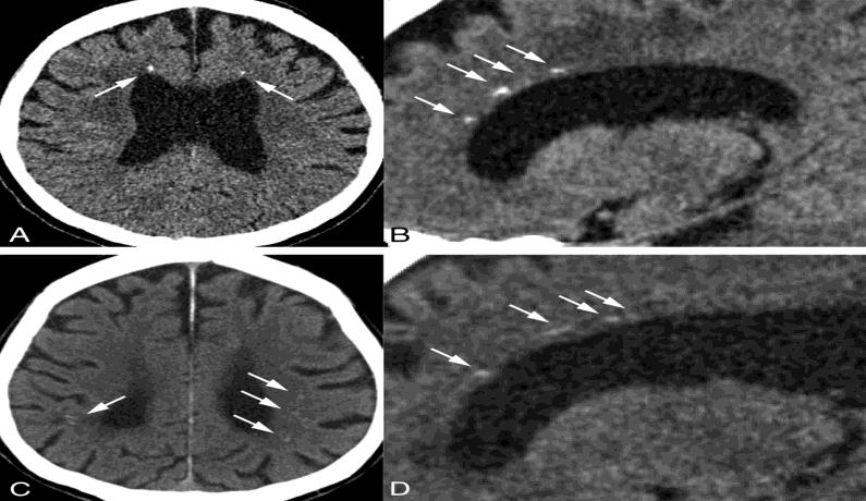
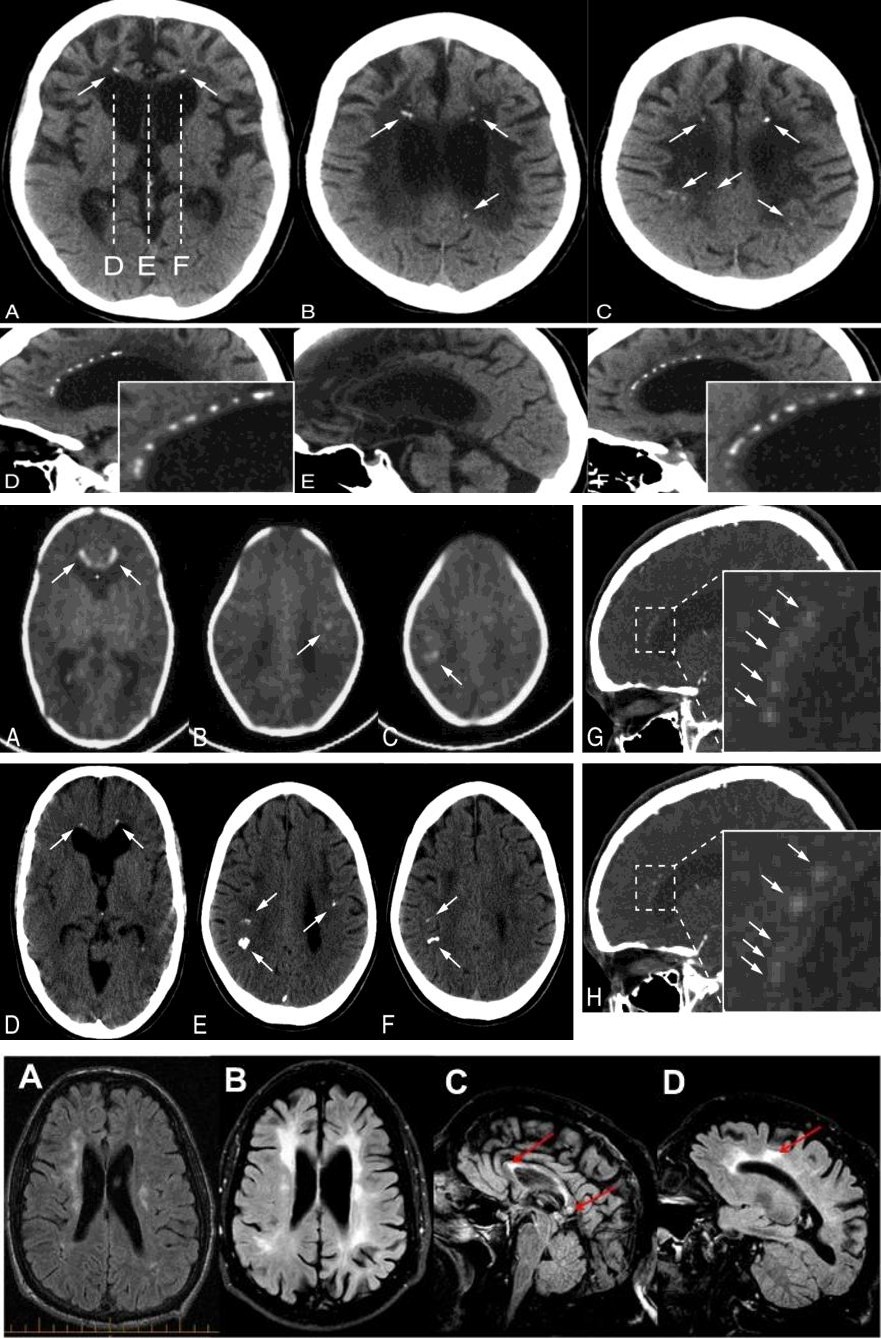
Adult-onset Leukoencephalopathy with Axonal Spheroids and Pigmented Glia Syndrome is a genetic disorder characterized by changes in specific brain regions. One of the signs of Adult-onset Leukoencephalopathy with Axonal Spheroids and Pigmented Glia Syndrome is leukoosfalopathy, which is a change in the type of brain tissue called white matter. The white matter contains neurons (axons) covered by a substance called myelin that insulates and protects the nerves. Axons spread from nerve cells (neurons) and transmit nerve pulses throughout the body. Limited damage to this brain tissue (white matter lesions) can be detected by magnetic resonance imaging (MRI). Another feature of Adult-onset Leukoencephalopathy with Axonal Spheroids and Pigmented Glia Syndrome is swelling in spheroids in brain axons, which can damage axons. In addition, in Adult-onset Leukoencephalopathy with Axonal Spheroids and Pigmented Glia Syndrome, glial cells usually have abnormal pigmentation. Glial cells are special brain cells that protect the neurons. Damage to myelin and neurons seems to play a role in many of the signs and symptoms of people with Adult-onset Leukoencephalopathy with Axonal Spheroids and Pigmented Gila Syndrome [1].
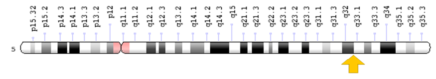
Symptoms of Adult-onset Leukoencephalopathy with Axonal Spheroids and Pigmented Glia Syndrome usually begin at age 40 and become worse over time. Personality changes such as depression and loss of social relationships are one of the first symptoms of Adult-onset Leukoencephalopathy with Axonal Spheroids and Pigmented Glia Syndrome. Damaged people may also experience memory loss and problem solving. Some people with Adult-onset Leukoencephalopathy with Axonal Spheroids and Pigmented Glia Syndrome have mild seizures and usually experience seizures that have symptoms of this syndrome. The progression of Adult- onset Leukoencephalopathy with Axonal Spheroids and Pigmented Glia Syndrome leads to a sharp decline in Adult-onset Leukoencephalopathy with Axonal Spheroids and Pigmented Glia Syndrome is caused by the mutation of the EPM2A and CSF1R genes (Figure 1), which is based on the long arm of chromosome number 5 as 5q32. This gene provides instructions for the synthesis of a protein called CSF-1 receptor found in the outer membrane of certain cells, including glial cells. The CSF-1 receptor controls the signaling pathways that control many important cellular processes, such as cell growth and cell division (proliferation) and cell differentiation [4].
thinking and the ability to reason (rational deterioration) [2].
Over time, motor skills are also affected, and people with Adult-onset Leukoencephalopathy with Axonal Spheroids and Pigmented Glia Syndrome may have difficulty walking. Many types of motor abnormalities, such as Parkinson's, which include unusual slow motion (bradycynzia), involuntary tremor and muscle stiffness (spasm), may also occur in people with Adult-onset Leukoencephalopathy with Axonal Spheroids and Pigmented Glia Syndrome. It is worth noting that the pattern of cognitive and motor problems varies even among individuals in a family. Almost all people with this syndrome are ultimately unable to walk, talk, and take care of themselves [3].
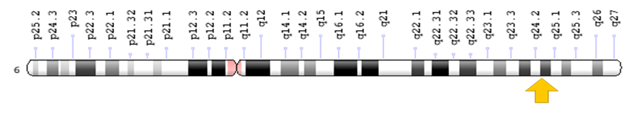
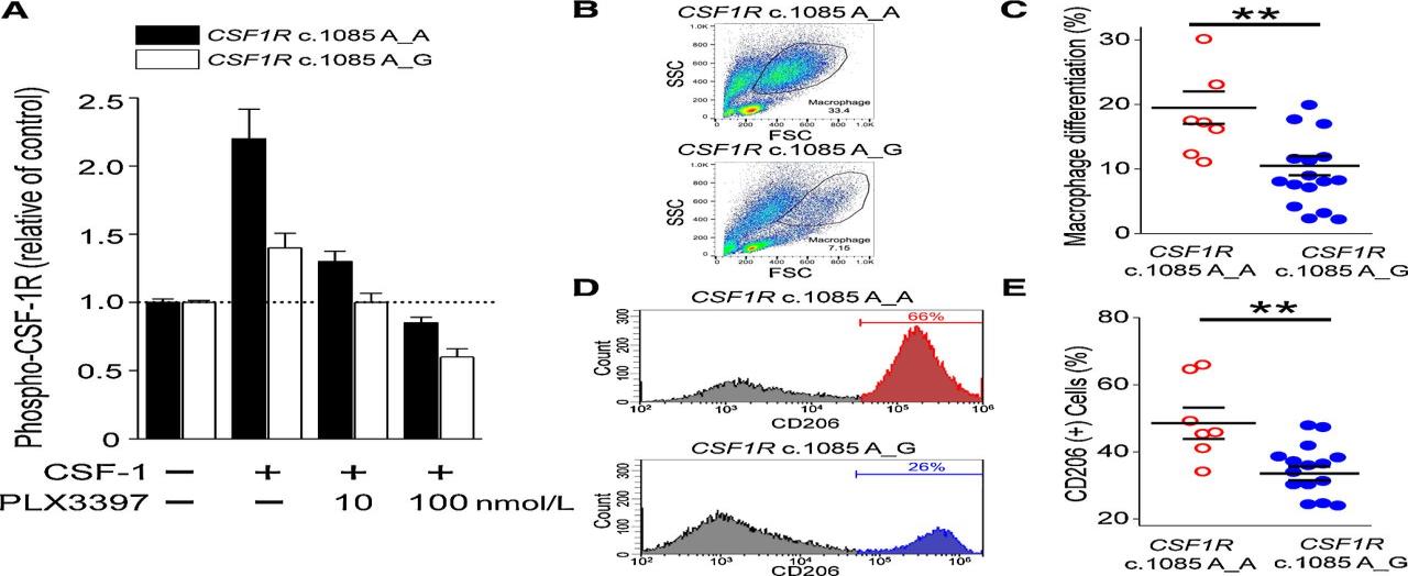
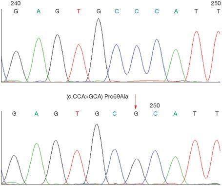
CSF1R and EPM2A genes mutations in the Adult-onset Leukoencephalopathy with Axonal Spheroids and Pigmented Glia Syndrome result in changes in the CSF-1 receptor protein, which may not be able to stimulate cellular signaling pathways. However, it is still unclear how these gene mutations lead to signs and symptoms associated with Adult-onset Leukoencephalopathy with Axonal Spheroids and Pigmented Glia Syndrome (Figure 2).
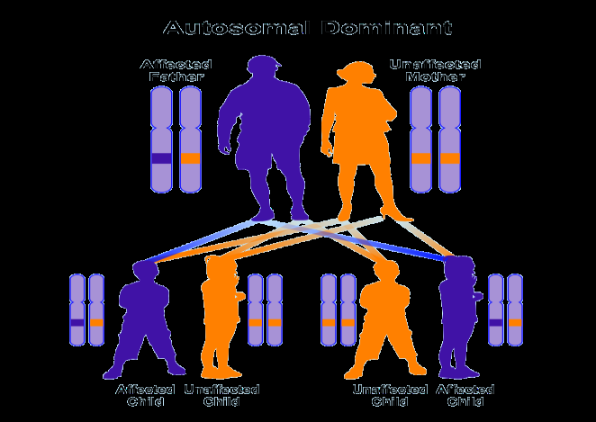
Adult-onset Leukoencephalopathy with Axonal Spheroids and Pigmented Glia Syndrome (Figure 3) follows the dominant autosomal inheritance pattern. Therefore, to produce this syndrome, a mutational version of the EPM2A and CSF1R genes (parent or parent) is required and the chance of having a child with this syndrome in the dominant autosomal state is 50% for each pregnancy. In most cases, Adult-onset Leukoencephalopathy with Axonal Spheroids and Pigmented Glia Syndrome is caused by new mutations without family history [4].
Adult-onset Leukoencephalopathy with Axonal Spheroids and Pigmented Glia Syndrome appears to be a rare genetic disorder, with the frequency of its prevalence uncertain in the world. Adult-onset Leukoencephalopathy with Axonal Spheroids and Pigmented Glia Syndrome is diagnosed based on the clinical and clinical findings of the patients and some pathological examinations. The most accurate method for detecting this syndrome is the molecular genetic test for the CSF1R and EPM2A genes to investigate the presence of possible mutations.
The treatment and management strategy for Adult- onset Leukoencephalopathy with Axonal Spheroids and Pigmented Glia Syndrome is symptomatic and supportive. Treatment may be done by a team of experts, including a neurologist, dermatologist, and other healthcare professionals. There is no definite treatment for this syndrome and all clinical interventions are designed to reduce the suffering of the sufferers. Using anticonvulsants such as phenobarbital or carbamazepine is also useful in controlling seizures. Genetic counseling is also needed for all parents who want a healthy baby [4].
Materials and Methods
In this study, 40 patients with Adult-onset Leukoencephalopathy with Axonal Spheroids and Pigmented Glia Syndrome and 60 healthy controls were studied. Peripheral blood samples from patients and parents with written permission control were prepared. After separation of serum, using Real Time-PCR technique of tRNA molecules were collected [4]. To isolate Neuroglial cells erythrocytes were precipitated from hydroxyethyl starch (HES) was used. At this stage, HES solution in ratio of 1 to 5 with the peripheral blood of patients and controls were mixed. After 70 minutes of incubation at room temperature, the supernatant was removed and centrifuged for 18 min at 600 Gera. The cells sediment with PBS (phosphate buffered saline), pipetting and slowly soluble carbohydrate ratio of 1to2 on ficole (Ficol) was poured in the 580G was centrifuged for 39 minutes. Mono nuclear Neuroglial cells also are included, has a lower density than ficole and soon which they are based. The remaining erythrocyte has a molecular weight greater than ficole and deposited in test tubes [5].
The supernatant, which contained the mononuclear cells, was removed, and the 600 Gera was centrifuged for 19 minutes. Finally, the sediment cell, the antibody and Neuroglial cells was added after 39 minutes incubation at 5 °C, the cell mixture was passed from pillar LSMACS. Then the cells were washed with PBS and attached to the column LSMACSS pam Stem cell culture medium containing the transcription genes EPM2A and CSF1R and were kept [6].
To determine the purity of Neuroglial cells are extracted, flow cytometry was used. For this purpose, approximately 5-7 × 10³ Neuroglial cells were transfer red to1.5ml eppendorf tube and then were centrifuged at 4000 rpm for 9 minutes a time. Remove the supernatant culture medium and there maining sediment, 100μl of PBS buffer was added. After adding 5-10μl CD34+ PE monoclonal anti body to the cell suspension for 70 min at 5°C incubated and read immediately by flow cytometry. For example, rather than control anti body Neuroglial cells PE, IgG1 negative control solution was used [7].
Total RNA Extraction Procedure Includes
a) 1ml solution spilled Qiazolon cells, and slowly and carefully mixed and incubated at room temperature for 7 minutes. Then 300μl chloroform solution to target mix, and then transfer the micro tubes was added, and the shaker well was mixed for 15 seconds. The present mix for 9 minutes at room temperature and then
Results
Figures 4-11
incubated for 30 minutes at 5°C was centrifuged at 15200 rpm era. Remove the upper phase products were transferred to a new microtube and to the one times the volume of cold ethanol were added. The resulting mixture for 24 hours at -25°C was incubated [8]. b) Then for 55 minutes at 4°C was centrifuged at 16000 rpm era. Remove the supernatant and the white precipitate, 1ml of cold 75% ethanol was added to separate the sediment from micro tubes were vortex well. The resulting mixture for 25 minutes at 6°C by the time we were centrifuged 16000 rpm. Ethanol and the sediment was removed and placed at room temperature until completely dry deposition. The precipitate was dissolved in 20μl sterile water and at a later stage; the concentration of extracted RNA was determined [9].
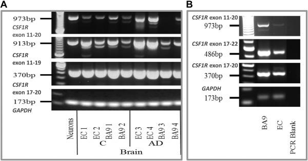
To assessment the quality of mi-RNAs, the RT-PCR technique was used. The cDNA synthesis in reverse transcription reaction (RT) kit (Fermentas K1622) and1μl oligoprimers18 (dT) was performed. Following the PCR reaction2μM dNTP, 1μg cDNA, Fermentas PCR buffer 1X, 0 / 75μM MgCl2, 1.25 U / μL Tag DNA at 95°C for 4min, 95°C for 30s, annealing temperature 58°C for 30s, and72 °C for30 seconds, 35 cycles were performed. Then 1.5% agarose gel, the PCR product was dumped in wells after electrophores is with ethidium bromide staining and colors were evaluated [10, 11, 12].
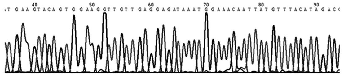
According to the results of sequencing the genome of patients with sleep disturbances, and the genetic mutations EPM2A and CSF1R genes found that about 100% of patients with Adult-onset Leukoencephalopathy with Axonal Spheroids and Pigmented Glia Syndrome, they have this genetic mutation. Patients with Adult-onset Leukoencephalopathy with Axonal Spheroids and Pigmented Glia Syndrome, unusual and frightening images in the process of Adult-onset Leukoencephalopathy with Axonal Spheroids and Pigmented Glia Syndrome, experience. Lot epigenetic factors involved in Adult-onset Leukoencephalopathy with Axonal Spheroids and Pigmented Glia Syndrome. But the most prominent factor to induce Adult-onset Leukoencephalopathy with Axonal Spheroids and Pigmented Glia Syndrome, mutations is CSF1R and EPM2A genes. This gene can induce the birth and can also be induced in childhood and adulthood. We suggest with this study that other neurological researchers are more concerned with the study of more genetic mutations in the development of Adult-onset Leukoencephalopathy with Axonal Spheroids and Pigmented Glia Syndrome, in order to identify more effective results for faster, more accurate diagnosis and more effective treatment.
1. Ali ZS, Van Der Voorn JP, Powers JM (2007) A
comparative morphologic analysis of adult onset leukodystrophy with neuroaxonal spheroids and pigmented glia--a role for oxidative damage. J Neuropathol Exp Neurol 66(7): 660-672.
2. Freeman SH, Hyman BT, Sims KB, Hedley WET,
Vossough A, et al. (2009) Adult onset leukodystrophy with neuroaxonal spheroids: clinical, neuroimaging and neuropathologic observations. Brain Pathol 19(1): 39-47.
3. Kleinfeld K, Mobley B, Hedera P, Wegner A, Sriram S,
et al. (2013) Adult-onset leukoencephalopathy with neuroaxonal spheroids and pigmented glia: report of five cases and a new mutation. J Neurol 260(2): 558- 571.
4. Mitsui J, Matsukawa T, Ishiura H, Higasa K, Yoshimura
J, et al. (2012) CSF1R mutations identified in three families with autosomal dominantly inherited leukoencephalopathy. Am J Med Genet B Neuropsychiatr Genet 159(8): 951-957.
5. Nicholson AM, Baker MC, Finch NA, Rutherford NJ,
Wider C, et al. (2013) CSF1R mutations link POLD and HDLS as a single disease entity. Neurology 80(11): 1033-1040.
6. Rademakers R, Baker M, Nicholson AM, Rutherford
NJ, Finch N, et al. (2011) Mutations in the colony stimulating factor 1 receptor (CSF1R) gene cause hereditary diffuse leukoencephalopathy with spheroids. Nat Genet 44(2): 200-205.
7. Sundal C, Wszolek Z (2012) Adult-Onset Leukoencephalopathy with Axonal Spheroids and Pigmented Glia. In: Pagon RA, et al. (Eds.), GeneReviews® [Internet]. Seattle (WA): University of Washington, Seattle; 1993-2017.
8. Wider C, Van Gerpen JA, DeArmond S, Shuster EA,
Dickson DW, et al. (2009) Leukoencephalopathy with spheroids (HDLS) and pigmentary leukodystrophy (POLD): a single entity? Neurology 72(22): 1953- 1959.
9. Lyon G, Valevski FA, Kolodny EH (2006) Leukodystrophies: clinical and genetic aspects. Topics in Magnetic Resonance Imaging 17(4): 219-242.
10. Santhosh K, Kesavadas C, Thomas B, Gupta AK,
Thamburaj K, et al. (2009) Susceptibility weighted imaging: a new tool in magnet resonance imaging of stroke. Clinical Radiology 64(1): 74-83.
11. Libon DJ, Price CC, Davis GK, Giovannetti T (2004)
From Binswanger's disease to Leukoaraiosis: What we have learned about subcortical vascular dementia. Clinical Neuropsychologist. 18(1): 83-100.
12. Davis GK, Ronald AC, Robert HP, Moser Dj, Malloy PF,
et al. (2004) Computer-mediated measurement and subjective ratings of white matter hyperintensities in vascular dementia: Relationships to neuropsychological performance. The Clinical Neuropsychologist 18(1): 50-62.
- A Review of Gene Therapy for Parkinson's Disease to Control Dopaminergic Neurons
- Late-Onset Myasthenia Gravis in a Patient with Recurrent Breast Cancer: A Case Report
- Covid-Induced Dystonia and Opsoclonus: A Case Report
- Generalized Tonic-Clonic Seizure in a Pediatric Patient with Sunflower Syndrome: A Case Report
- Comparison of Doppler Guided Seldinger Technique Versus Classic Palpatory Seldinger Technique for Radial Artery Cannulation-an Open Label Randomized Controlled Trial
- Brown Sequard Syndrome: Understanding the Complexities of Spinal Cord Injury