Occlusal Trauma: A Threat
A 20 year old female patient reported to the department of Oral Medicine and Radiology with a complaint of broken teeth in upper front tooth region since 6 months associated with pain on chewing food.
Case Report
A 20 year old female patient reported to the department of Oral Medicine and Radiology with a complaint of broken teeth in upper front tooth region since 6 months associated with pain on chewing food. Medical and dental history was not significant .Patient gave no history of deleterious habits. Intraoral examination revealed attrition in relation to all the upper and lower anteriors (Figure 1 A). Wear facet with pulp exposure was seen in relation to maxillary left central incisor and lateral incisor (Figure 1B). Maxillary left central incisor no response was elicitated on electric pulp testing. Provisional diagnosis was given as Primary occlusion Trauma in relation to upper and lower anteriors. Patient was referred to the department of endodontics for root canal treatment, followed by endodontic evaluation.
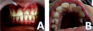
Figure 1A: Reveals attrited Maxillary and Mandibular teeth; 1B: Reveals Pulp exposure i.e. Maxillary left central and lateral incisors.
Discussion
Occlusal trauma can be classified as primary or secondary. Primary occlusal trauma refers to excessive force applied to a tooth or teeth with normal supporting structures. Secondary occlusal trauma refers to when normal occlusal forces become excessive because of loss of attachment [1]. In its generalized form, occlusal trauma is characterized by severe attrition, exposed dentin, sensitive teeth, and tooth mobility. The maxillary anterior teeth are particularly susceptible to labial flaring and often exhibit obvious fremitus when the teeth are occluded, in our reported case lingual flaring is seen. The development wear facets can also be evident in the reported case. Erosion may accelerate tooth loss initiated by attrition or abfraction [2]. The signs and symptoms associated with progressive pulpal and periapical disease can give a reasonable indication of the likely state of an inflamed pulp, that is whether it is reversibly or irreversibly damaged [3]. In our reported case, we could appreciate the exposure of pulp chamber, for which endodontic evaluation was advised and with prosthetic rehabilitation was planned, considering the occlusion of the patient.
Conclusion
Occlusal trauma can result is esthetic concern to the patients. Evaluating the patient, ruling out the possible cause and providing them with the best possible treatment would serve to be valuable.
References
-
David K, Paul LA (2015) Dental Secrets (4th edn).
-
Samuel Paul N (2007) Treatment Planning in Dentistry (Second Edition), 2007.
-
Dummer PM, Hicks R, Huws D (1980) Clinical signs and symptoms in pulp disease. Int Endod J 13(1): 27-35.
- Diagnosis and Management of Mental Nerve Paresthesia Secondary to Apical Periodontitis of Mandibular Second Premolar: A CBCT Based Case Report
- A Randomized, Double Blinded Clinical Trial to Compare the Effect of Oral Premedication (Diclofenac Potassium or Dexamethasone) on Post-Operative Pain Following Pulpectomy
- Modified Lip Repositioning Technique for the Management of Excessive Gingival Display
- Integral Role of Non-Dental Providers and Fluoride Dissemination
- Root Canal Treatment Rate in Deciduous Teeth Among 6-Year- Olds in the Era of Discontinuing Water Fluoridation - Historical Cohort Study
- The Impact of the Notch1 on the Migratory Capacity and the Expression of E-Cadherin and CyclinD1 in Ameloblastoma Cells