Implant Materials: Topography and Their Influence in Osseointegration
With advancements in science and technology, edentulousness has been provided with a permanent solution in the form of dental implants. Implant materials have been evolving in various aspects to enhance their property of Osseointegration since their discovery. With research revealing the intense process of bone formation and biology around osseointegration, scientists have been experimenting with various compositions of alloys, allotropic forms, and commercially pure (cp) forms of titanium. Topographical modifications and their effect on new bone formations will be dealt with in this article.
Introduction
The skeletal system is a complex system in the body with a sensitive hard tissue bone formed and resorbed by a subtle equilibrium through an intricate process. The balance maintained by osteoblasts and osteoclasts is dictated by several metabolic and local factors which eventually lead to the structure and organization. While an average bone formation and remodeling process range from 3 to 6 months, acute bone loss is not so uncommon in dentistry. With research advancing in the biology associated with bone formation and resorption, Implantology has progressed in many challenging cases.
Methods and Materials
In this article, we have conducted a literature review to analyze the importance of modifications of certain regions of dental implants to improve the success rate and long-term stability of the implants placed.
Search Criteria
This includes articles between 2011 to 2021 for a period of 11 years, published in English, and includes substantial information relevant to the process of surface modifications of dental implants in electronic databases like Medline PubMed, Scopus, and The Cochrane. The search terms include ‘dental’ + ’ Implant’ + ’surface modifications’ or ‘implant topography’ or ‘topography modifications’.
Selection of Studies
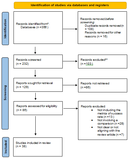
The initial search including the terms and the period of 11 years between 2011 to 2021 brings about 356 articles. Duplicates were eliminated after the initial filter. They were refined for English and it came down to 232. When filtering
Prisma
for the content and the relevance of the article to the theme of the review we had to cut down the search to 129 articles. When the articles were filtered for the availability of the full text of the article we were restricted to a total of 32 articles Figure 1.
This article mainly focuses on the efficiency of osseointegration of surface-modified implants in the patients with at least edentulous spaces and receiving implants. This article evaluates if surface modification of implant surfaces helps in osseointegration better when compared to smooth or unmodified implant surfaces.
Process of Osseointegration
The process of osseointegration is an extensive process initiated with the surgical insertion of an implant and extends till it is unified in the underlying bone. The immediate steps after insertion include blood clot formation around the implant followed by absorption of the cells onto the implant surface. The cells on the implant surface will then form granulation tissue which later translates into a provisional matric which forms a base for the cells to be embedded. These cells later initiate the mineralization phase which extends up to 6 months based on the systemic conditions of the recipient. During this phase, new bone formation and remodeling of the formed bone occur [1] (Figures 2 & 3).
![Figure 2: Events following implant placement: Blood clot formation, Granulation tissue formation Provisional matrix formation, Mineralization, Remodeling [2].](/fulltextimages/8685/fig_2.png)
![Figure 3: Implant healing [3].](/fulltextimages/8685/fig_3.png)
Research revealed the importance of implant surface in the initial stages facilitating osseointegration. Several factors like charge, roughness, hydrophilicity, and surface characterizations have been identified to play a major role in enhancing cell adhesion to the implant surface [4].
Implants have been classified into 3 major categories based on the roughness of the surface. 1. Smooth: These were the initial implants designed during the Brane mark period in implantology. 2. Moderately rough: These were the second-generation implants and included moderately rough surfaces which could enable optimal osseointegration. 3. Extremely rough: These implants are designed using advanced technology like nano and have a bioactive surface. These are aimed to increase the osseointegration and hydrophilicity of the cells but high susceptibility to microbial growth has been documented through the cause is not completely claimed to be associated with the topography, the surgical procedure also needs to be accounted for [5].
The surface texture has been playing a major role in recent age implants. There are several techniques and procedures to design and manipulate the topography of the Implant surface. The various types of the implant modifications are classified mainly into the following categories [6, 7, 8, 9, 10, 11, 12, 13].
Additive Method 1. Coatings a) Plasma spray, b) Antibiotic coatings, c) osteogenic agents like BMP, Growth factors d) nano coatings.
Photo functionalization Subtractive method a) Sandblasting b) Acid etching c) Laser Ablation Both 1. Electrolytic addition/reduction
Additive Method
This includes enhancing the surface with substances that could help in osteointegration from a biological perspective or physical interlocking for initial stability.
- The addition of plasma spray and osteogenic agents involves adding a layer of plasma proteins obtained from centrifugation of blood leading to a prepared surface layer for better surface interaction for blood cells [13, 14, 15, 16].
- The antibiotic coating is a preventive layer that aids in inhibiting bacterial growth in the early stages of implant placement hence promoting a healthy healing phase which is vital for implant success [17, 18].
- Nano coating of implant surface results in providing a more hydrophilic surface which promotes cell adherence to implant surface after placement. Nano treatment of a surface can be tricky as it leaves a charged surface which can also work in an opposite way to the desired outcome [19, 20, 21, 22, 23].
Subtractive Method
This includes removing surface material from the implant surface to increase surface area to provide a greater area for the cells to interact with the implant body. a) Sandblasting includes subjecting the implant surface to gritting agents sprayed on to them at high speeds leading to the removal of fine particles. Commonly used abrasives can be alumina and titanium dioxide [24, 25]. b) Acid etching involves the chemical removal of substances with acids like hydrochloric acid, sulphuric acid, hydrofluoric acid, and nitric acid. This is mostly done to remove the surface impurities after sandblasting process. c) Laser Ablation is a process of treating the surface with a high-intensity laser beam to augment the texture of the implant surface [26, 27].
The electrolytic method includes addition or subtraction of the implant surface and selectively opting to place the implant at the cathodic or the anodic ends. This is technique sensitive procedure, and the material can be added or subtracted.
The success of the implants also depends on the shape of the design of the implant surface, Bone Implant contact, and hygiene habits of the recipient [28]. The surface with a high contact angle attracts fewer bacteria but it does not favor cell migration and adhesion to the surface of the implant. While there are also the darker sides associated with surface modifications like corrosion and modification of mechanical properties like friction due to biofilms. Also, there are higher chances of implant failure in extremely rough implant surfaces as they form lodging sites for bacterial growth [5, 18, 19, 29, 30, 31, 32, 33, 34, 35].
Recent advances in Surface Modified Implants
a) 3d printed implants using nanotechnology are a theoretical concept that aims at accommodating stem cells obtained from the recipient of implants and embedding osteoblasts and osteocytes on the implant surface in the slots allotted to them by the nano printer [36, 37]. b) Addition of functional groups on the surface of the implants can increase the surface wettability and increase the adhesion of stem cells to increase bone formation [38, 39, 40].
Conclusion
Modern implantology aims to provide good initial stability with great tissue integration and rapid bone healing. In the process of improving the implant success researchers have adapted several surface improvisations to improve the final outcomes. The clinicians need to select the implant surface modification based on the patient’s requirements and health status.
References
-
Hotchkiss KM, Ayad NB, Hyzy SL, Boyan BD, Olivares Navarrete R (2017) Dental implant surface chemistry and energy alter macrophage activation in vitro. Clinical Oral Implants Res 28(4): 414-423.
-
Pochet M, Jeanmart H, Contino F (2020) A 22: 1 compression ratio ammonia-hydrogen HCCI engine: combustion, load, and emission performances. Front Mech Eng 6: 43.
-
Sukumaran A, Anand P, Alghamdi H, Jansen JA (2011) Dental implant surface enhancement and osseointegration. Implant Dent.
-
Ito H, Sasaki H, Saito K, Honma S, Yajima Y, et al. (2013) Response of osteoblast-like cells to zirconia with different surface topography. Dent Mater J 32(1): 122- 129.
-
Al Radha ASD, Dymock D, Younes C, O Sullivan D (2012) Surface properties of titanium and zirconia dental implant materials and their effect on bacterial adhesion. J Dent 40(2): 146-153.
-
Alla RK, Ginjupalli K, Upadhya N, Shammas M, Ravi RK, et al. (2011) Surface Roughness of Implants: A Review. Trends Biomater Artif Organs 25(3): 112-118.
-
Annunziata M, Guida L (2015) The effect of titanium surface modifications on dental implant osseointegration. Front Oral Biol 17: 62-77.
-
Kumar PS, KS Sateesh K, Grandhi VV, Gupta V (2019) The effects of titanium implant surface topography on osseointegration: literature review. JMIR Biomedical Engineering 4(1): e13237.
-
Demircioglu P (2014) Estimation of surface topography for dental implants using advanced metrological technology and digital image processing techniques. Measurement 48: 43-53.
-
Ogle OE (2015) Implant surface material, design, and osseointegration. Dent Clin North Am 59(2): 505-520.
-
Narsimha Rao G, Pampana S, Yarram A, Sajjan SMC, Ramaraju AV, et al. (2019) D Surface Modifications of Dental Implants: An Overview. Int J Dent Mater 1(1): 17- 24.
-
De Bruyn H, Christiaens V, Doornewaard R, Jacobsson M, Cosyn J, et al. (2017) Implant surface roughness and patient factors on long‐term peri‐implant bone loss. Periodontol 2000 73(1): 218-227.
-
Bruschi M, Steinmüller Nethl D, Goriwoda W, Rasse M (2015) Composition and modifications of dental implant surfaces. Journal of Oral Implants 2015.
-
Duske K, Koban I, Kindel E, Schröder K, Nebe B, et al. (2012) Atmospheric plasma enhances wettability and cell spreading on dental implant metals. J Clin Periodontol 39(4): 400-407.
-
Kundu R, Rathee M (2014) Effect of platelet-rich-plasma (PRP) and implant surface topography on implant stability and bone. J Clin Diagn Res 8(6): 26-30.
-
Furuhashi A, Ayukawa Y, Atsuta I, Rakhmatia YD, Yasunami N, et al. (2013) Influence of titanium surface topography on peri-implant soft tissue integration. Key Engineering Materials pp: 529-530.
-
Yin C, Zhang Y, Cai Q, Li B, Yang H, et al. (2017) Effects of the micro–nano surface topography of titanium alloy on the biological responses of osteoblast. Journal of Biomedical Materials Research Part A 105(3): 757-769.
-
Milleret V, Lienemann PS, Gasser A, Bauer S, Ehrbar M, et al. (2019) Rational design and in vitro characterization of novel dental implant and abutment surfaces for balancing clinical and biological needs. Clin Implant Dent Relat Res 21(1): 15-24.
-
Han A, Tsoi JK, Rodrigues FP, Leprince JG, Palin WM (2016) Bacterial adhesion mechanisms on dental implant surfaces and the influencing factors. International Journal of Adhesion and Adhesives 69: 58-71.
-
Ajami E, Mahno E, Mendes V, Bell S, Moineddin R, et al. (2014) Bone healing and the effect of implant surface topography on osteoconduction in hyperglycemia. Acta Biomater 10(1): 394-405.
-
Damiati L, Eales MG, Nobbs AH, Su B, Tsimbouri PM, et al. (2018) Impact of surface topography and coating on osteogenesis and bacterial attachment on titanium implants. Journal of Tissue Engineering 9: 2041731418790694.
-
Thakral G, Thakral R, Sharma N, Seth J, Vashisht P (2014) Nanosurface–the future of implants. J Clin Diagn Res 8(5): 7-10.
-
Pachauri P, Bathala LR, Sangur R (2014) Techniques for dental implant nanosurface modifications. J Adv Prosthodont 6(6): 498-504.
-
Cha JK, Paeng K, Jung UW, Choi SH, Sanz M, et al. (2019) The effect of five mechanical instrumentation protocols on implant surface topography and roughness: A scanning electron microscope and confocal laser scanning microscope analysis. Clin Oral Implants Res 30(6): 578-587.
-
Yeo IS (2014) Reality of dental implant surface modification: a short literature review. The Open Biomed Eng J 8: 114-119.
-
Almas K, Smith S, Kutkut A (2019) What is the best micro and macro dental implant topography? Dental Clinics 63(3): 447-460.
-
Ramakrishnaiah R, Mohammad A, Divakar DD, Kotha SB, Celur SL, et al. (2017) Preliminary fabrication and characterization of electron beam melted Ti–6Al–4V customized dental implant. Saudi Journal of Biological Sciences 24(4): 787-796.
-
Vivan Cardoso M, Vandamme K, Chaudhari A, De Rycker J, Van Meerbeek B, et al. (2015) Dental Implant Macro‐Design Features Can Impact the Dynamics of Osseointegration. Clin Implant Dent and Relat Res 17(4): 639-645.
-
Lai Y, Chen J, Zhang T, Gu D, Zhang C, et al. (2013) Effect of 3D microgroove surface topography on plasma and cellular fibronectin of human gingival fibroblasts. Journal of Dentistry 41(11): 1109-1121.
-
Naves MM, Menezes HHM, Magalhaes D, Ferreira JA, Ribeiro SF, et al. (2015) Effect of Macrogeometry on the Surface Topography of Dental Implants. Int J Oral Maxillofac Implants 30(4): 789-799.
-
Ren B, Wan Y, Liu C, Wang H, Yu M, et al. (2021) Improved osseointegration of 3D printed Ti-6Al-4V implant with a hierarchical micro/nano surface topography: An in vitro and in vivo study. Materials Science and Engineering C 118: 111505.
-
Noro A, Kaneko M, Murata I, Yoshinari M (2013) Influence of surface topography and surface physicochemistry on wettability of zirconia (tetragonal zirconia polycrystal). J Biomed Mater Res B Appl Biomater 101(2): 355-363.
-
Toffoli A, Parisi L, Tatti R, Lorenzi A, Verucchi R, et al. (2020) Thermal-induced hydrophilicity enhancement of titanium dental implant surfaces. J Oral Sci 62(2): 217-
-
Glon F, Flys O, Lööf PJ, Rosén BG (2014) 3D SEM for surface topography quantification–a case study on dental surfaces. Journal of Physics: Conference Series.
-
Arnhart C, Dvorak G, Trefil C, Huber C, Watzek G, et al. (2013) Impact of implant surface topography: a clinical study with a mean functional loading time of 85 months. Clinical Oral Implants Research 24(9): 1049-1054.
-
Wang H, Zhang X, Wang H, Zhang J, Li J, et al. (2018) Enhancing the osteogenic differentiation and rapid osseointegration of 3D printed Ti6Al4V implants via nano-topographic modification. J Biomed Nanotechnol 14(4): 707-715.
-
Laird NZ, Acri TM, Chakka JL, Quarterman JC, Malkawi WI, et al. (2021) Applications of nanotechnology in 3D printed tissue engineering scaffolds. European Journal of Pharmaceutics and Biopharmaceutics 161: 15-28.
-
Liu Q, Ding J, Mante FK, Wunder SL, Baran GR (2002) The role of surface functional groups in calcium phosphate nucleation on titanium foil: a self-assembled monolayer technique. Biomaterials 23(15): 3103-3111.
-
Webb K, Hlady V, Tresco PA (1998) Relative importance of surface wettability and charged functional groups on NIH 3T3 fibroblast attachment, spreading, and cytoskeletal organization. J Biomed Mater Res 41(3): 422-430.
-
Arima Y, Iwata H (2007) Effect of wettability and surface functional groups on protein adsorption and cell adhesion using well-defined mixed self-assembled monolayers. Biomaterials 28(20): 3074-3082.
- Diagnosis and Management of Mental Nerve Paresthesia Secondary to Apical Periodontitis of Mandibular Second Premolar: A CBCT Based Case Report
- A Randomized, Double Blinded Clinical Trial to Compare the Effect of Oral Premedication (Diclofenac Potassium or Dexamethasone) on Post-Operative Pain Following Pulpectomy
- Modified Lip Repositioning Technique for the Management of Excessive Gingival Display
- Integral Role of Non-Dental Providers and Fluoride Dissemination
- Root Canal Treatment Rate in Deciduous Teeth Among 6-Year- Olds in the Era of Discontinuing Water Fluoridation - Historical Cohort Study
- The Impact of the Notch1 on the Migratory Capacity and the Expression of E-Cadherin and CyclinD1 in Ameloblastoma Cells