Extensive Partial Obturador Oropharyngeal in Class IV Aramany after Oral Adenocarcinoma
When a patient is interrupted by velopharyngeal closure or defects in the hard palate, air escapes inappropriately through the nose in nasal speech sounds. The obturator prosthesis with extension to the oropharynx is currently the best indication for maxillary defects, but it also represents a challenge for manufacturing and oral adaptation. The aim of this study was to report a clinical case of successful prosthetic rehabilitation of a 65-year-old woman who presented with a defect in the maxilla, soft palate, tonsil, and oropharynx after treatment for polymorphous low-grade adenocarcinoma. With her considerable loss of structure and tissue, she reported difficulties with impaired functions such as chewing, swallowing, and speaking and difficulty interacting with people. The oral rehabilitation was performed with an oropharyngeal obturator prosthesis with an extension to the oropharynx and a total mandibular denture. This case demonstrates that the rehabilitation with an oropharyngeal obturator prosthesis with an extension to the oropharynx and a total mandibular denture provides speech, chewing, and phonetics, directly interfering with her social life in the mutilated patient.
Introduction
Oral and oropharyngeal cancer have a significant incidence among the neoplasms that affect the head and neck region [1]. Polymorphous low-grade adenocarcinomas (PGLA) were initially described by Evans and Batsakis in 1984 as distinctive salivary gland neoplasms with an almost exclusive propensity to arise from minor salivary glands [2, 3, 4]. Most patients of PLGA occur in the sixth and seventh decade and with an apparent female predilection with a 2:1 ratio. Complete surgical excision is an appropriate therapy for these patients [2].
The functional and esthetic rehabilitation of individuals who underwent complex surgical procedures constitutes one of the biggest challenges and requires a multidisciplinary team [5]. The maxillary resection and the oropharyngeal deficiencies can cause alterations in regulating speech emission and resonance and in oral activities, such as swallowing, whistling, blowing, and sucking [6]. The speech alterations are caused by voice hypernasality, oronasal regurgitation, and oropharyngeal dysphagia [7]. Defects of the maxilla and oropharynx substantially affect the individual’s quality of life [6, 7, 8, 9].
The maxillofacial prosthesis promotes the restoration of lost stomatognathic structures, rehabilitating compromised functions (chewing, swallowing, and phonetics) [7, 8]. The dentist must observe difficulties in performing stomatognathic functions during adaptation to the prosthesis, and recognizing the repercussions caused by using the prosthesis helps the speech therapist create a more effective therapeutic direction when necessary [7]. The oropharyngeal obturator prosthesis is commonly made with acrylic resin. However, achieving sufficient functional recovery with obturator prostheses is often tricky when the cavity is vast. Some clinical researchers reported that silicone soft reliners could also be an option for functional recovery after maxillectomy [10].
Considering the great importance of oral rehabilitation in patients with polymorphous low-grade adenocarcinoma who underwent marginal maxillectomy and the restoration of oral function, swallowing, and enabling intelligible speech, this study aims to present a clinical report of reconstruction with a maxillofacial prosthesis by palate and oropharyngeal obturator manufacturing using gradual additions.
Clinical Report
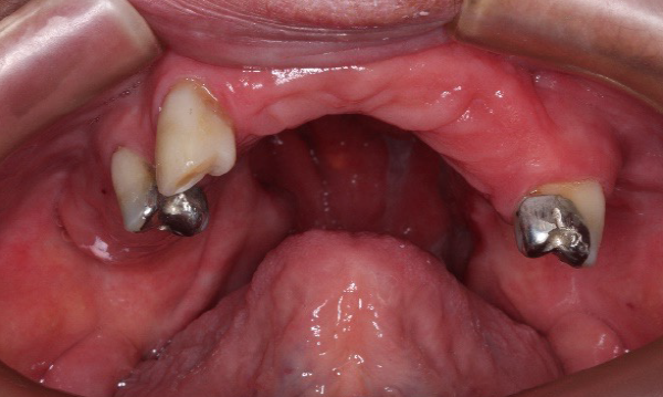
A 65-year-old ex-smoker patient with an intra-oral defect in the maxilla, soft palate, tonsil, and oropharynx due to maxillectomy surgery to remove polymorphous low-grade adenocarcinoma. The patient was referred to the maxillofacial prosthetic rehabilitation service by the Department of Maxillofacial Prosthodontics, Federal University of Minas Gerais. Due to treatment, the patient reported difficulties with impaired functions such as chewing, swallowing, and speaking and difficulty interacting with people. Clinical examination revealed oroantral communication, partially edentulous, good surgical wound healing, and good condition of adjacent tissues (Figure 1). The proposed treatment plan was the construction of an oropharyngeal obturator prosthesis with an extension to the oropharynx and a total mandibular denture.
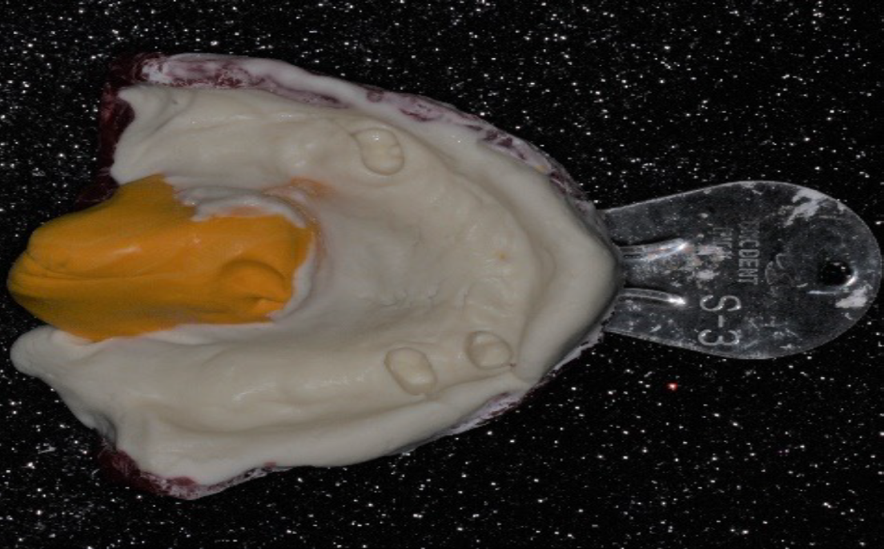
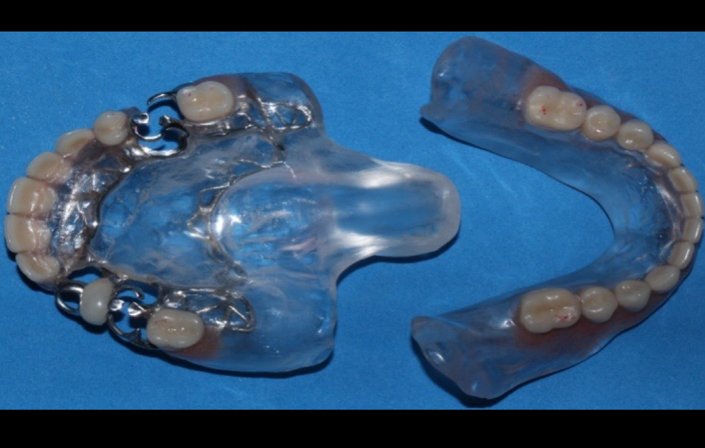
The first immediate impression was taken using the irreversible hydrocolloid (Jeltrate Plus, Dentsply Sirona, New York, USA) to create special acrylic resin trays. The impression’s great challenge was copying the defect faithfully to achieve a seal with the obturator prosthesis. The upper tray was made with an extension in the oropharyngeal area to support the impression material and to obtain an accurate copy of the defect region. The oropharyngeal obturator prosthesis was designed to prepare support niches of the metal structure after that with niches already prepared in the teeth. Supporting niches of pillar teeth 13, 15, and 25 were made with straight T-type retaining clips and corrugated palatal bars to reduce the weight of the metallic structure. For the final impression, at the upper edge, an impression with polyvinyl siloxane (Express XT, 3M Espe, Maplewood, USA) was first obtained only in the oroantral communication region to copy and compress the soft tissue that delimits the communication. Then, an irreversible hydrocolloid (Jeltrate Plus, Dentsply Sirona, New York, USA) was used to complete the impression (Figure 2).
The lower impression was performed with thermoplastic molding material (Godibar; Lyzanda Produtos Odontologicos Ltda, São Paulo, São Paulo, Brazil), and the molding material used was zinc oxide eugenol paste (ZOE) (Lyzanda; Lyzanda Produtos Odontologicos Ltda, São Paulo, São Paulo, Brazil). After the impression, stone casts (Durone IV, Dentsply Sirona, New York, USA) were obtained.
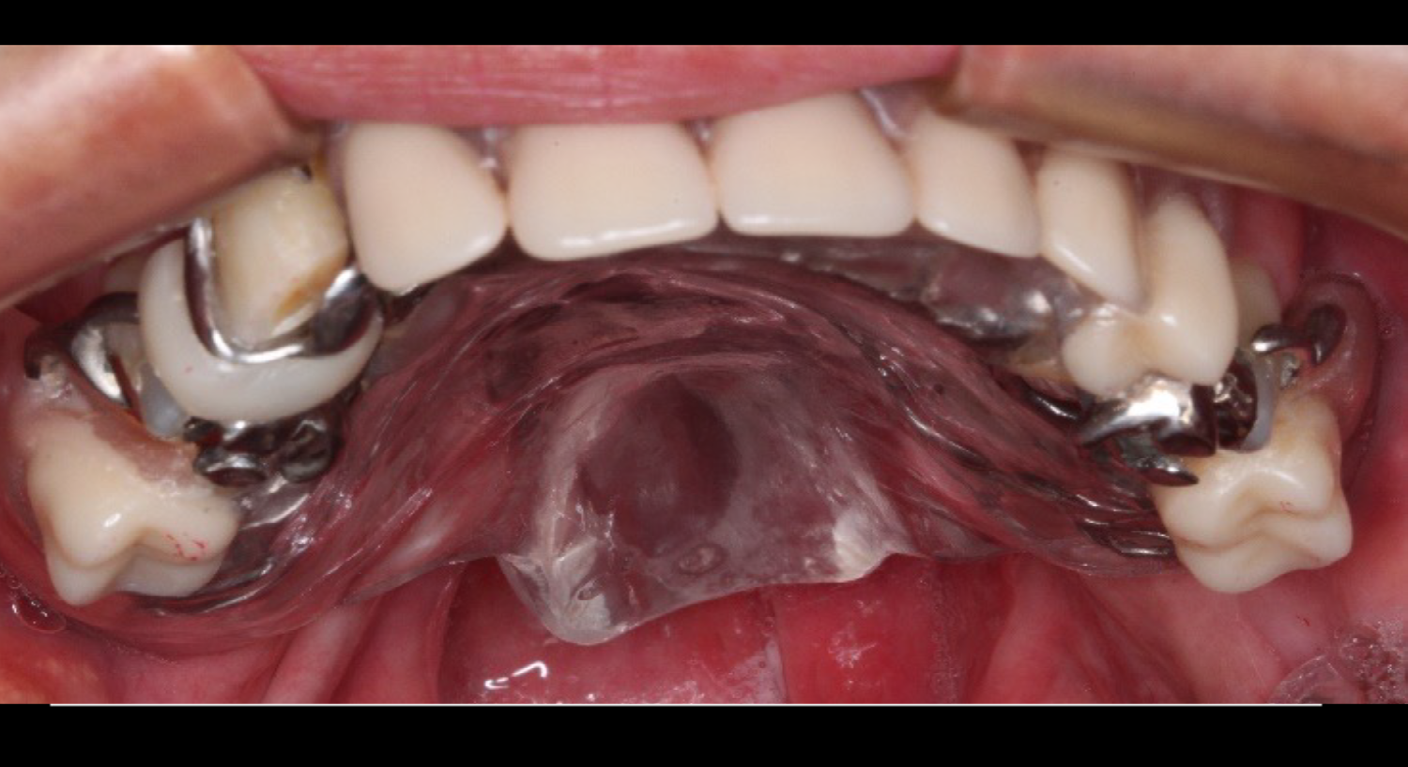
The metallic structure and record bases were placed on the patient’s mouth to determine the vertical dimension of occlusion, the smile height, and the position of the median line. The record bases were transferred to a semi‐adjustable articulator for later tooth assembly. Teeth were registered in wax, and tests were made about teeth adaptation and sealing of defect areas. Subsequently, the prosthesis was acrylic, and final adjustments were made (Figure 3). After installing the prosthesis, occlusal adjustments were performed. It was possible to observe an immediate improvement in phonation, swallowing, and, therefore, in the patient’s well- being (Figures 4 and 5).
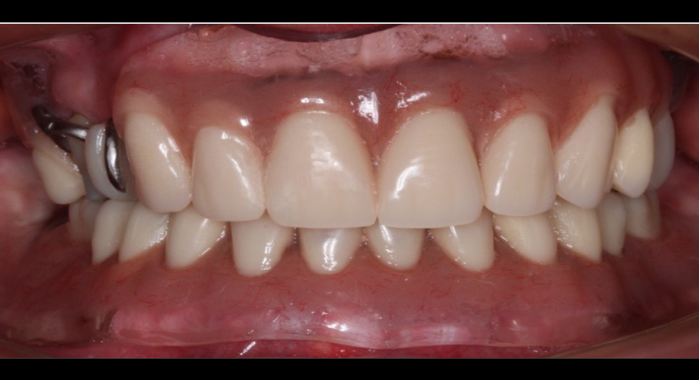
After the entire process, the patient was satisfied with her smile. Therefore, it is crucial to highlight a psychological aspect of this patient, who is significantly affected by visual appearance and all oral functions by speech, chewing, and swallowing. Also, it is essential to note that this type of treatment with an obturator prosthesis is to return all these functions in a lower traumatic way.
Discussion
Polymorphous low-grade adenocarcinoma (PGLA) occurs more frequently in female patients and presents clinically as a mass lesion. The palate is the most frequent anatomic site of occurrence [2–4]. Complete surgical excision is an appropriate therapy for these patients, with an excellent long-term prognosis [2]. In this case, the tumor affects the maxilla, soft palate, tonsil, and oropharynx. However, the defect that was generated signified a big challenge for rehabilitation.
The treatment choice after maxillectomy depends on each case and the location and extent of the defect. After maxillectomy for malignant tumors, treatment includes rehabilitation with an obturator prosthesis or reconstructive surgery with an autogenous tissue graft [10]. In this study, the advantages of oropharyngeal obturator prosthesis after maxillectomy are that the surgical site recurrence may be easily detected and that functional recovery can be obtained by acquiring occlusion early after surgery. Maxillary defects predispose patients to undesirable effects such as hypernasal speech, current intraoral secretion, and difficulties swallowing and chewing [7]. Reconstruction with maxillofacial prostheses by palatal and oropharyngeal obturator is the first choice in several centers of oral rehabilitation because it is the most viable reconstructive option financially and functionally acceptable in cases of extensive surgical interventions. In this context, several factors influence treatment success with the proposed rehabilitation technique’s choice: the defect’s size, availability of hard and soft tissues in the area, proximity to vital structures, systemic conditions, and the patient’s ability to adapt dental prosthesis [7].
Oropharyngeal obturator prosthesis for patients with oral and pharyngeal defects arising from tumor resection is challenging since each individual has specific anatomical characteristics. After rehabilitation with the prostheses, the patient reported improved speech, chewing, and phonetics, directly interfering with her social life. The treatment should be planned and executed individually with almost artisanal care.
References
-
INCA (2022) Estimativa 2023: Incidência de câncer no Brasil / Instituto Nacional de Câncer.
-
Castle JT, Thompson LDR, Frommelt RA, Wenig BM, Kessler HP (1999) Polymorphous low grade adenocarcinoma: A clinicopathologic study of 164 cases. Cancer 86(2): 207-219.
-
Evans HL, Batsakis JG (1984) Polymorphous low‐grade adenocarcinoma of minor salivary glands a study of 14 cases of a distinctive neoplasm. Cancer 53(4): 935-942.
-
Gottlieb JB, Joachim M, Leiser Y, Abdelraziq M, Abu El- Naaj I (2019) Polymorphous low-grade adenocarcinoma: A proposed reconstruction protocol based on past surgical experience. J Craniofac Surg 30(4): 1228-1230.
-
Ragbir M, Brown JS, Mehanna H (2016) Reconstructive considerations in head and neck surgical oncology: United Kingdom National Multidisciplinary Guidelines. J Laryngol Otol 130(S2): S191-S197.
-
Akshayalingam M, Malar K, Lenapriya A (2022) Palatopharyngeal obturator prosthesis - A substitute for a dynamic separator: A technique. J Indian Prosthodont Soc 22(2): 200-204.
-
Revoredo ECV, Gomes A de OC, Ximenes CRC, Oliveira KGSC de, Silva HJ da, et al. (2022) Oropharyngeal Geometry of Maxilectomized Patients Rehabilitated with Palatal Obturators in the Trans-surgical Period: Repercussions on the Voice. J Voice S0892-1997(22): 00072-8.
-
Akin H, Coskun ME, Akin EG, Ozdemir AK (2012) Multidisciplinary approach for esthetic, functional, and quality-of-life outcome in soft palate cleft patient. Cleft Palate Craniofac J 49(5): 622-625.
-
Goiato MC, Pesqueira AA, da Silva CR, Gennari H, dos Santos DM (2009) Patient satisfaction with maxillofacial prosthesis. Literature review. J Plast Reconstr Aesthet Surg 62(2): 175-180.
-
Yanamoto S, Soutome S, Murata M, Kawakita A, Yamaguchi E, et al. (2020) Efficacy of silicone soft reliner on the obturator prosthesis after maxillectomy for oral malignant tumors: A single-arm prospective interventional study. Clin Exp Dent Res 6(6): 612-617.
- Diagnosis and Management of Mental Nerve Paresthesia Secondary to Apical Periodontitis of Mandibular Second Premolar: A CBCT Based Case Report
- A Randomized, Double Blinded Clinical Trial to Compare the Effect of Oral Premedication (Diclofenac Potassium or Dexamethasone) on Post-Operative Pain Following Pulpectomy
- Modified Lip Repositioning Technique for the Management of Excessive Gingival Display
- Integral Role of Non-Dental Providers and Fluoride Dissemination
- Root Canal Treatment Rate in Deciduous Teeth Among 6-Year- Olds in the Era of Discontinuing Water Fluoridation - Historical Cohort Study
- The Impact of the Notch1 on the Migratory Capacity and the Expression of E-Cadherin and CyclinD1 in Ameloblastoma Cells