Concomitant Existence of Tooth Agenesis (Agenesis of Four Second Premolars) and Supernumerary Tooth (Dens Distomolar)-Report of a Rarest Case
“Distomolar” is also called as ‘fourth molar’ is the supernumerary tooth occurring distal to the third molar. Occurrence of distomolar is a rare phenomenon. Along with this condition congenital agenesis which is a contrast phenomenon compared to extra teeth is still a rarer entity. Therefore, the aim of this paper is to present occurrence of fourth molar in the mandibular arch and congenital agenesis of all second premolars (four premolars) involving both maxillary and mandibular quadrant in a non-syndromic Indian female patient. Such combination of two different anomalies representing two different dental phenomenon is not reported till date according to author’s best of knowledge.
Introduction
“Fourth molars” also referred by various synonyms as ‘distomolars,’ ‘distodens,’ ‘retromolars’ or ‘dens distomolar’ is a supernumerary tooth that is seen distally to a wisdom tooth or third molar and when it fully erupted extends the dental arch. They may occur unilateral or bilateral either in the maxilla or mandible or both and can be erupted, partially erupted or impacted [1, 2]. Most of the time, they remain unerupted and only detected accidentally following a routine radiographic examination performed for other purpose. In case of erupted distomolars, the crown morphology appears similar to third molars except for the size, however authors have also found that they may appear as tuberculated, molariform, conical or multi-cusps forms. On radiographs, fourth molars can appear as clear osteoslerotic foci either having short single root or a rudimentary root structure [3, 4, 5].
Congenital agenesis of human dentition is most commonly observable finding in clinical practice. The frequently missing teeth are the third molars, followed by mandibular second premolars and maxillary lateral incisors. Agenesis of second premolars is more common in the mandible with a reported prevalence of 4.4% compared to its occurrence in the maxilla of only 1.7% [6, 7]. Congenital agenesis of first premolars ranges from about 0.1% in the mandible and about 0.2% in the maxilla. An epidemiological study performed by Stritzel [6] revealed that, among 176 white European patients evaluated with specifically pertaining to agenesis of second premolars and found that mandible was affected more than maxilla and also observed the absence of one or two second premolars in 75% of the cases. Occurrence of all second premolars including both maxillary and mandibular arch is an extremely uncommon finding. Literature shows hardly one or two number of publications on congenital agenesis of all four second premolars [7].
In the research evidence, there are isolated reports describing either any one of the above-mentioned dental anomalies [8, 9, 10, 11, 12]. Concomitant occurrence of two different dental phenomenon involving formation of extra tooth and agenesis of some teeth is an interesting question mark to the existing literature and strongly implies for more research including genetic studies in the future. Therefore, keeping research-oriented hypothesis, the current article is put forward to the genetic researchers or research anatomists to explore more evidence pertaining to this interesting dental phenomenon.
Case Report
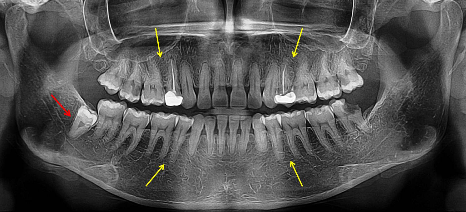
A 31-year-old female patient reported to a private dental clinic complaining of decayed tooth in the lower left back region which often causes pain. Physical examination was carried out which revealed normal built and moderately nourished. There was no signs and symptoms of any metabolic, systemic or syndromic disroders. Intraoral examination was carried out, which showed presence of grossly destructed mandibular left third molar due to dental caries. All permanent teeth erupted including all four third molars in the maxilla and mandible except for the missing of all four second premolars including both maxilla and mandible. The maxillary right and left first premolars were found with porcelain crowns. Patient was asked about any previous history of teeth extraction performed in the upper and lower arch which resulted no such incidence of tooth extraction being performed. Patient did not take any orthodontic treatment too which got to know upon eliciting the history. Therefore, to know the condition of the destructed tooth, and also to confirm the agenesis of all four second premolars patient was subjected to radiographic examination. On the radiograph, (orthopantomograph) slight periapical changes were noticed in relation to the mandibular left third molar with deep dental caries leading to destruction of the distal part of the crown (Figure 1). On careful examination of the radiograph, author also noticed presence of extra tooth in the mandibular right quadrant located distal to the third molar (Table 1). The tooth was smaller in size compared to third molar having short single root and was impacted (vertical impaction). Further examination of the radiograph showed missing of all four second premolars in both the maxillary and mandibular arch. The space of this tooth in the upper and lower arch was nicely adjusted with the eruption of the all third molars in the dental arch. The maxillary right and left first premolars have been treated with root canal treatment and porcelain crown placement which was evident on the radiograph (Figure 1). No other dental findings observed. Therefore, based on clinical features and radiological findings, the present case was diagnosed as idiopathic concomitant occurrence of agenesis of all four second premolars (congenital tooth agenesis) and a fourth molar or distomolar (a supernumerary tooth). Patient was explained about the presence of different conditions. As the patient did not have any discomfort and pain associated with the impacted fourth molar, extraction of left third molar was advised and keeping the patient under observation for development of any discomfort or pathology associated with the impacted distomolar.
| Patient Age | Gender | Ethnicity | Dental Anomalies present | Related Dental Phenomenon | Treatment provided |
|---|---|---|---|---|---|
| 31 years | Female | India | Presence of Fourth molar (Distomolar/Retromolar/Distodens/Dens Distomolar) in the mandibular right quadrant, located distal to the third molar, impacted in vertical position. | Supernumerary teeth | |
| 31 years | Female | India | Congenital agenesis of all four second premolars in both maxillary and mandibular arch (both right and left). | Tooth agenesis | Not required |
Table 1: Patient details with two dental anomalies of different dental phenomenon (Concomitant occurrence).
Discussion
Fourth molars also called as ‘retromolars’ or ‘distomolars’ as they are located distally or posterior location with respect to third molar due to alterations occurring in the process of odontogenesis. Shahzad, et al. [13] in their literature review has classified fourth molars into two types as heteromorphic and eumorphic variety and their description is shown in Table 2.
| Types | Description |
|---|---|
| Heteromorphic | Also called as rudimentary or dismorphic, cone-shaped (conical crown and rudimentary root) or tuberculated (crown with tubercles and single curved root) |
| Fourth molar possessing atypical morphology. | |
| Eumorphic | These appear similar to normal teeth. |
| They also receive the name of inculomism the following can be observed. | |
| Infundibular (funnel shaped with invaginations in the crown) | |
| Molariform (shaped as premolar or molar) |
Table 2: Types of fourth molars.
In the case described here, the distomolar appeared smaller in size compared to the third molar. Although it was impacted, on radiograph its crown morphology appeared similar to the third molar (Eumorphic type), with single and short root with blunt closed apex and single root canal.
Sometimes they found fused with third molars, and characterized by a tubercle appended to its crown in the distal-lingual area. These teeth are usually called in the literature by a term as ‘distomolar tubercle’. Some reports showed conical shaped distomolars with malformed apex in the short single root. There are also other reports showing it appended to the third molar roots most commonly involving palatal root as reported in the case published by Gay Escoda [14] and Duarte and Azevedo [15]. Grimanis, et al. [3] did epidemiological survey on supernumerary molars and showed that majority (79.7%) occurred in the maxilla, and only 23.9% occurred bilaterally. Whereas Leco, et al. [5] found the incidence of mandibular distomolars of about only 4.8% in their investigation. In 2017, Choontharu, et al. [4] found four fourth molars occurring both right and left quadrant of the maxilla and mandible.
Clinically with the erupted distomolars some food lodgement or plaque accumulation can be noticed. The ratio of erupted distomolars and impacted distomolars varies from 1 to 5 [17]. Most of the distomolars remain asymptomatic, however, they may cause odontogenic inflammation, periodontal diseases, primordial or odontogenic cysts and neuralgic pains. As most of the distomolars remain asymptomatic without causing any complications and no clinical signs conservative treatment including only observation as a preventive measure is the treatment of choice. But these cases should be kept under periodic radiographic evaluation for the development of any pathology associated with it. Surgical extraction is carried out in such condition like inflammatory complications and chronic pain or due to orthodontic purpose. When surgery is planned, clinician should consider some of the factors like potential complications or problems anticipated during their removal, complications which may occur during or following the surgery, its location and access to the teeth during surgery. This unusual dental phenomenon is more interesting especially considering the fact that in contemporary homo-sapiens missing third molars are becoming a common observation. There are also reports showing occurrence of fifth molars in the literature. Therefore, occurrence of extra teeth (molars either fourth or fifth) is still a questionable domain to the dental evolution in humans [18, 19].
Agenesis of permanent teeth has been reported in the literature [20, 21, 22] with most commonly missing teeth found are third molars followed by maxillary lateral incisors and mandibular second premolars. Although there are numerous publications on agenesis of mandibular second premolars but agenesis of all four second premolars including both maxillary and mandibular is an extremely rare dental phenomenon. Only one case report was noticed in the literature contributed from India in 2015 [7].
The true etiology behind the occurrence of congenital agenesis of teeth is not known. It is stated that it may be due to the expression or misexpression of particular genes at certain times during the development of a tooth germ. It is also explained that although there is normal inhiation of the developing tooth germ, there will be abnormal apoptosis leading to involution of the developing tooth. In addition to this, the genes which cause programmed cell death are inadvertently expressed causing the body to start resorbing the developing tooth germ [7, 20, 21, 22].
Clinically agenesis of premolars is associated with retained primary molars [7]. Therefore, presence of retained primary second molar beyond its exfoliation age, or when there are primary molars seen in adults, that signifies the absence of its permanent successors. In such cases, clinician always suspect the missing permanent teeth and go for detailed evaluation of the patient including radiographic examination. Sometimes agenesis of premolar leads to spacing and also cause supra-eruption of opposing teeth resulting in occlusion variation and its subsequent sequel such as pain in the tooth, attrition, food lodgement and plaque accumulation. But in the present case, no such clinical features were observed and there was proper alignment of the missing space by the eruption of adjacent teeth and also due to the proper eruption of the third molars. The space was nicely closed with the first premolar and first molars in both upper and lower quadrants. In a case published by Nirmala, et al. in [7] 2015 (India), the affected patient was a17-year-old male patient showing retained primary second molars (with evident full length of the roots and absence of root resorption) in the maxillary left region and mandibular right and left quadrant. On right side of the maxillary arch the primary second molar was exfoliated with existing single tooth space due to the absence of second premolar. In the left side of the mandibular arch the primary second molar was pulp treated showing radiopaque root filling material on the radiograph. Other than this, no other dental anomalies were found in this case. In the current case the affected patient is again Indian but a 31-year-old female patient. Along with agenesis of all four second premolars presence of a supernumerary fourth molar or distomolar was observed which is not reported till date.
Treatment of tooth agenesis depends on some factors such as age of the patient, amount of space present and type of tooth missing. Proper planning including various protocols should be considered at appropriate time to close the edentulous space [7]. The existing space because of missing premolars can be corrected by closing space using orthodontic treatment. If this cannot be carried out then removable or fixed prosthesis can be considered to restore the existing space. Recently due to advancements in the treatment options, implant can be placed as it provides the fixed and permanent solution and also restores normal functioning and esthetic purpose of the missing tooth [20, 21, 22]. In the case presented here, as there was no any existing space present, and all teeth were in normal alignment, occlusion and function no treatment was considered.
Conclusion
Knowledge about interesting cases like congenital agenesis of all four second premolars in association with presence of a supernumerary tooth structure like mandibular distomolar or fourth molar is more often essential for all clinicians as diagnosis and treatment varies in such cases. Therefore, author made an effort to present an uncommon occurrence of two different anomalies occurring in a patient of Indian ethnicity.
References
-
Jaiyeoba OO, Ifesanya JU (2018) Mandibular and maxillary distomolars in the orthodontic child patient: A report of 3 cases. Ann Ib Postgrad Med 16(2): 177-180.
-
Rahnama M, Szyszkowska A, Pulawska M, Szczerba- Gwozdz JA (2004) rare case of retained fourth molar teeth in maxilla and mandible, Case report. Curr Issues Pharm Med Sci 27(2): 118-120.
-
Grimanis GA, Kyriakides AT, Spyropoulos ND (1991) A survey on supernumerary molars. Quintessence Int 22(12): 989-995.
-
Choontharu MM, Prasad R, Pandya KM, Pradeep S (2017) Four Fourth Molars. J Contemp Dent 7(3): 166-168.
-
Berrocal MIL, Morales JFM, Gonzalez JMM (2077) an observational study of the frequency of supernumerary teeth in a population of 2000 patients. Med Oral Patol Oral Cir Bucal 12(2): 134-138.
-
Stritzel F, Symons AL, Gage JP (1990) Agenesis of second premolar in males and females: distribution, number and sites affected. J Clin Pediatr Dent 15(1): 39-41.
-
Nirmala SVSG, Tharay N, Kolli NKR, Dasarraju RK, Tirupathi SP (2015) Agenesis of second premolars in maxilla and mandible - A rare case report. Journal of Biomedical Sciences 2(2): 12-16.
-
Nagaveni NB, Sreedevi B, Praveen BS, Praveen B, Vidyullatha BG, et al. (2010) Survey of mesiodens and its characteristics in 2500 children of Davangere city, India. Eur J Paediatr Dent 11(4): 185-188.
-
Nagaveni N, Shashikiran N, Reddy VS (2009) surgical management of palatal placed, inverted, dilacerated and impacted mesiodens. Int J Clin Pediatr Dent 2(1): 30-32.
-
Nagaveni NB (2023) Inversion of impacted mesiodens: Report of Case series with literature review. Glob J Res Dent Sci 3(5): 7–12.
-
Nagaveni NB, Umashankara KV, Radhika NB, Praveen B, Manjunath S (2010) Maxillary paramolar: report of a case and literature review. Arch Orofac Sci 5(1): 24-28.
-
Nagaveni NB, Umashankar KV (2023) Report of a rare Odonto-Stomatologic Anomaly – Maxillary Paramolar. Series Clin Med Case Rep Rev 1(6): 1-3.
-
Shahzad KM, Roth LE (2012) Prevalence and management of fourth molars: a retrospective study and literature review. J Oral Maxillofac Surg 70(2): 272-275.
-
Escoda GC, Berini AL (2004) Tratado de cirugía bucal. Editorial Ergon.
-
Duarte MD, Azevedo SL, Sousa MS, Queiroz FD (2014) unusual fusion of a distomolar with a third molar assessed by conebeam computed tomography. Stomatos 20(38): 12-17.
-
Berrocal MI, Martin MJ, Martinez GJ (2007) Estudio observacional sobre la frecuencia de dientes supernumerarios en una población de 2000 pacientes. Medicina oral, patología oral y cirugía bucal, Ed. Española 12(2): 96-100.
-
Nirmala SVSG, Tirupathi SP (2016) rare combination of developing unerupted paramolar and distomolar in maxilla: a case report and review of literature. J Interdiscipl Med Dent Sci 4(4): 1-6.
-
Kokten G, Balcioglu H, Buyukertan M (2003) Supernumerary fourth and fifth molars: a report of two cases. J Contemp Dent Pract 4(4): 67-76.
-
Nagaveni NB (2023) Migration of mandibular supernumerary premolar in association with multiple anomalies – A rarest case report with literature review. Glob J Res Dent Sci 3(6): 1-6.
-
Nagaveni NB, Umashankara KV (2009) congenital bilateral agenesis of permanent mandibular incisors: case reports and literature review. Arch Orofac Sci 4(2): 41-46.
-
Nagaveni NB (2023) a rare combination of tooth agenesis in association with anomalous supernumerary tooth: Report of a rare case. Oral Health Dent 6(1): 18-21.
-
Nagaveni NB (2023) bilateral agenesis of maxillary second premolars and bilateral ectopic eruption of mandibular first molars – A rare case report. Glob J Res Dent Sci 3(5): 16-20.
- Diagnosis and Management of Mental Nerve Paresthesia Secondary to Apical Periodontitis of Mandibular Second Premolar: A CBCT Based Case Report
- A Randomized, Double Blinded Clinical Trial to Compare the Effect of Oral Premedication (Diclofenac Potassium or Dexamethasone) on Post-Operative Pain Following Pulpectomy
- Modified Lip Repositioning Technique for the Management of Excessive Gingival Display
- Integral Role of Non-Dental Providers and Fluoride Dissemination
- Root Canal Treatment Rate in Deciduous Teeth Among 6-Year- Olds in the Era of Discontinuing Water Fluoridation - Historical Cohort Study
- The Impact of the Notch1 on the Migratory Capacity and the Expression of E-Cadherin and CyclinD1 in Ameloblastoma Cells