Elemental and Micromorphological Analysis of Ion Releasing Restorative Materials Subsequent to Finishing and Polishing Procedures: In Vitro Study
Purpose: The purpose of this study was to evaluate the surface morphology and composition of of two ion-releasing materials) submitted to finishing and polishing (F/P). Methodology: Disc-shaped specimens (10 mm × 2 mm) were made from both ion-releasing materials restorative materials Giomer (Beautifil II, Shofu Inc.), and Alkasite based material (Cention N, Ivoclar Vivadent, Schaan, Lichtenstein) submitted to F/P with [Super-Snap X-Treme Technique Kit, Shofu Inc. and Astropol, Ivoclar Vivadent, Schaan, Lichtenstein)]. Filler particles morphology and composition were examined by scanning electron microscopy (SEM) and energy dispersive X-ray spectroscopy (EDX), respectively. Data were analysed by one-way analysis of variance and Tukey’s test (α = 0.05). Results: The smoothest surfaces were obtained for the control (unfinished) subgroups of all specimens. Regarding to F/P groups, Giomer specimens presented a relatively smooth surface after F/P with Super-Snap X-Treme Technique Kit. The EDX spectra showed no elemental transference from F/P tool to material surface by F/P procedures. Conclusion: The effect of F/P systems on surface roughness was dependent on the particle type and size of both F/P system and resin based restorative material.
Introduction
Over years, tooth-colored restorative materials such as resin composites are widely used in the field of operative dentistry due to their main benefits such as excellent esthetics, bioactivity, conservative tooth preparation, and good retention to tooth structures [1, 2]. Studies have been carried out to create aesthetic restorative materials that improve remineralization and stop caries from progressing. Both the F/P process and the composition of resin-based products have a major impact on surface hardness and roughness [3].
Giomer, which contains pre-reacted glass ionomer filler particles within a resin matrix, was introduced as the genuine hybridization of glass ionomer and composite resin. Giomer combines the esthetics, physical and handling properties of composite resin with the fluoride release of Glass ionomer [4]. Recently, alkasite-based restorative material (Cention N) has been introduced to the market. It is called “Alkasite” because it has alkaline glass filler that releases ions of calcium, fluoride to stop demineralization during acid attacks and produce remineralization. Also, it releases hydroxide ions to regulate PH [5]. Because it contains acyl phosphine oxide initiator and photoinitiator Ivocerin, Cention N can be self- cured with optional additional light-curing [6]. Nevertheless, Cention N satisfies the minimum ISO 4049 value in both self- cured and light-cured modes, making it a capable material in stress-bearing areas [7]. Both Alkasite-based restorative material and Giomer are used in management of carious lesions because of their ability to release fluoride that helps in caries inhibition [8].
Smooth surface is one of the ideal qualities for a satisfying, long-lasting tooth-colored restoration. Surface roughness of dental restorations related to improper F/P of dental restorations increases bacterial adhesion, secondary caries and subsequent restoration replacement [8, 9]. To increase the esthetics and durability of restorations, proper F/P process of the restorative material is crucial. Finishing is the process of sculpting the restoration to give it anatomical shapes and removing extra material from the surface. Polishing is done subsequently to finishing for obtaining a high shine surface and an enamel-like texture [10]. Finishing and polishing systems come in a wide range of forms, including silicone discs, aluminum oxide discs, carbide burs, and rubber cups that are available in single-step, dual-step, or multi- step polishing processes [11]. The ion-releasing restorative materials have been reported in previous literatures and these materials supposed to play an important role in the progression of de- and remineralization of dental hard tissues [12, 13]. However, the effect of F/P tools on their elemental analysis, has not been completely elucidated yet.
Thus, this study aimed to evaluate the relative surface topography and elemental changes of resin-based dental restorative materials before and after F/P with two finishing systems aluminum oxide based tools (Super-Snap X-Treme) and silicon dioxide based tools (Astropol). The null hypothesis of this study was that surface topography and elemental analysis were not influenced by dental F/P systems.
Materials and Methods
Sample Size Calculation
The sample size was calculated using G Power version 3.1.9.4 software. Test family is t tests. Type of power analysis is A priori: Compute required sample size-given power and effect size. A total sample of 54 was the enough required sample to detect a standardized effect size of 0.5, statistical power = 95% [14] and at a significance level of 0.05. The total sample size will be increased to 60 samples: 30 samples for each material (10 samples per subgroup).
Specimens Preparation
Alkasite based material (Cention N, Ivoclar Vivadent, Schaan, Lichtenstein) and Beautifil-II,Shofu Inc, Kyoto, Japan) with an A2 shade were employed in this study in Table 1. A total of 60 specimens (30 of each material) were produced in a half-split round Teflon mold with dimensions of 10 mm in diameter and 2 mm in thickness. A 1000 mW/cm2 LED device (Bluephase C8, Ivoclar Vivadent, AG, Schaan, Liechtenstein) was used to photocure Beautifil II specimens against a Mylar matrix and glass slab for 20 seconds. Periodically, the light intensity was checked using a radiometer (Ivoclar Vivadent).
Degrading Cention N, two scoops of powder and two drops of liquid resin were dispensed and manually blended on a mixing pad to a smooth consistency. Half of the powder was first thoroughly moistened with the liquid before the remaining powder was gradually added. The duration of the mixing process was no longer than 60 seconds. The paste was then squeezed to a flat surface, covered with a Mylar matrix and glass slab, and adapted into the mould using a spatula. Then, it was left undisturbed for 2 minutes then photocured for 20 seconds utilizing the same LED light curing machine to ensure setting by using the dual-cure method according to manufacturer’s instructions [15]. Details on the materials used were shown in table 1. In order to generate samples with flat bubble-free surfaces, transparent matrices were positioned between the glass slab and the moulds. The discs were removed from the mould after setting and examined for obvious voids with magnifying loupe (magnification × 3). Each material discs were separated randomly into three treatment subgroups based on the F/P system used: Subgroup 1: Ten samples from each material group were kept without F/P system after the Mylar strip was removed to serve as a baseline (control). Subgroup 2: The specimens were finished and polished using Super-Snap X-Treme Technique Kit, in which first the Course (black) and Medium (violet) polishing discs were used on the composite samples followed by the Fine (20 μm) and Super fine (7μm ) polishing discs. Subgroup 3: The specimens were finished and polished with Astropol. Astropol F (45 μm) was used first followed with Astropol P (1 μm) then Astropol HP (0.3 μm). The F/P process was carried out in accordance with the manufacturer’s instructions (10 seconds for each phase) and timed using a stopwatch; and used water spray and a slow-speed hand piece rotating at 10,000 rpm. The operator utilized a gentle pressure of 40grs
(calibrated every 10 samples) to achieve standardization in all groups [14]. To prevent differences between the treated surfaces, all samples were prepared, finished, and polished by the same operator (H.S.). After the polishing process, the samples were rinsed with distilled water for 20 seconds to remove any surface deposits, and they were then dried with air spray for 5 seconds.
Scanning Electron Microscopy and Microanalysis Energy-Dispersive X-ray Spectroscopy (EDX) (SEM and EDX)
Before using Scanning electron microscope (SEM), the specimens were metallized with silver as a conductive material. SEM used a focused electron beam to scan the surface of the sample to produce high-quality image of the surface topography. One sample of each subgroup was subjected to SEM examinations. The material surfaces were scanned by SEM (JSM-6510LV, JEOL, USA) at accelerating voltage of 5 KV in Mansoura Microscopy Center, Faculty of Agriculture and Mansoura University. Photographs of representative areas of the material surfaces were taken at 1000× magnifications. EDX is used for elemental analysis of a sample, providing information about the chemical composition. EDX is often coupled with SEM to analyze the characteristic X-rays emitted from the sample when it is bombarded with the electron beam. Each element emits X-rays at characteristic energy levels, permitting the quantification and identification of elements present in the sample. The output of EDX analysis is an elemental spectrum showing the presence and intensity of characteristic X-ray peaks for different elements. EDX produces spectra showing peaks corresponding to different elements present in the sample, allowing for qualitative and quantitative elemental analysis.
Statistical Analysis
Statistical investigation was done using a SPSSxsoftware program (SPSS; V17, Chicago, Illinois, United States). Firstly, the homogeneity of variances and the normal distribution of errors and were checked by Shapiro–Wilk’s test and Levene’s test. Based on these primary analyses, data of each test were separately analyzed using the one-way analysis of variance (ANOVA) and Tukey’s honestly significant difference post hoc test.
| Material | Manufacturer | Constituents | Filler Loading | |
|---|---|---|---|---|
| Weight | Volume | |||
| Beautifil II restorative | Shofu Inc, Kyoto, Japan | Fillers: S-PRG filler formed of fluoroboroaluminosilicate glass and nano filler Resin: Bis-GMA, TEGDMA, Camphorquinone | 83.30% | 69% |
| Cention N | Ivoclar vivadent | Fillers : Br-Al-Si glass filler, ytterbium trifluoride, Isofiller(copolymer), a calcium barium aluminium fluorosilicate glass filler and a calcium fluorosilicate (alkaline) glass filler Resin: UDMA, PEG-400 DMA | 78.40% | |
| Astropol | Ivoclar Vivadent | 3-Step finishing and polishing system: Astropol F: Silicon carbide particles and color pigments (coarse grey) : 45 μm; Astropol P: Silicon carbide particles and color pigments (Fine green ) :1 μm; Astropol HP: Diamond particles, aluminum oxide, titanium oxide, and iron oxide (extrafine pink) : 0.3 μm | ||
| Super-Snap X-Treme | Shofu, Japan | Course (black) Medium (violet) Fine (green) : 20 μm; Super fine (red) polishing discs: 7 μm |
Table 1: Materials used in the study.
Results
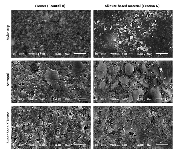
SEM micrographs of the surface topography of both Alkasite and Giomer groups, including control, Astropol, and Super-Snap X-Treme subgroups) were shown in Figure 1. SEM observations showed that, specimens of control groups had lower superficial degradation and fewer scratches. Besides that, F/P procedures promoted superficial alterations on specimens. SEM observation showed that an Alkasite-based restorative material specimen has spherical fillers with lower volumetric filler content than Giomer under investigation. All the specimens in Alkasite-based restorative material showed more surface irregularities than in Giomer. SEM analysis showed more destructive surface alteration using Astropol in Giomer and alkasite groups under investigation.
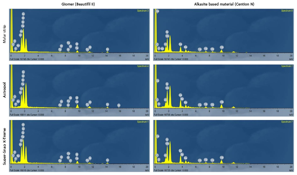
Comparison between the surface composition of Alkasite-based restorative material and Giomer groups was shown in figure 2. The EDX analysis showed that there was no significant difference among all experimental groups (P <0.05) (Tables 2 & 3). Filler particles of Giomer were mainly composed of silicon, Strontium, aluminum, barium, and small amount of Fluorine. Size and morphology of fillers and composition of the tested composites influenced their surface characterization. EDX analysis of Alkasite-based restorative material (Cention N) showed high amount of silica, followed by barium, and ytterbium followed with calcium with small traces of aluminum.
| Elements | Control | Astropol | Super-Snap X-Treme | P-VALUE |
|---|---|---|---|---|
| C (Carbon) | 18.038±1.776 | 17.297±2.330 | 17.540±1.739 | 0.695 |
| O (Oxygen) | 19.242±1.019 | 19.100±2.715 | 19.843±2.939 | 0.7626 |
| F (Fluorine) | 4.219±0.070 | 4.222±0.814 | 4.697±0.570 | 0.1209 |
| Na (Sodium) | 0.841±0.026 | 0.875±0.048 | 0.878±0.034 | 0.0653 |
| AL (Aluminum) | 10.117±0.731 | 10.048±0.511 | 10.600±0.854 | 0.1877 |
| Si (Silicon) | 11.457±1.725 | 11.135±1.013 | 11.192±0.981 | 0.8375 |
| Sr (Strontium) | 32.129±1.768 | 33.173±3.857 | 31.357±3.345 | 0.437 |
| Ta (Tantalum) | 1.535±0.312 | 1.485±0.453 | 1.325±0.271 | 0.3958 |
| W (Tungesten) | 2.422±0.534 | 2.948±0.648 | 2.749±0.617 | 0.162 |
Table 2: Effect of finishing and polishing systems on Element weigth percentage of Giomer (Beautifil II).
| Elements | Control | Astropol | Super-Snap X-Treme | P value |
|---|---|---|---|---|
| C (Carbon) | 42.953±2.063 | 42.907±1.604 | 42.850±1.448 | 0.9911 |
| O (Oxygen) | 8.936±1.039 | 8.951±1.290 | 8.926±0.998 | 0.9987 |
| Na (Sodium) | 0.232±0.210 | 0.120±0.187 | 0.145±0.186 | 0.4148 |
| AL (Aluminum) | 1.462±0.332 | 1.433±0.472 | 1.380±0.475 | 0.9116 |
| Si (Silicon) | 18.088±1.373 | 18.058±1.519 | 18.273±1.465 | 0.938 |
| Ca (calcium) | 8.167±0.903 | 8.377±0.679 | 8.259±0.686 | 0.8278 |
| Ba (barium) | 10.202±0.509 | 10.082±0.538 | 10.121±0.838 | 0.9143 |
| Yb (ytterbium) | 10.189±0.480 | 10.103±0.598 | 10.081±0.623 | 0.905 |
Table 3: Effect of finishing and polishing systems on surface characterization of Alkasite-based restorative material (Cention N)
Discussion
Successful restoration should mimic the human enamel’s smooth surface. The present study compared the surface topography and surface elements characteristics of two ion releasing resin restorative materials; before and after F/P with different systems. Two restorative materials were selected based on their ion releasing property. Previous studies revealed that ion releasing materials might have more surface roughness due to its high water sorption as compared to other conventional nanohybrid composites [16], which is common among fluoride-releasing materials, as they have to permit a certain amount of water diffusion in order to release fluoride [16, 17]. Thus, this study compared the surface topography and surface characteristics of Giomer and Alkasite-based restorative material before and after F/P with two different F/P systems with different abrasive materials. The polishing systems investigated in this study were Astropol which is silicon oxide based F/P tool and Super-Snap X-Treme Technique Kit which is aluminum oxide based F/P tool.
These tools were selected according to their different composition to compare and evaluate the effectiveness of silicon dioxide polishers compared to aluminum oxide polishers and to evaluate their effect on surface characteristics of tested materials. SEM was used to provide detailed images of the surface morphology, while EDX was used to analyze the elemental composition of a sample. When combined, SEM and EDX offer a powerful tool for both imaging and chemical analysis of materials. Photomicrography analysis in SEM and microanalysis were qualitative, so a descriptive statistic method of images was utilized in this research.
In this study, all F/P procedures were standardized according to application time of each disc (10 s), pressure (40grs), positioning of the F/P disc in relation to specimens’ surface, and the movement employed. Nevertheless, superficial scratches were detected on specimens by SEM related to the contact of polishing discs with surface of composite resin [18]. In this experiment, Giomer yields slight better surface quality with less surface irregularities than Alkasite-based restorative material in SEM analysis, this may be attributed to the fact that Giomer has more filler loading (83% by weight) than Alkasite-based restorative material (78.4 by weight) [19, 20]. It should be highlighted that the control subgroups of both Giomer and Alkasite- based restorative material showed lower surface roughness than the tested subgroups in SEM analysis. These results were in accordance with other studies since utilizing Mylar strips without finishing or polishing produced the smoothest material surfaces [10, 21]. However, the removal of the material surface is still necessary because this smooth surface contains a resin-rich layer or oxygen-inhibition layer [10].
This study found that Super-snap X-Treme finished specimens produced smoother surface than Astropol finished specimens. This result is in accordance with a recent study which reported that Super-snap X-Treme formed of aluminum oxide abrasives with small particle. The use of aluminum oxide discs is best optional due to their malleability endorses a homogenous abrasion of the fillers and the resin matrix. Aluminum oxide presents higher Vickers hardness than all the evaluated materials [22]. Previous study reported that the aluminum oxide is one of the most important components used to reach a smooth surface [1].
Regarding the EDX analysis, the filler particles composition of Alkasite-based restorative material (Cention N) in this study are in accordance with the composition described by the manufacturer, except for the absence of fluoride, which was beyond the detection power of EDXS tool. Previous study revealed that Cention- N contains a lesser amount of fluoride ions [23]. According to inorganic composition of each resin based tested materials, no change was identified by EDX in each groups after finishing procedures. The finished specimens were homogenous and did not reveal any embedded particles from finishing tools. The reasons for the obtained results could attribute to using proper F/P techniques. Additionally, using F/P tools under wet condition that could decreases the friction between restorative surfaces and F/P tools, thus protect the surface from heat production which may cause microcracks and elemental transmission from F/P tools to materials surfaces. Both F/P systems that were used in this study didn’t cause surface degradation of the materials and didn’t precipitate any particles on the material surfaces. SEM and EDX analysis revealed that size and morphology of fillers and composition of the tested composites influenced their topograghy when samples were submitted to F/P.
Limitations of this study can be that the tests were performed on two restorative materials; henceforth the effect of varied filler types and sizes on surface topography and characterization could not be established. Further studies are recommended to test more F/P tools such as diamond based or zirconium oxide based F/P tools. The samples were polished under wet conditions. Hence, a comparative analysis between the effects of dry and wet polishing could not be made to detect the elemental analysis of the restorative materials. To conclude, recognizing the limitations of the present in vitro study, the Super-Snap X-Treme and Astropol F/P systems exhibit a comparable effect on surface qualities for Alkasite-based restorative material and Giomer. The effect of F/P systems on surface roughness was dependent on the particle type and size of both F/P system and resin based restorative material. Giomer and restorative materials based on alkasite appear to be compatible with the Super- Snap X-Treme polishing system.
Authors’ contributions
Hanan A. Soliman and Naglaa Rizk Elkholany: Study Design, preparation of samples, testing procedures and writing the manuscript. Nazem A: Performing statistical analysis of this lab work. Noha El-Wassefy: Manuscript editing and revision.
References
-
Jaramillo CR, Lopez GEJ, Latorre CF, Agudelo SAA (2021) Effect of Polishing Systems on the Surface Roughness of Nano-Hybrid and Nano-Filling Composite Resins: A Systematic Review. Dent J 9(8): 95.
-
Tepe H, Erdılek AD, Sahın M, Efes BG, Yaman BC (2023) Effect of Different Polishing Systems and Speeds on the Surface Roughness of Resin Composites. J Conserv Dent 26(1): 36-41.
-
Pala K, Tekce N, Tuncer S, Serim ME, Demirci M (2016) Evaluation of the Surface Hardness, Roughness, Gloss and Color of Composites after Different Finishing/Polishing Treatments and Thermocycling using a Multitechnique Approach. Dent Mater J 35(2): 278-289.
-
Francois P, Fouquet V, Attal JP, Dursun E (2020) Commercially Available Fluoride-Releasing Restorative Materials: A Review and a Proposal for Classification. Materials 13(10): 2313.
-
Meshram P, Meshram V, Palve D, Patil S, Gade V, et al. (2019) Comparative Evaluation of Microleakage around Class V Cavities Restored with Alkasite Restorative Material with and without Bonding Agent and Flowable Composite Resin: In Vitro Study. Indian J Dent Res 30(3): 403-407.
-
Naz F, Khan AS, Kader MA, Gelban LOS, Hakeem AS, et al. (2021) Comparative Evaluation of Mechanical and Physical Properties of a New Bulk-fill Alkasite with Conventional Restorative Materials. Saudi Dent J 33(7): 666-673.
-
Francois P, Remadi A, Goff SL, Abdel-Gawad S, Attal JP, et al. (2021) Flexural Properties and Dentin Adhesion in Recently Developed Self-adhesive Bulk-fill Materials. J Oral Sci 63(2): 139-144.
-
Ruengrungsom C, Burrow MF, Parashos P, Palamara JE (2020) Evaluation of F, Ca, and P Release and Microhardness of Eleven Ion-Leaching Restorative Materials and the Recharge Efficacy using a New Ca/P Containing Fluoride Varnish. J Dent 102: 103474.
-
Kozmos M, Virant P, Rojko F, Abram A, Rudolf R, et al. (2021) Bacterial Adhesion of Streptococcus Mutans to Dental Material Surfaces. Molecules 26(4): 1152.
-
Pietrokovski Y, Zeituni D, Schwartz A, Beyth N (2022) Comparison of Different Finishing and Polishing Systems on Surface Roughness and Bacterial Adhesion of Resin Composite. Materials 15(21): 7415.
-
Kemaloglu H, Karacolak G, Turkun LS (2017) Can Reduced‐step Polishers be as Effective as Multiple‐step Polishers in Enhancing Surface Smoothness? J Esthet Restor Dent 29(1): 31-40.
-
Albelasy EH, Hamama HH, Chew HP, Montaser M, Mahmoud SH (2022) Secondary Caries and Marginal Adaptation of Ion-releasing Versus Resin Composite Restorations: A Systematic Review and Meta-analysis of Randomized Clinical Trials. Sci Rep 12(1): 19244.
-
Daabash R, Alqahtani MQ, Price RB, Alshabib A, Niazy A, et al. (2023) Surface Properties and Streptococcus Mutans Biofilm Adhesion of Ion-Releasing Resin-Based Composite Materials. J Dent 134: 104549.
-
Carrillo MA, Salazar CG, Castro RL, Ladera CM, Lopez GC, et al. (2022) The Microhardness and Surface Roughness Assessment of Bulk-fill Resin Composites Treated with and without the Application of an Oxygen-Inhibited Layer and a Polishing System: An in Vitro Study. Polymers 14(15): 3053.
-
Bahari M, Kahnamoui MA, Chaharom MEE, Kimyai S, Sattari Z (2021) Effect of Curing Method and Thermocycling on Flexural Strength and Microhardness of a New Composite Resin with Alkaline Filler. Dent Res J 18: 96.
-
Gonulol N, Ozer S, Sen Tunc E (2015) Water Sorption, Solubility, and Color Stability of Giomer Restoratives. J Esthet Restor Dent 27(5): 300-306.
-
McCabe JF, Rusby S (2004) Water Absorption, Dimensional Change and Radial Pressure in Resin Matrix Dental Restorative Materials. Biomaterials 25(18): 4001-4007.
-
Soliman HA, Elkholany NR, Hamama HH, El-Sharkawy FM, Mahmoud SH, et al. (2020) Effect of Different Polishing Systems on the Surface Roughness and Gloss of Novel Nanohybrid Resin Composites. Eur J Dent 15(2): 259-265.
-
Verma V, Mathur S, Sachdev V, Singh D (2020) Evaluation of Compressive Strength, Shear Bond Strength, and Microhardness Values of Glass-Ionomer Cement Type IX and Cention N. J Conserv Dent 23(6): 550-553.
-
Yazkan B, Celik E, Recen D (2021) Effect of Aging on Surface Roughness and Color Stability of a Novel Alkasite in Comparison with Current Direct Restorative Materials. Oper Dent 46(5): E240-E250.
-
Zhang L, Yu P, Wang X-Y (2021) Surface Roughness and Gloss of Polished Nanofilled and Nanohybrid Resin Composites. J Dent Sci 16(4): 1198-1203.
-
Mierzejewska Z, Lukaszuk K, Choromanska M, Zalewska A, Borys J, et al. (2024) Influence of Different Polishing Methods on Surface Roughness and Microhardness of Dental Composites. Advances in Science and Technology Research Journal 18(1): 268-279.
-
Todd JC (2016) Scientific Documentation: Cention N Ivoclar-Vivadent Press, Schaan, Liechtenstein 2016: 1-58.
- Diagnosis and Management of Mental Nerve Paresthesia Secondary to Apical Periodontitis of Mandibular Second Premolar: A CBCT Based Case Report
- A Randomized, Double Blinded Clinical Trial to Compare the Effect of Oral Premedication (Diclofenac Potassium or Dexamethasone) on Post-Operative Pain Following Pulpectomy
- Modified Lip Repositioning Technique for the Management of Excessive Gingival Display
- Integral Role of Non-Dental Providers and Fluoride Dissemination
- Root Canal Treatment Rate in Deciduous Teeth Among 6-Year- Olds in the Era of Discontinuing Water Fluoridation - Historical Cohort Study
- The Impact of the Notch1 on the Migratory Capacity and the Expression of E-Cadherin and CyclinD1 in Ameloblastoma Cells