Fabrication of a CAD-CAM Custom Modified Anatomic Healing Abutment based on Gingival Profile Measurement in an Instructional Dentition Model - A Dental Technique
This report introduces a new modified anatomic healing abutment designed to shape the gingival contour peri-implants. Experimental implants were placed in a dentition model after extracting a maxillary central incisor and a mandibular first molar. Scanning procedures were performed on the model. The reduction in gingival contour dimensions at the neck of the implants guided the development of the modified abutment. CAD/CAM technology was used to manufacture a titanium-based abutment, reducing chairside workflow time and minimizing costs. The present report presents the evolutionary progression of a modified anatomic healing abutment through its incorporation within a dentition model, thereby showcasing an avantgarde technology paradigm and methodological. The modified healing abutment has been intricately fashioned to cater to delayed implantation circumstances, in stark contrast to its conventional counterpart employed in immediate implantation scenarios. Its primary objective resides in the meticulous cultivation of an exquisitely defined gingival contour, thereby augmenting the aesthetic visage of the anterior dental region.
Introduction
In recent years, the field of dental implant restoration has witnessed a growing emphasis on esthetic restoration. The concept of esthetic dental implant restoration entails achieving a seamless integration between the prosthesis and the surrounding soft tissue after implantation, such that it becomes visually indistinguishable from the natural dentition. The key to accomplishing this esthetic outcome lies in maximizing the preservation or reconstruction of the soft tissue surrounding the implant.
To ensure the stability of the soft tissue surrounding the implant, esthetic dental implant restoration begins with the placement of a healing abutment. These healing abutments typically possess a circular cross-section and are predominantly composed of titanium [1, 2]. To achieve an optimal soft tissue profile around the implant, the shape of the healing abutment should closely resemble that of the natural tooth’s cervical region [3]. However, circular healing abutments lack the contour characteristic of a natural tooth, resulting in an imperfect peri-implant soft tissue profile when employed for shaping purposes. Consequently, a subsequent surgical intervention is required to reshape the soft tissue [4].
To address this issue, implantologists have implemented personalized healing abutments in clinical settings [5, 6, 7, 8, 9]. Personalized healing abutments facilitate early restoration of the gingival contour curve, eliminating the need for a secondary procedure and reducing the risk of premature detachment [5, 6, 7, 8]. These abutments are custom-made to match the natural tooth profile, preserving the original gingival structure and providing insulation for the bone graft material at the extraction site during immediate implantation [5, 7]. Numerous researchers have proposed different techniques for creating customized healing abutments to maintain gum contour [8, 9, 10, 11, 12]. We hypothesize that anatomic healing abutments that closely emulate the shape of the tooth neck are more effective for immediate implantation. Therefore, this study focuses on the mandibular first molar as exemplary cases, aiming to design and manufacture improved anatomic healing abutments based on the amount of alveolar bone absorption after tooth extraction. These abutments will be utilized to shape the pre-implant gingival contour. This innovative design methodology holds promising prospects for successful implementation in clinical trials.
Technique
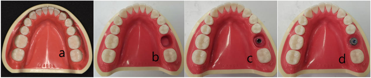
- The single mandibular first molars were extracted from the dental models (Shanghai Xinmanke Teaching Equipment Co., LTD., China) Figure 1a to expose the neck cross-section of tooth (Figure 1b).
- Experimental implants (Beijing Daqing Biotechnology Co., LTD., China) were inserted into the vacant cross- sections using red wax (Figure 1c). A scanning abutment (Beijing Daqing Biotechnology Co., LTD.,) China was placed alongside implant within the cross-section (Figure 1d).
- The dental models were scanned using a Shining 3D Scanner (oralscan2, SHINING 3D Technology Co., LTD., China).
- A digital caliper (Mitutoyo 500-196-30, Shenzhen Sanjian Precision Measuring Instrument Co., LTD., China) was employed to measure the cervical cross-sectional dimensions of the mandibular first molar on the dental models, as depicted in Figure
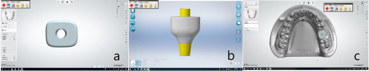
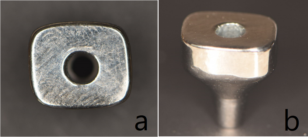
![Figure 2: 5. According to the findings of a previous investigation [13], the horizontal resorption of the alveolar bone on the buccal aspect exhibited the following percentages: 19.4 ± 9.4% at the mesial point, 39.1 ± 10.4% at the midpoint, and 20.3 ± 10.7% at the distal level, within a six-month period post-extraction of a single tooth. Taking into account this data, alongside the acquired gingival contour information of the mandibular first molar, the lengths of lines B, C, and D were reduced by 20% in order to design a modified anatomical healing abutment. The fabrication of this healing abutment involved CAD-CAM casting of a base made of grade 5 titanium. 6. Make a 3D digital model intraoral scan impression: the digital model is the transverse section of the modified anatomic healing abutment for the mandibular first molar was designed in the form of an round rectangle (Figure 3a). 7. The design parameters for the modified healing abutments are presented in Table 1. The lateral profile of each modified anatomic healing abutment by Computer aided design equipment and software design (EXOCAD2019, Exocad, Germany) (Figure 3b). 8. Placing the modified anatomic healing abutments designed in the model (Figure 3c). A central screw hole was positioned within each modified anatomic healing abutment. 9. Send the design for the CAD-CAM metal modified healing abutment for printing to 3D numerical control processing equipment (B51, Shenzhen Colide Medical Technology Co., LTD., China) 10. Proceed with the CAD-CAM fabrication Figures 4a & b these modified healing abutments were fabricated using a grade 5 titanium (JZ-V98, Xi ‘an Jiuzhou Biomaterials Co., LTD., China).](/fulltextimages/12714/fig_2.png)
Figure 2: Gingival contour measurement of mandibular first molar. Line A is the distance between the proximal and distal of the gingival contour, line B is the distance of the buccolingual of the gingival contour, line C is the distance from the proximal buccal of gingival contour to the distal lingual gingival contour through the focal points of AB, and line D is the distance from the proximal lingual of gingival contour to the distal buccal gingival contour through the focal points of AB.
| Modified Anatomical Healing Abutment Parameters | Parameter Function | ||
|---|---|---|---|
| Angle | 0 degrees | Ensure stress distribution after restoration | |
| Intragingival height | 2 or 3 mm | Form cuff according to gingival thickness | |
| Cross-sectional data of mandibular first molar | A | 9.9 mm | According to different tooth positions, different sizes of gingiva are formed |
| B | 7.6 mm | ||
| C | 9.3 mm | ||
| D | 9.4 mm | ||
| Transverse morphology of mandibular first molar | Round rectangle | Form different shapes according to different tooth positions | |
| Overall height | 5 or 6 mm | Suitable for different restoration heights |
Table 1: Design Parameters of Modified Anatomic Healing Abutment.
Discussion
The report introduced a modified anatomic healing abutment characterized by a slightly concave profile, resembling the contour of the tooth neck. Implementation of this type of healing abutment has the potential to enhance peri-implant gingival esthetics and contour, thereby yielding superior final esthetic outcomes when compared to circular healing abutments [4, 6, 14, 15, 16]. The utilization of personalized healing abutments is progressively being documented in clinical practice, encompassing both esthetic and non-esthetic contexts [5, 6, 7, 8]. For instance, Raheem IMA, et al. [11] employed X-ray film to measure the distance from the implant shoulder to the proximal and distal root of the tooth, as well as the crest of the buccal and lingual alveolar crest. After adjusting the acquired data, a CAD/CAM system was employed to design a customized healing abutment. Some researchers have utilized CAD/CAM systems or manual methods in conjunction with silicone impressions or oral scans to fabricate personalized healing abutments [1, 2, 8, 17]. These techniques have proven effective and are gaining popularity among both dentists and patients. However, they necessitate additional equipment, such as CAD/CAM, and involve chair-side fabrication, which heightens the risk of patient infection, prolongs chair-side operation times, and incurs supplementary costs.
The dentition model manufactured by Shanghai Xinman Science Teaching Equipment Co., Ltd. in China provides the opportunity to acquire the dimensions of the tooth neck cross-section (lines A, B, C, D). For this investigation, the mandibular first molars were chosen as representative examples. Based on the neck morphology of the model teeth and the buccal bone plate absorption subsequent to tooth extraction, the cross-section of the modified anatomic healing abutment for the mandibular first molar was designed as a rounded rectangle. The goal was to closely replicate the natural tooth’s neck shape. The enhanced anatomical form of the healing abutment offers improved shaping of the peri- implant soft tissue profile. Generally, the majority of healing abutments are fabricated from biocompatible titanium [1, 2]. However, some researchers [9] have explored the use of zirconia ceramics for healing abutments due to their superior soft tissue esthetic characteristics [18] and reduced bacterial adhesion to zirconia ceramic surfaces [19]. In the subsequent clinical trial, a Datsing implant with a tapered connection was employed for the abutment. Shota [20] discovered that zirconia-based abutments exhibited lower fracture resistance compared to titanium-based abutments in cone-connected implants. Therefore, in this study, grade 5 titanium was chosen as the raw material for producing the modified anatomic healing abutment. Furthermore, since the healing abutment serves as a temporary tool for shaping the gingiva, aesthetic requirements are minimal.
Acknowledgement
This report received support from the Program for Young Talents of Science and Technology in Universities of Inner Mongolia Autonomous Region (grant no. NJYT 23064), the First-class Scientific Research Fund Project of Stomatology School of Chifeng University (grant no. KQYXYYLXK 202101), and the Chifeng University Affiliated Hospital research project (grant no. FSYY2022-20).
Competing Interest
The authors declare no competing interests.
References
-
Proussaefs P (2016) Custom CAD-CAM Healing Abutment and Impression Coping Milled from a Poly(Methyl Methacrylate) Block and Bonded to a Titanium Insert. J Prosthet Dent 116(5): 657-662.
-
Alshhrani WM, Amri MDAl (2016) Customized CAD- CAM Healing Abutment for Delayed Loaded Implants. J Prosthet Dent 116(2): 176-179.
-
Schoenbaum TR, Swift EJ (2015) Abutment Emergence Contours for Single-Unit Implants. J Esthet Restor Dent 27(1): 1-3.
-
Schwarz F, Mihatovic I, Becker J, Bormann KH, Keeve PL, et al. (2013) Histological Evaluation of Different Abutments in the Posterior Maxilla and Mandible: an Experimental Study in Humans. J Clin Periodontol 40(8): 807-815.
-
Su H, Gonzalez-Martin O, Weisgold A, Lee E (2010) Considerations of Implant Abutment and Crown Contour: Critical Contour and Subcritical Contour. Int J Periodontics Restorative Dent 30(4): 335-343.
-
Steigmann M, Monje A, Chan HL, Wang HL (2014) Emergence Profile Design Based on Implant Position in the Esthetic Zone. Int J Periodontics Restorative Dent 34(4): 559-563.
-
Akin R (2016) A New Concept in Maintaining the Emergence Profile in Immediate Posterior Implant Placement: The Anatomic Harmony Abutment. J Oral Maxillofac Surg 74(12): 2385-2392.
-
Hartman MJ (2021) A Workflow to Design and Fabricate a Customized Healing Abutment From a Dynamic Navigation Virtual Treatment Plan. Compend Contin Educ Dent 42(2): 86-92.
-
Teslak M, Ziemlewski A, Foltyn I, Ordyniec-Kwasnica I, Drogoszewska B (2021) Development of Custom Anatomic Healing Abutment Based on Cone-Beam Computer Tomography Measurement on Human Teeth Cross-Section. Materials (Basel) 14(16): 4570.
-
Berkei G, Vag J (2022) Peri-Implant Soft Tissue Conditioning with Prefabricated Titanium Anatomic Healing Abutment Compared with Conventional Circular Healing Abutment: A Case Letter. J Oral Implantol 48(6): 523-532.
-
Raheem IMA, Hammad IA, Kader SHA, Fahmy RA (2022) Fabrication of a CAD-CAM Custom Healing Abutment Guided by a Conventional Dental Radiograph for Delayed Loaded Dental Implants: A dental Technique. J Prosthet Dent 127(1): 49- 54.
-
Vag J, Freedman G, Szabo E, Romanszky L, Berkei G (2022) Cervical Tooth Anatomy Considerations for Prefabricated Anatomic Healing Abutment Design: A Mathematical Formulation. J Prosthet Dent 127(6): 852- 859.
-
Covani U, Ricci M, Bozzolo G, Mangano F, Zini A, et al. (2011) Analysis of the Pattern of the Alveolar Ridge Remodelling Following Single Tooth Extraction. Clin Oral Implants Res 22(8): 820-825.
-
Doliveux S, Jamjoom FZ, Finelle G, Hamilton A, Gallucci GO (2020) Preservation of Soft Tissue Contours Using Computer-Aided Design/Computer-Assisted Manufacturing Healing Abutment with Guided Surgery in the Esthetic Area: Case Report. Int J Oral Maxillofac Implants 35(1): e15-e20.
-
Orozco VA, Arroyo CG, Martinez FR, Jimenez CE (2015) Biometric Analysis of the Clinical Crown and the Width/ Length Ratio in the Maxillary Anterior Region. J Prosthet Dent 113(6): 565.e2-570.e2.
-
Scoble HO, White SN (2014) Compound Complex Curves: The Authentic Geometry of Esthetic Dentistry. J Prosthet Dent 111(6): 448-454.
-
Giovanni-Battista MF, Crespi R, Toti P, Crespi G, Rubino L, et al. (2020) A 3-Year Retrospective Study of Fresh Socket Implants: CAD/CAM Customized Healing Abutment vs Cover Screws. Int J Comput Dent 23(2): 109-117.
-
Naveau A, Rignon-Bret C, Wulfman C (2019) Zirconia Abutments in the Anterior Region: A Systematic Review of Mechanical and Esthetic Outcomes. J Prosthet Dent 121(5): 775-781.
-
Hanawa T (2020) Zirconia Versus Titanium in Dentistry: A Review. Dent Mater J 39(1): 24-36.
-
Watanabe S, Nakano T, Ono S, Yamanishi Y, Matsuoka T, et al. (2022) Fracture Resistance of Zirconia Abutments with or without a Titanium Base: An In Vitro Study for Tapered Conical Connection Implants. Materials (Basel) 15(1): 364.
- Diagnosis and Management of Mental Nerve Paresthesia Secondary to Apical Periodontitis of Mandibular Second Premolar: A CBCT Based Case Report
- A Randomized, Double Blinded Clinical Trial to Compare the Effect of Oral Premedication (Diclofenac Potassium or Dexamethasone) on Post-Operative Pain Following Pulpectomy
- Modified Lip Repositioning Technique for the Management of Excessive Gingival Display
- Integral Role of Non-Dental Providers and Fluoride Dissemination
- Root Canal Treatment Rate in Deciduous Teeth Among 6-Year- Olds in the Era of Discontinuing Water Fluoridation - Historical Cohort Study
- The Impact of the Notch1 on the Migratory Capacity and the Expression of E-Cadherin and CyclinD1 in Ameloblastoma Cells