A Rare Case of Mucinous Cystadenoma Presenting as Unilateral Proptosis at Birth
Mucinous cystadenoma is a rare cause of unilateral proptosis at birth. A two day old female baby presented with non-pulsatile, transiliuminantaxial proptosis right eye. On CT scan cystic lesion was seen behind the globe in retrobulbar space. Gross total excision of cyst was done through trans-frontal craniotomy approach. Histopathological examination revealed mucinous cystadenoma. This case has been reported because of rarity of this condition
Introduction
Proptosis at birth is very rare. Various causes of proptosis at birth include birth trauma, teratoma, encephelocoel [1]. Other rare cases reported are of congenital myofibroma, congenital rhabdoidtumor [2]. Mucinous cystadenoma is a benign tumor affecting the ovary, pancreas, appendix and sometimes liver [3]. Because of its malignant potential, cystadenoma is treated by total surgical excision. Published English literature however does not mention mucinous cystadenomaas a cause of proptosis at birth. The authors present a rare case of proptosis at birth caused by mucinous cystadenoma of the orbit.
Case Report
A two day old female baby, born of an uneventful pregnancy and vaginal delivery, was examined at the Oculoplasticsand Orbit unit, Department of Ophthalmology, King George’s Medical University, Lucknow. She presented with proptosis since birth. On examination Right eye showed axial proptosis that was non-pulsatile, compressible, reducible and brilliantly transilluminant. There was inadequate eye closure, congested and chemosed conjunctiva and dry lustreless cornea (Figure 1). No bruit was present on auscultation. Pupillary reaction was sluggish. Fundus details were not clearly visible. Left eyeball and orbit were unremarkable.
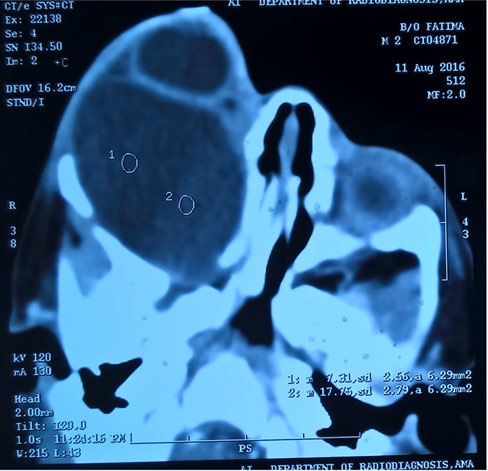
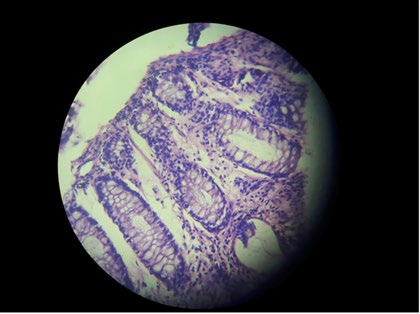
Tarsorrhaphy was done to prevent further exposure keratitis. CT scan showed a well-defined single large cystic lesion present in the right retrobulbar space. The cyst wall mildly enhanced on contrast. Signs of orbit expansion and thinning of roof and lateral wall were seen. No orbital structure was identifiable (Figures 2A & 2B). Pre-operative diagnosis of orbital meningocele was made. Surgical excision of cyst through right transfrontal craniotomy was performed under general anesthesia. Histopathological examination revealed mucinous cystadenoma (Figure 3).
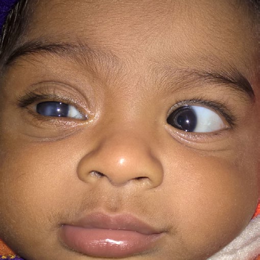
Figure 2A: Coronal view of CT scan showing enlarged orbit with homogenous hypodense single large cystic lesion in orbit (Right side).
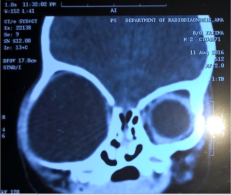
Figure 2B: Axial view of CT scan showing retrobulbar hypodense lesion.
At six month follow-up patient had residual corneal opacity and accurately followed light (Figure 4).
Discussion
Extensive search of literature (pubmed and google scholar) does not report a single case of mucinous cystadenoma as a cause of proptosis at birth. Mucinous cystadenomas usually an epithelial origin ovarian benign tumor with malignant potential occur as a large multi- loculated cystic mass with mucus-containing fluid. These tumors occur most commonly in women in their twenties to forties [4]. Few cases of congenital cystadenoma has been reported in pancreas [5]. Among the differential diagnosis of unilateral proptosis at birth mucinous cystadenoma is one of the rarest causes. Other causes which include orbital meningocele which present with pulsatile proptosis and bony defect on CT scan [6] and teratoma which are cystic lesions and sometimes tooth or hairs are seen. They originate from pluripotent germ cells [7]. Other cause includes birth trauma which can be ruled out with history of use of forceps during delivery or marks of instrumentation can be seen on temporo-parietal area of baby [1]. It is important to know the probable cause because management of each condition is different and most of these cystic swelling are have malignant potential including this case of mucinous cystadenoma. Proptosis at birth is rare but, its presence usually reflects a serious problem. Most of these conditions are fast growing. With early intervention eye and life salvage can be aimed.
References
-
Preece JM, Cornette L, El Hindy N (2005) Simple management of isolated proptosis at birth. Arch Dis Child Fetal Neonatal Ed 90(3): 234.
-
Bloom RI, Schwarcz RM, Zhang C, Rosenberg JB (2013) A case of congenital myofibroma of the orbit presenting at birth. Orbit 32(1): 33-35.
-
Brown J, Frumovitz M (2014) Mucinous tumors of the ovary: current thoughts on diagnosis and management. Curr Oncol Rep 16(6): 389.
-
Hart WR, Norris HJ (1974) Borderline and malignant mucinous tumors of the ovary. Histologic criteria and clinical behavior. Obstetrical &Gynecological Survey 29(2): 160-163.
-
Gentimi FE, Papandreou E, Tzovaras AA, Antoniou D (2011) Pancreatic cystic lesion in an infant. J Indian Assoc Pediatr Surg 16(2): 72-74.
-
Consul BN, OP Kulshrestha (1965) Orbital meningocele. Br J Ophthalmol 49(7): 374-376.
-
Baidya KP, Ghosh S, Datta A, Mukhopadhyay S, Bhaduri G (2014) Huge congenital teratoma containing tooth in a three-day-old neonate. Oman J Ophthalmol 7(1): 13-15.
- Screening of Hospital Staff During World Glaucoma Week in a Tertiary Eye Care Centre
- Angioid Streaks with Macular Neovascularization: Clinical Insights from Two Cases
- Giant Kissing Naevus: An Oculoplastic Challenge
- Why Freedom of Vision Should Not Cost the Freedom of Feeling - LASIK in the Climate of Change
- Asymmetric Optic Nerve with Small Disc and Large Cup: A Rare and Challenging Case of Unilateral Optic Nerve Hypoplasia
- Large Angle Exotropia in a Child: A Case Report