Retinitis Pigmentosa with Bilateral Ectopia Lentis
Retinitis pigmentosa (RP), a degenerative disorder characterized by a diverse array of ocular and systemic manifestations, was initially reported in 1857. Despite over a century of research, zonular weakness in RP still remains unsolved. We report a late presentation of bilateral inferior subluxation of lens, with visible zonular fibres. The patient exhibited no systemic abnormalities suggestive of a syndromic presentation. This appears to be a rare instance of bilateral concurrent spontaneous ectopia lentis (EL) associated with RP, with no syndromic association. It serves as a wake-up call for urgent need of further research in understanding the aetiology of RP.
Introduction
Retinitis pigmentosa (RP), a misnomer that despite the term “retinitis” implies no role of inflammation in its pathology, is a genetically heterogenous group of inherited disorder characterized by progressive dysfunction of rod photoreceptors, leading to subsequent degeneration of cones and retinal pigment epithelium (RPE). It manifests as night blindness, visual field loss and abnormal electro retinography findings [1]. RP is believed to be associated with zonular weakness, presenting as varying degrees of pre-operative and intraoperative phacodonesis or delayed post-operative lens dislocation. It also is linked to a wide spectrum of ocular abnormalities, including cataracts, glaucoma, keratoconus and refractive errors [2].These if present in majority of cases is associated with syndromes like marfans, ushers syndrome or Bardet Biedl syndrome [3].
We report a case of bilateral spontaneous subluxation of lens in a middle-aged male without any syndromic associations. Such incidental occurrences instigate the need of detailed systemic evaluations of such individuals and also emphasize the necessity for further research to enhance our understanding of the etiology of these occurrences.
An elderly male in his sixties with no systemic co- morbidities presented to our outpatient clinic, with complaints of reduced vision in both eyes over the past 2 years. It was gradual and painless in nature. The patient gave no history of ocular trauma, watering or episodic redness of eyes. Blood investigations revealed normal values.
After obtaining consent, a thorough examination was conducted. On, Ocular examination, visual acuity in both eyes was 2/60 not improving with pin hole. Intraocular pressure in both eyes was 16 mmm Hg (Millimetres of Mercury) when measured with a non-contact tonometer. Pupils showed sluggish reaction to light, and no relative afferent pupillary defect was observed on swinging torch light examination. Slit lamp examination revealed irregular depth of the anterior chamber in both eyes, with bilateral inferior subluxation of lens (Figures 1a & 1b) with superior zonular dialysis extending for roughly 4 clock hours (Figures 2a & 2b). Retro- illumination disclosed a few elongated superior zonular fibres, with the lens just hanging on these fibres.
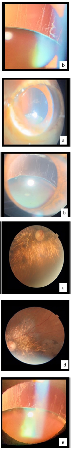
Figure 1a: Anterior Segment of right eye.
Figure1b: Anterior Segment of left eye.
Figure 2a: Zonules visualized in the right eye.
Figure 2b: Zonules visualized in the left eye.
Dilated fundus examination revealed pale waxy disc with generalised arterial attenuation and absent foveal reflex (Figures 1c & 1d). Pericentral intraretinal pigmentation in form of spicules was observed on a grossly tessellated background.
Figure 1c: Fundus Photograph of right eye.
Figure 1d: Fundus Photograph of left eye.
Clinically retinitis pigmentosa was suspected to be the most probable diagnosis and confirmed when similar history of nyctalopia was given by his younger sibling.
On detailed systemic examination, he was moderately built and nourished, exhibited no visible systemic abnormalities on gross examination. His arm span, upper to lower segment ratio were within normal limits. No hyperextensibility of joints, skin or polydactyly were noted. ECG and echocardiography were performed to rule out cardiovascular anomalies and both turned out normal. Pure tone audiometry indicated mild senile hearing loss.
Upon reviewing the ocular findings, system examination and blood investigations, syndromes associated with ectopia lentis and retinitis pigmentosa were ruled out. Thus, this appeared to be a rare occurrence of bilateral spontaneous ectopia lentis due to RP.
Discussion
RP is an inherited degenerative disorder affecting photoreceptors and retinal pigment epithelial cells (RPEs), resulting in diverse ocular manifestations. The global prevalence of this disease is estimated to be 1 in 4000 [4]. It is commonly associated with complicated cataracts, notably posterior subcapsular cataracts (PSC) being the most prevalent. This is attributed to the altered intraocular microenvironment caused by the inflammatory reaction associated with RP [5]. In about 10 percent of RP cases, zonular weakness is reported and its extent can vary. The suggested cause of this weakness is postulated to be scattered zonular loss and liquefied vitreous although the precise reason remains elusive.
EL, characterized by lens dislocation can result from various causes such as trauma, pseudoexfoliation syndrome, spontaneous dislocations and familial diseases like marfan’s syndrome and homocystinuria. While occasional reports mention zonular weakness, instances of bilateral spontaneous EL are rarely report. Absence of any cause of EL and no syndromic associations of RP and EL following comprehensive systemic and ophthalmic evaluations, raises concerns about a potential incidental association which must be investigated.
The pathogenesis of subluxation and the necessity for frequent follow-ups in individuals diagnosed with RP to facilitate timely lens extraction remains unanswered. Additionally, our case emphasizes the importance of thorough preoperative assessment in individuals with RP highlighting the need for specialized implants, such as capsular tension rings, during cataract surgery.
Addressing these concerns will enhance the outcomes of surgical intervention in patients with retinitis pigmentosa.
References
-
Sato H, Wada Y, Abe T, Kawamura M, Wakusawa R, et al. (2002) Retinitis Pigmentosa Associated With Ectopia Lentis. Arch Ophthalmol 120(6): 852-854.
-
Kwon YA, Bae SH, Sohn YH (2007) Bilateral Spontaneous Anterior Lens Dislocation in a Retinitis Pigmentosa Patient. Korean J Ophthalmol 21(2): 124-126.
-
Ferrari S, Di Iorio E, Barbaro V, Ponzin D, Sorrentino FS, et al. (2011) Retinitis Pigmentosa: Genes and Disease Mechanisms. Curr Genomics 12(4): 238-249.
-
Dikopf MS, Chow CC, Mieler WF, Tu EY (2013) Cataract extraction outcomes and the prevalence of zonular insufficiency in retinitis pigmentosa. Am J Ophthalmol 156(1): 82-88.
-
Hong Y, Li H, Sun Y, Ji Y (2020) A Review of Complicated Cataract in Retinitis Pigmentosa: Pathogenesis and Cataract Surgery. J Ophthalmol 6699103.
- Screening of Hospital Staff During World Glaucoma Week in a Tertiary Eye Care Centre
- Angioid Streaks with Macular Neovascularization: Clinical Insights from Two Cases
- Giant Kissing Naevus: An Oculoplastic Challenge
- Why Freedom of Vision Should Not Cost the Freedom of Feeling - LASIK in the Climate of Change
- Asymmetric Optic Nerve with Small Disc and Large Cup: A Rare and Challenging Case of Unilateral Optic Nerve Hypoplasia
- Large Angle Exotropia in a Child: A Case Report