Paratesticular Masson’s Hemangioma Case Report and Review of the LiteratureÂ
<p>Masson’s hemangioma or intravascular papillary endothelial hyperplasia is a rare intravascular benign lesion. It may involve a wide variety of organs, most frequently the skin and subcutaneous tissue. Regarding the urogenital system, it has been described in the bladder and the renal vein.  <br />To our knowledge, this is the first case report of a Masson’s Hemangioma in the male genital organs.  </p> <p> </p>
Introduction
Vascular tumors and malformations may be challenging to diagnose. Masson’s Hemangioma is a rare intravascular benign lesion consisting of an intravascular papillary endothelial hyperplasia described by Pierre Masson in 1923 [1].
Masson’s hemangioma is may involve a variety of organs most frequently skin and subcutaneous tissue [2]. Regarding the urogenital system involvement of the renal vein and bladder have been described [3, 4].
To our knowledge this is the first case report of Masson’s hemangioma located in paratesticular tissue.
A 38 year old male is referred to the Urology department due to a painless right testicular mass. Physical examination revealed a paratesticular cyst independent from the testicle or epididymis. Tran illumination was doubtful.
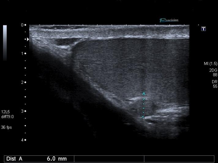
A testicular ultrasound was performed showing a 6 mm mass with minimum Doppler enhancement (Figure 1). The mass was independent from the testicle or the epididymis.
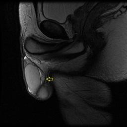
MRI described a 6 mm nodule adjacent to the body of the epididymis. The nodule was enhanced with MRI contrast in a heterogeneous manner (Figure 2).
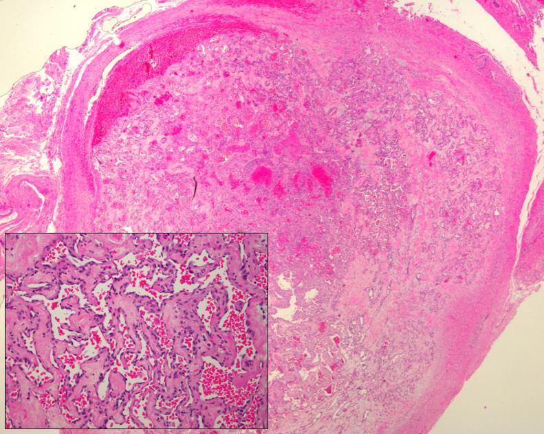
The patient underwent excision of the nodule with no perioperative complications. The mass was not fixed to the epididymis or testis and was easily excised. No intraoperative biopsy was performed due to the benign appearance. The pathology report rendered intravascular papillary endothelial hyperplasia (Masson’s Hemangioma) (Figure 3).
Follow up was made with testicular ultrasound and physical examination. Due to the benign course of the pathology the patient was discharged after 1 year.
Discussion Masson’s Hemangioma (MH) was first described in 1923 in a man with a painful, ulcerated hemorrhoid. It was characterized by enlarged papillae covered by endothelium whose tips contained capillaries and fibrin. These formations became elongated and ramified in an unorganized fashion that gradually obstructed the vein. It is different from a hemangioma because the process remained within the vessel. [1, 5].
MH represents approximately 2% of benign and malignant tumors of the skin and subcutaneous tissues. It commonly appears between the third and fourth decade with a slight greater prevalence in women. It is also described in head and neck region, oral cavity, thyroid, gastrointestinal tract, brain, cervix and liver [2,6 ].
Regarding the genitourinary tract it has been described in a few cases. Renal vein involvement is described in eight cases. Most of them present hematuria and loin pain as first sign. Due to the rarity of the pathology preoperative suspicion is not usually made and nephrectomy is generally performed [3, 6, 7]. In our case radical surgery was not performed due to benign appearance on imaging studies and intraoperative findings.
It is also described in the bladder. Tavora et al. reported three cases in a large series of vascular tumors of the bladder. All three cases associated radiation therapy for prostate, bladder and endometrium malignancies respectively. The tumors were restricted to the submucosa and there was no evidence of recurrence. It is important to differentiate MH from angiosarcoma. Although both lesions show minimal cytologic atypia in cells lining poorly formed and fused vascular channels, papillary endothelial hyperplasia is confined to a localized area of the thrombus, whereas angiosarcoma tend to invade into surrounding tissue [4].
To our knowledge this is the first case report of a MH located in the male genital organs. The course of the disease seems similar to other locations with no recurrence or local complications.
References
-
Masson P (1923) Hemangioendotheliome vegetant intravasculaire. Bull soc Anat (Paris) 93: 517.
-
Anderson JC, Brown KK (2013) Masson tumor arising in a congenital vascular anomaly. Pediatr Dermatol 30(6): 745-747.
-
Akhtar M, Aslam M, Al-Mana H, Bamefleh H, Al- Khateeb SS, et al. (2005) Intravascular papillary endothelial hyperplasia of renal vein: Report of 2 cases. Arch Pathol Lab Med 129(4): 516-519.
-
Tavora F, Montgomery E, Epstein JI (2008) A series of vascular tumors and tumor like lesions of the bladder. Am J Surg Pathol 32(8): 1213-1219.
-
Steffen C (2003) The man behind the eponym: C. L. Pierre Masson. Am J Dermatopathol 25(1): 71-76.
-
Rizza V, Coletti G, Di Cocco P, Mazzotta C, Famulari A, et al. (2009) Serious renal hemorrhage in masson tumor. Transplant Proc 41(4): 1402-1404.
-
Pelosi G, Sonzogni A, Viale G (2011) Intravascular papillary endothelial hyperplasia of the renal vein. Int J Surg Pathol 19(4): 518-520.
- Results of 6-Month Follow-Up of Patients After B-Turp and Thulep
- The Effect of Drinking Water with a High Content of Antimony and Arsenic on the Dynamics of their Distribution in the Kidneys and the Renal Excretory Function in Rats
- Effectiveness and Safety of Tansurethral Thulium Laser Enucleation of the Prostate in the Treatment of BPH: Review
- A Systematic Review on Molecular Pathophysiology Involved in Chronic Kidney Disease and the Role of Animal Models in Drug Discovery to Manage in Chronic Kidney Disease - An Update
- Functional Development of Kidneys in Human Ontogenesis
- Testicular Metastasis: Uncommon Prostate Cancer Case Report