Heat-Shock Proteins and their Role in the Adaptation of Living Organisms to the Environment Temperature
The adaptation mechanisms of animal and plant organisms to the temperature are considered. The polyamines, heatshock proteins and their synthesizing genes actively participate in it, by means of which as a result of their aggregation the endurance of living organisms to the temperature has been formed, which prevents the damage to the proteins in the nucleus and other organelles of the cell. The adaptation to high or low temperatures promotes the normal functioning of the peripheral and central nervous systems. In connection with this problem, we studied the thermo-stabilizing proteins of rats brain and found that this is neurolectin pH3 (40), the content of which was measured in the rat brain cerebral cortex, as well as its dependence on the temperature to which the animals were exposed to outdoors during 1 year. In May, the specific activity of neurolectin pH3 (40) in the cerebral cortex was 4 units, whereas in December, it was increased more than 37-times. The perspective of therapeutic use of heat-shock proteins in the treatment of malignant tumors is also considered.
Introduction
The adaptability of the living organisms to the temperature still remains one of the actual problems in social neurology. It’s known that the psycho-emotional condition and behavioral reactions of humans are fundamentally changing depending on the temperature. For example, under the conditions of a high temperature the aggressive behavior of humans significantly changes, as well as the number of murder and suicide cases increases [1, 2, 3]. Due to it to produce a thermal energy as a result of the formation of adaptation syndrome, the specific thermoregulation mechanisms have been formed in humans and plants, among which the synthesis of heat- Perspective shock proteins in stress conditions have a special interest [4, 5, 6, 7, 8, 9, 10].
The maintenance of the temperature at constant level has a crucial significance in the normal functioning of the central nervous system, in particular of the brain. In this connection taking into account the thermal stability of proteins the question of a high temperature impact on the biological systems has been raised. One thing is clear that the proteins of the organisms adapted to a high temperature lose their functional activity on a much higher temperature as compared with the proteins of animals un-adapted to the temperature.
The thermal stability is determined by amino acid specific content of the proteins, as well as by the number of hydrogen and electrostatic bonds, which rather strengthens the ability of proteins thermal stability (free energy makes up 20.9-41.8 kJ calculated per mol). The distribution of hydrophilic and hydrophobic groupings is also changeable, which has a crucial importance in the endurance to a high temperature. As a rule, in the depth of thermostable protein the hydrophobic groupings are more than on its surface, which provides the high structural organization of proteins with minimal entropy. The change of several aminoacids in the protein may significantly alter its primary structure and accordingly its thermal stability. It has been experimentally corroborated that polyamines and heat-shock proteins (chaperones) are actively involved in the thermal stability of the apparatus synthesizing the protein. Without them, already at 50oC a thermal denaturation of proteins is observed. In response to temperature stress the synthesis of heat-shock proteins increases (heat-shock protein – hsp), which strengthens the ability of biochemical adaptation of animals to a high temperature [11].
The study of heat-shock proteins began since 1962, when on the giant chromosomes of Drosophila worms salivary glands F. Ritossa reported on chromatin loosening and the formation of “puffs” in the active areas of the genome during the temperature increase from 20°C to 37°C [12]. As a result, the gene of heat shock proteins was discovered. The heat-shock proteins are expressed by the indicator of molecular mass and kDa. For example, the heat-shock protein with molecular mass 70 kDa is marked as kDa70, etc.
Five minutes after the impact of temperature stress, due to the nucleus reprogramming a messenger RNA (m- RNA) and as a result of it the biosynthesis of heat-shock proteins (Drosophila) begins. At molecular level the similar mechanism was revealed also at animal tissue level in vitro conditions, when in the environment only the temperature changed. It has been established that the process begins from the nucleus (the synthesis of m-RNA); the proteins synthesized in the cytoplasm are moving into target organelles. They are concentrated in the nucleolus and save the cell genome from stress damage. After the temperature shock removal, the biosynthesis of the typical proteins characteristic of the organism begins rapidly.
There is an opinion that the mitochondria sensitive to the temperature appears to be a primary target of stress agents. It was experimentally shown that under the temperature impact on the isolated mitochondria a non- dialyzed factor was synthesized, which at the administration into Drosophila salivary gland led to the creation of “puff”. Such a reaction was not noted in the genome without mitochondria. Hence, it is believed that both the nucleus and the mitochondria are actively involved in reversible adaptive processes to thermal stress [13].
This is of great interest how real is the relation between the synthesis of heat-shock proteins and the temperature shock. According to the results obtained in the carried out experiments dealt to this issue, the following was revealed [14]:
- Against the background of temperature shock during the inhibition of protein synthesis, the heat-shock protein synthesis is inhibited;
- As a result of preliminary thermal impact the inductive synthesis of heat-shock proteins enhances;
- As a result of the change in gene structure by means of mutation, when the biosynthesis of heat-shock proteins is violated, the ability to adapt to the temperature is lost; The confirmatory material on a special role of heat-shock proteins was also obtained on the example of fibroblasts in tissue culture;
- At preliminary addition of proteins, as a result of temperature shock the cells die.
Now there are the evidences that under the influence of heat-shock proteins the endurance of plants to the temperature is due to the stabilization of the proteins in aggregation conditions, when protein damage is protected.
In kDa70-ADP complex the proteins are in expanding, inspiralized condition, during the replacement of ADP by ATP this complex is expanded, and kDa70-ATP – a stable form of kDa70-ATP is obtained. After the removal of stress-factor, as a result of ATP destroys by ATPase, the reaction is reversible and kDa70-ADP is again obtained – an unstable form of heat-shock protein.
Thus, the resistance to stress shock of different types in the animal and plant organisms appears to be the expression of general biological adaptation and is implemented at the level of genetic apparatus, which indicates the enhancement of adaptation ability of the living organisms to the stress of heat-shock proteins. On the other hand, during the lasting exposition of the organism, in conditions of a high temperature, the non- reversible organisms, which have new structural- functional features and a great reproductive and a high ability to adapt to the environment are created (the adaptation delayed in time).
The adaptation of the organisms to a low temperature is principally unlike the mechanisms having an adaptation to a high temperature. Unfortunately, such mechanisms are not yet studied properly, though it is known that animal behavioral reactions and emotional mood significantly change at a low temperature.
Unlike those animals which are not characterized by hibernation, the pretty different results were obtained in the conditions of a low temperature. Because of the adaptation, such animals relatively easily tolerate a hypothermic condition at the expense of the metabolism activation. It has been established that as a result of the hypothermia of warm-blooded animals a total number of proteins reduces as compared to normothermic animals, which, on the one hand, is due to the activation of enzymes breaking down the proteins and, on the other hand, due to the reduction of their renewal speed [15].
The hormones also impact the thermogenesis; in particular, in conditions of thyroid gland hypothyroidism, one of the characteristic symptoms is a constant feeling of cold. Under the influence of hormone - thyroxine, such a reaction remains within the limits of norm. The evaluation of the ecological position of these phenomena leads to the conclusion that the formation of endothermic reaction, as the ecological adaptation, takes place after the formation of strong aerobic systems of the energy production. For the adaptation to a low temperature a quite original reaction has been revealed in insects, which is performed by means of enhanced synthesis of the substances having antifreese feature (Table 1).
It is believed that a grey fatty tissue in the animals appears to be as one of the significant sources of the energy, the main purpose of which is the production of heat. 75% out of the generated energy is used for the thermoregulation, 25% is accumulated in ATP, which at the critical moment, can be again used for the thermoregulation.
The fatty tissue is characteristic of only the mammalians, including the newborns, which have no other way for the thermoregulation. The aerobic processes, the decay of triglycerides intensively take place in the fatty tissue and are distinguished by a high activity of oxidative enzymes. The released entire energy is used for the thermoregulation. In grey fatty tissue the transport chain of electrons is shunted and as a result of it, the protons move the ATP-synthase bypass. Because of it in the various compartments of mitochondria the energy released during protons movement, completely is dissipated in the form of heat. At such a time the ATP decays to ADP, 30.5 kJ heat energy is released, a coordinated action of muscle is inhibited and only the movements of small amplitude, i.e. shiver are observed with energy release.
| Molecular | ||||||||
|---|---|---|---|---|---|---|---|---|
| Weights | Eukaryotic Proteins | The Function | ||||||
| (kDa) | ||||||||
| 20-30 kDa | Seven members of Hsp group including Hsp27 | Promotes the thermoregulation processes | ||||||
| 40 kDa | Hsp40 | Cofactor of Hsp70 | ||||||
| 60 kDa | Hsp60 | Participates in the formation of protein spiral structure after the posttranslational transport in mitochondria and сhloroplasts | ||||||
| 70 kDa | HspA, Hsp, Hsp71, Hsp70, Hsp72 | Provides the thermal stability of the cell in the conditions of protein stress, contradicts to protein spiralization in the mitochondria after the posttranslational transport | ||||||
| 90 kDa | HspC, Hsp group, including Hsp90 | Provides the stability of steroid receptors and transcriptional factor | ||||||
| 100 kDa | Hsp104, Hsp110 | Provides the tolerance of proteins to stress temperature |
Table 1: The heat shock proteins (Hsp) are divided into six conservative classes with small Hsp members.
Thus, the adaptation to high or low temperatures promotes the normal functioning of the peripheral and central nervous systems and the psycho-emotional condition of organisms. The adaptation is an amarelle including the adjustment to physical/natural, mental and social environment in universe and as a global / integrated behavior, this behavior represent one of the most important aspect and criteria for mental health. In
connection with this problem, we studied the thermo- stabilizing proteins of rat brain and found that this is neurolectin with hemagglutination activity, which in the cerebral cortex was found to be membrane bound and its concentration changes depending on the environment temperature. Bearing in mind that extraction of the protein was carried out at and at 40 percent saturation with ammonium sulfate, therefore, the detected lectin we called neurolectin pH3 (40). After investigation of some physic-chemical characteristics of neurolectin, we measured the content of neurolectin in the rat brain and its dependence on the temperature, to which the rats were exposed to outdoors during 1 year. It has been established that endothermic animals - rats, easily adapt to low temperature, which was seen as increased synthesis of membrane-bound neurolectin pH3 (40). In May, the specific activity of neurolectin pH3 (40) in the cerebral cortex was 4 units, whereas in December, more than a 37-fold increase was noted (Figure 1). We believe that neuroleptic pH3 (40) is involved in thermoregulation of the brain [16].
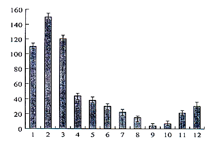
Figure 1: The dependence of changes in the Specific activity of neurolectin pH3 (40) in the cerebral cortex on environmental temperature during a 1-year exposure. Abscissa axis: months: 1, January; 2, December; 3, November; 4, October; 5, September; 6, August; 7, July; 8, June; 9, May; 10, April; 11, March; 12, February. Ordinate axis: specific activity of neurolectin.
The fact discovered by an American immunologist Pramod Srivastava has arisen a special interest to heat- shock proteins [17]. He reported that one of the fractions of protein revealed an anti-carcinogenic ability after the processing of mice suffering from malignant tumor with the extracts of separate fractions. This protein appeared to be heat-shock proteins, which are considered as the vaccines, by means of which in vivo the stabilization and transport of polypeptide native structures take place in the form of non-covalent ensembles. As it turned out later, the complexes of heat-shock proteins and peptides impact as the antigens and because of this their recognition and a complete destruction with antibodies become easy.
After the discovery of such a fact since 1994 Pramod Srivastava began the collaboration with the biotechnological companies in the city of Lextone for the creation of immunotherapeutic individual oncophages for treatment of malignant tumors. The first anti- carcinogenic vaccine was produced and tested on the example of brain glioblastoma in Russia in 2008. At the same time the action of the above-said preparation was studied at 40 carcinogenic centers of the USA. Based on the results obtained on the tumor vaccines, Pramod Srivastava has reported that in the nearest future the development of tumor will be suspended and the full cure of the patients with oncophages take place. For today the “individual vaccines” for the treatment of various tumors (fibrosarcoma, hepatoma, melanoma, lung carcinoma, lymphoma, prostate cancer etc.) are already produced. The specification of technologies of vaccines production and the results of their large-scale clinical testing in carcinogenic immunology will probably be known in the nearest future.
References
-
Aleksidze N (2014) The Basics of Psychological Biology. Tbilisi.
-
Lindquist S (1986) The heat-shock response. Annu Rev Biochem 55**:** 1151-1191.
-
Lindquist S, Craig EA (1988) The heat-shock proteins. Annu Rev Genet 22**:** 631-677.
-
Boston RS, Viitanen PV, Vierling E (1996) Molecular chaperones and protein folding in plants. Plant Mol Biol 32(1-2): 191-222.
-
Bukau B, Horwich AL (1998) The Hsp70 and Hsp60 chaperone machines. Cell 92(3): 351-366.
-
Frydman J (2001) Folding of newly translated proteins in vivo: the role of molecular chaperones. Annu Rev Biochem 70**:** 603-647.
-
Hong SW, Vierling E (2000) Mutants of Arabidopsis thaliana defective in the acquisition of tolerance to high temperature stress. Proc Natl Acad Sci USA 97(8)**:** 4392-4397.
-
Hong SW, Ung L, Vierling E (2003) Arabidopsis hot mutants define multiple functions required for acclimation to high temperatures. Plant Physiol 132(2): 757-767.
-
Morimoto RI (1998) The regulation of the heat shock transcriptional response: cross talk between a family of heat shock factors, molecular chaperones, and negative regulators. Genes Dev 12(24): 3788-3796.
-
Waters ER, Lee GJ, Vierling E (1996) Evolution, structure and function of the small heat shock proteins in in plants. Evolution, structure and function of the small heat-shock proteins in plants. J Exp Bot 47(296): 325-338.
-
Easton DP, Kaneko Y, Subjeck JR (2000) The Hsp110 and Grp170 stress proteins: newly recognized relatives of the Hsp70s. Cell Stress Chaperones 5(4): 276-290.
-
Ritossa F (1962) A new puffing pattern induced by temperature shock and DNP in Drosophila. Experientia 18(12)**:** 571-573.
-
Kulaeva NO, Mikulovich TP, Khokhlova VA (1991) Stress proteins of plants. Sovr Probl Biokhimii, Moskva, Nauka, pp: 173-190.
-
Baraboi VА, Brekman VG, Golotin IV, Kudriashov B (2010) Peroksidacia i stress. Ecofiziologia stressa. Mariiski Gos Universitet.
-
Lee-Yoon D, Easton D, Murawski M, Burd R, Subjeck JR (1995) The identification of a major subfamily of large Hsp70-like proteins through the cloning of the mammalian 110-kDa heat shock protein. J Bio Chem 270(26)**:** 15725-15733.
-
Aleksidze NG (2012) The involvement of lectin pH3 (40) in the thermoregulation of the rat brain. Neurochemical Journal 6(4): 265-267.
-
Pramod Srivastava. heroes/hc-hometown-hero- pramod-srivatava-20160122-story.html.
- Occupational Stress and Mental Health Outcomes Among Police Officers: A Mini Review
- The Experience of Counterproductive Leadership on Mental Health and Impact on Retention in U.S. Marines: A Phenomenological Study
- Nomophobia in the Digital Age: A Study on College and University Students
- Emotional Regulation in Children with Autism and Learning Disabilities
- Antisemitism on American College Campuses and Its Impact on Jewish Students
- Exploring the Role of Empathy in the Associations of Family Functioning and Purpose in Life with Attitude towards Abortion among Undergraduates: A Moderation Analysis