Physiotherapeutic Intervention with Biofotododynamic Therapy for the Reduction of Pseudomonas Aeruginosa
Pressure ulcers are injuries that can cause bacterial proliferation, and when infected are associated with increased morbidity and mortality. Thus, photodynamic therapy becomes an adjunct to the treatment with medications because it has the bactericidal capacity. In this way, this study aims to analyze in vitro the bactericidal effect of methylene blue by associating it with the low-level laser in the wavelength of830nm to determine which therapeutic doses present better efficacy in the treatment of bacteria pseudomonas aeruginosa. The microbiological material was collected by an aseptic swab and the antimicrobial activity was verified by disc diffusion method using sterile filter paper. Methylene blue was used at concentrations of 0,001% and 0,005% and the low-level laser of 830nm at the intensities of 4J/cm², 8J/cm², 10J/cm² and14J/cm², being tested in three plates for each dose, as well as the low-level laser and the methylene blue, were tested separately. The results were statistically analyzed by SPSS through the "t-test for dependent samples". It was verified that there was reduction of the bacterial activity with the application of the low-level laser in doses of 10J/cm² (p=0,00) and 14J/cm² (p=0,03) added to them ethylene blue in 0.005% concentration, however, there was a greater reduction of bacterial proliferation in the application of 10J/cm². Thus, it was concluded that the photodynamic therapy applied in the concentration of 0.005% of the methylene blue combined with the low-level laser of 830nm of 10J/cm² and 14J/cm² of intensity, present a reduction in the amount of bacterial proliferation.
Introduction
The pressure ulcer is considered a public health problem, defined as an injury caused by friction or shearing on the cutaneous tissue, due to the state of immobilization of an individual [1, 2]. This disease is easily contaminated by bacteria, resulting in a significant loss of functional capacity and quality of life of the individuals [3]. Once the bacteria multiply in the pressure ulcer, new clinical signs and symptoms are observed, worsening the clinical prognosis of the individual and developing greater wound degradation. This fact makes the treatment of infected pressure ulcers difficult, and in several cases, the bacteria are resistant to the effects of antibiotics, so it is necessary to add supporting treatments with different health professionals so that it is possible to fight infection and minimize this disease [4, 5]. In physiotherapy, there are photodynamic therapies that are showing significant results in reducing microbial activity [6]. This therapy uses the low-level laser associated with photosensitizers that when stimulated by light react chemically producing reactive oxygen and free radicals that have the potential to destroy bacteria by modifying their cell wall [7, 8]. When applied in an infected area, such as a pressure ulcer, this therapy destroys the infected cells, conserving the ones without alterations present in the wound, because the microorganisms perform more metabolic work being more likely to receive the chemical substrate produced by the photodynamic agents inducing bacterial death [9, 10]. With the scientific advance, the clinical effectiveness of this technique became better known and in this way, photodynamic therapy becomes promising for the treatment of infected pressure ulcers [11]. However, the current literature is still very restricted on the use of different wavelengths and doses of low-level laser in the treatment of this disease, allowing clinical errors during the therapeutic decision of the physiotherapist, on which resource and dosage to choose. Thus, this study aims to analyze the bactericidal effect of the methylene blue associated with the low-level laser with the wavelength of 830nm in different intensities so that it is possible to identify the best doses to combat the microbial agent pseudomonas aeruginosa, isolated from a pressure ulcer. As a hypothesis, this technique is expected to reduce the bacterial proliferation of the microorganism pseudomonas aeruginosa.
Material and Methods
Study Design
This self-paired experimental cross-sectional study was developed at the Microbiology Laboratory of UNIFAFIBE University Center-Bebedouro, SP, Brazil, from the collection of biological material carried out in an institutionalized patient at the “Lar do Idoso” Elderly Center in Bebedouro, SP. It was approved by the Research Ethics Committee (CAAE number:
67741017.4.0000.5387) of the University Center UNIFAFIBE - Bebedouro, SP.
Collection of Biological Material and Microbiological Analysis
The pressure ulcer was previously cleaned and the biological material was collected aseptically using an alginate and sterilized swab. The material was inoculated into BHI (Brain Heart Infusion) broth and was transported and immediately incubated at 35 ± 2°C for 24 or 48 horas in the laboratory of Microbiology of the University Center UNIFAFIBE - Bebedouro, SP [12, 13]. Materials that showed turbidity after incubation for 24 or 48 hours were seeded and stored in MacConkey and Mannitol Agar. The developed colonies were submitted to morphological and tannic analyzes through Rugai Tubes with Lysine and Gram staining, in which it was possible to identify the pseudomonas aeruginosa as the bacterium isolated from the pressure ulcer [14, 15].
Preparation of Methylene Blue at Concentrations of 0.001% and 0.005%
The methylene blue photo sensitizer prepared with spatula and beaker was used in the study to carry out the weighing in a Bel micro-processed digital analytical balance with automatic calibration - model M214AiH.The already weighed material was dissolved in 1000ml of distilled water using volumetric flask and funnel. After this step, the produced material was stored and identified according to its concentration in a non-transparent plastic tube which was kept covered to prevent contact of the light until its use.
Determination of Bactericidal Activity and Low- Level Laser Application
Three petri dishes were used for each clinical application condition, all of which received the diffusion disc method, which consisted of using 6mm diameter Cecon® sterile filter paper disks to determine the bactericidal activity in methylene blue and low-level laser applications [16]. Bacteria were pre-seeded using the aseptic swab on the surface of Mueller Hinton Agar and then the disks moistened with 20μL of methylene blue at the concentrations of 0,001% and 0,005% with the aid of a sterile forceps. After this step, the plates were kept in the oven and incubated at 37°C/24 h. Each plate, measuring 60mm x 15mm, received two sterile disks with the pre- established concentration. The tests were performed in triplicate and the results expressed in mm by the diameter arithmetic average of the growth inhibition halos formed around the discs. The presence of growth inhibition halos ≥ 8 mm in diameter were considered active [17, 18]. In this study, the plaques were divided into groups, being identified as: plates that received methylene blue in the different concentrations, being 0,001% and 0,005% associated with low-level laser, plates that received only methylene blue, plates that received only the laser of low- level in the wavelength of 830nm at different intensities and three control boards for each type of clinical condition tested. The low intensity laser was placed in the center of the petri plates with a support, made by the authors of the research, in the intensities of 4J/cm2, 8J/cm2, 10J/cm2 and 14J/cm2. The device used was the Laser pulse model of Ibramed (Table 1).
| Physical | Galium-aluminum-arsenide | ||||
|---|---|---|---|---|---|
| Characteristics | (GaAlAs) | ||||
| Output power (mW) | 30Mw | ||||
| Wave-length (nm) | 830nm | ||||
| Energy (j/cm²) | 4J/cm², 8J/cm², 10J/cm² and 14J/cm² | ||||
| Time per point (s) | 16s, 32s, 40s and 56s | ||||
| Contact area (cm²) | 0,11cm² | ||||
| Power Density (W/cm²) | 272W/cm² |
Table 1: Physical characteristics of low-level laser in the wavelength of 830nm.
Count of Colonies
To determine the reduction or proliferation of bacterial colonies in this study, a digital colony counter was used to aid in the counting of all petri dishes that occurred visually from small demarcations indicating that they had already been counted [19, 20]. Subsequently a second evaluator, who did not have contact with the first one re- counted and then was compared to ascertain the accuracy of the samples.
Statistical Analysis
The results obtained from counting of the colonies were tabulated in the SPSS program version 22.0 and analyzed statistically from the “t-test for paired sample” (p≤0,05) to compare the bacterial proliferation between the control plates, the plates that received the application of the low-level laser with methylene blue, the ones that received only the laser application and the ones with the application of methylene blue only.
Results
In the application of low-level laser inthe wavelength of830nm was observed that there was a reduction of colonies for all clinical conditions tested. The results were statistically significant for the clinical conditions with 0,005% methylene blue associated with the low-level laser at the intensities of 10J/cm² and 14J/cm² (Table2).
Pairing P ValueAverage Standard
Error AM0,001% and 4J/cm² -
Control 0,06 ns -48,66 ±12,78
AM0,001% and 8J/cm² -
Control 0,31 ns -31,33 ±23,83
AM0,001% and 10J/cm² -
Control 0,51 ns -21,00 ±27,00
AM0,001% and 14J/cm² -
Control 0,21 ns -26,00 ±14,57
AM0,005% and 4J/cm² -
Control 0,19 ns -14,33 ±7,44
AM0,005% and 8J/cm² -
Control 0,22 ns -9,33 ±5,36
AM0,005% and 10J/cm² -
$$ 5 \% \text {and} 1 0 \mathrm {J} / \mathrm {c m} ^ {2} - \mathrm {Control} \quad 0, 0 0 ^ {* *} \quad - 4 2, 6 6 \quad \pm 3, 3 3 $$ AM0,005% and 14J/cm² - $$ 5 \% \text {and} 1 4 \mathrm {J} / \mathrm {c m} ^ {2} - \mathrm {Control} \quad 0, 0 3 ^ {*} \quad - 3 3, 0 0 \quad \pm 6, 5 0 $$
4J/cm² - Control
0,32 ns -22,00
±17,00
8J/cm² - Control
0,59 ns -19,00
±30,23
10J/cm² - Control
0,44 ns -23,33
±24,63
14J/cm² - Control
$$ \begin{array}{l} 0, 7 5 \mathrm {n s} - 8, 6 6 \pm 2 4, 6 0 \\ 0. 7 0 - 4. 0 0 \pm 4. 0 4 \\ \end{array} $$
AM0,001% - Control
$$ 0, 7 3 \mathrm {n s} - 4, 0 0 \pm 1 0, 1 1 $$
AM0,005% - Control
0,24 ns -24,33
±15,02 ns – non-significant values (p≤0,05); * - significant values (p≤0,05); ** - significant values (p≤0,01) Table 2: Values of the difference of colony numbers between the paired samples of each application performed with the 830nm pen (paired t-test, p≤0,05).
Discussion
Pressure ulcers represent a decline in the physical and physiological status of the individual's integument tissue, leading to a worsening of his clinical prognosis by increasing hospitalization time. When infected, they increase the incidence of clinical complications and impair tissue and functional recovery, leading to death due to septicemia. Thus, it is important to immediately determine the microorganism present in the pressure ulcer and the initiation of specific treatment to resolve the infection and assist in the healing of this disease [21, 22].
In this study, photodynamic therapy was applied to reduce the bacterial proliferation of the microbial agent pseudomonas aeruginosa, as this bacterium is easily found in pressure ulcers and is resistant to several antibiotics, making the treatment of pressure ulcers complex for health professionals [23]. According to Aspiroz, et al. This bacterium becomes resistant to antibiotics because it is able to develop a biofilm formed of bacterial communities covered by an extracellular matrix of polysaccharides that resist the drug action [24]. Therefore, we believe that the search for supporting therapies that may be able to increase the bacterial reduction in the pressure ulcer is fundamental to improve the clinical prognosis, reducing the time of tissue recovery and softening the suffering of the individual. When comparing the control and the proliferated colonies plates after application of the low-level laser in the wavelength of 830nm it was observed that there was a significant reduction in the number of proliferated colonies in the clinical conditions with 0,005% methylene blue associated to the low-level laser at 10J/cm² and 14J/cm² for the pseudomonas aeruginosa. Our findings do not corroborate with the in vitro study conducted by Street et al. That used the same bacterium and observed a reduction in the number of colonies proliferated with low-level laser in the wavelength of 670nm, with intensities of 1,7J/cm², 5,2J/cm², 10,3J/cm², 15,5J/cm² e 20,6 J/cm² associated with methylene blue in the concentration 0,01% [25]. Our results are justified according to Carvalho, et al. Which indicate that the greater absorption of light emitted by the laser of low- level in the blue of methylene occurs in wavelengths larger than 600nm [26]. Thus, we believe that our results occurred because the exposure of a photo sensitizer to the light emitted by a low-level laser causes chemical reactions capable of causing the destruction of proteins, lipids, nucleic acids and other cellular components, resulting in the destruction of microbial cells [27]. Another important fact in our study was the observation of the reduction in the number of proliferated colonies in all the clinical conditions tested with both low- level lasers associated with methylene blue and isolated. These findings corroborate with Nussbaum, Lilge, Mazzulli which observed that the low-level laser in the wavelength of 810nm at 1J/cm², 2J/cm², 5J/cm², 10J/cm², 20J/cm², 30J/cm², 40J/cm², 50J/cm², 60J/cm², 70J/cm² and 80 J/cm² presented the decrease in the amount of bacterial proliferation of the microorganism pseudomonas aeruginosa. Asnaashari, et al [28]. Also showed results that agree with our study, since they verified that the methylene blue in the concentration 0,01% associated to the application of low-level laser in the wavelengths of 810nm generate a reduction in the bacterial proliferation in the intensity of 0.2J [29]. Thus, we believe that these results occurred because the wavelength of 830nm is capable of stimulating the production of reactive oxygen that is responsible for the destruction of bacterial cells, justifying the reduction of colonies in applications with low-laser intensity associated or not with methylene blue [30]. It was also observed in this study that the methylene blue at the concentrations of 0,001% and 0,005% applied in the bacteria without being associated with the low- level laser showed a reduction in the number of proliferated colonies. We believe that this fact occurred because this chemical compound acted as a toxic agent to the bacterium pseudomonas aeruginosa interfering in its natural physiological reactions. If this hypothesis is correct, we observed that the low-level laser acted as a chemical enhancement of the reaction between the methylene blue and the bacterium, increasing its elimination [31].
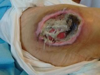
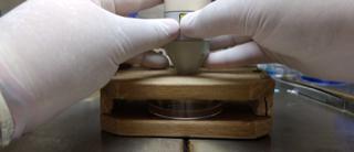
Figure 2: Applied low-level laser in the wavelength of 830nm at different intensities. This study demonstrated that photodynamic therapy is able to reduce the proliferation of pseudomonas aeruginosa. This is a great clinical relevance fact since health professionals will be able to count on this therapy associated to the treatment with medicines to favor the recovery of the individuals with an infected pressure ulcer, reducing the costs and the time of treatment, improving the clinical prognosis and positively impacting their quality of life. In this way, it was concluded that the application of the low-level laser in the wavelength of 830nm at intensities of 10J/cm² and 14J/cm², associated to the methylene blue in the concentration of 0,005% present a reduction of the microbial activity of the bacterium pseudomonas Aeruginosa.
Acknowledgement
We thank the Department of Physical therapy and Study Center and Research on Regional Development (CEPed) of University Center UNIFAFIBE – SP.
References
-
González-Méndez MI, Lima-Serrano M, Martín- Castaño C, Alonso-Araujo I, Lima-Rodríguez JS (2017) Incidence and risk factors associated with the development of pressure ulcers in an intensive care unit. J Clin Nurs 1-10.
-
Auiwattanakul1 S, Ungpinitpong W, Yutthakasemsunt S, Buranapin S, Chittawatanarat K (2017) Prevalence of Pressure Ulcer and Nutritional Factors Affecting Wound Closure Success in Thailand. Mater Sociomed 29(3): 196-200.
-
Dana AN, Bauman WA (2015) Bacteriology of pressure ulcers in individuals with spinal cord injury: What we know and what we should know. The Journal of Spinal Cord Medicine 38(2): 147-160.
-
Aftab S, Tarik M, Siddique A, Yusuf A (2014) Clinical and Microbiological Aspect of Wound Infection: A Review Update. Bangladesh Journal of Infectious Diseases 1(2): 32-37.
-
Braga IA, Brito CS, Filho AD, Filho PP, Ribas RM (2017) Pressure ulcer as a reservoir of multiresistant Gram-negative bacilli: risk factors for colonization and development of bacteremia. Braz j infect dis 21(2): 171-175.
-
Stojmenski1 S, Merdzanovski1 I, Gavrilovski1 A, Pejkova S, Dzokic G, et al. (2017) Treatment of Decubitis Ulcer Stage IV in the Patient with Polytrauma and Vertical Share Pelvic Fracture, Diagnosed Entherocollitis and Deep Wound Infection with Clostridium Difficile with Combined Negative Pressure Wound Therapy (NPWT) and Faecal Management System: Case Report. Open Access Maced J Med Sci 5(3): 349-351.
-
Pérez-Laguna V, Pérez-Artiga L, Lampaya-Pérez V, Garcia-Luque I, Ballesta, S, et al (2017) Bactericidal Effect of Photodynamic Therapy, Alone or in Combination with Mupirocin or Linezolid, on Staphylococcus aureus. Frontiers in Microbiology 8: 1002.
-
Shafirstein G, Bellnier D, Oakley E, Hamilton S, Postasek M, et al. (2017) Interstitial Photodynamic Therapy-A Focused Review. Cancers 9(2): E12.
-
Livesley NJ, Chow AW (2002) Infected Pressure Ulcers in Elderly Individuals. Clin Inf Diaseases 35(11): 1390-1396.
-
Lin X, Yan SZ, Qi SS, Xu Q, Han SS, et al. (2017) Transferrin-Modified Nanoparticles for Photodynamic Therapy Enhance the Antitumor Efficacy of Hypocrellin A. Front Pharmacol 8: 815.
-
Morimoto K, Ozawa T, Awazu K, Ito N, Honda N, et al. (2014) Photodynamic Therapy Using Systemic Administration of 5-Aminolevulinic Acid and a 410nm Wavelength Light-Emitting Diode for Methicillin- Resistant Staphylococcus aureus-Infected Ulcers in Mice. PLoS ONE 9(8): e105173.
-
Asahi MG, Chon AT, Gallemore E, Gallemore RP (2017) Photodynamic therapy combined with antivascular endothelial growth factor treatment for recalcitrant chronic central serous chorioretinopathy. Clin Ophthalmol 11: 2051-2056.
-
Gardner SE, Frantz RA, Saltzman CL, Hillis SL, Park H, et al. (2006) Diagnostic validity of three swab techniques for identifying chronic wound infection. Wound Repair Regen 14(5): 548-557.
-
Huang L, Dai T, Hamblin MR (2010) Antimicrobial Photodynamic Inactivation and Photodynamic Therapy for Infections. Methods Mol Biol 635: 155- 173.
-
Pereira PR, Paula JB, Cielinski J, Polinetto M, Bahten LCV (2014) Effects of low intensity laser in vitro bacterial culture and in vivo infected wounds. Rev Col Bras Cir 41(1): 49-55.
-
Pereira HC, Gomes DO, Hirano LQL, Santos ALQ, Lima AMC (2017) Oral microbiota in healthy Bothropsatrox (Serpentes: Viperidae) and in snakes with stomatitis. Acta Veterinaria Brasilica 11(3): 180- 183.
-
CLSI (2012) Performance Standards for Antimicrobial Disk Susceptibility Tests; Approved Standard- Eleventh Edition. Clinical and Laboratory Standards Institute 32(1).
-
Wong-Leung YL (1988) Antibacterial activities of some Hong Kong plants used in Chinese medicine. Fitoterapia 69(1): 11-16.
-
Naovi SA, Khan MS, Vohora SB, Naqvi S (1991) Antibacterial, anti-fungal and anthelmintic investigations on Indian medicinal plants. Rev Fitoterapia 62: 221-228.
-
Sundaram M, Nayak UA, Ramalingam K, Reddy V, Rao AP, et al. (2013) A comparative evaluation of Oratest with the microbiological method of assessing caries activity in children. J Pharm Bioallied Sci 5(1): S5-9.
-
Yadav M, Kaushik M, Roshni R, Reddy P, Mehra N, et al. (2017) Effect of Green Coffee Bean Extract on Streptococcus mutans Count: A Randomised Control Trial. J Clin Diagn Res 11(5): ZC68-ZC71.
-
Carvalho Pde T, Marques AP, Reis FA, Belchior AC, Silva IS, et al. (2006) Photodynamic inactivation of in vitro bacterial cultures from pressure ulcers. Acta Cir Bras 21(4): 32-35.
-
Rai MK, Deshmukh SD, Ingle AP, Gade AK (2012) Silver nanoparticles: the powerful nanoweapon against multidrug-resistant bacteria. Journal of Applied Microbiology 112(5): 841-852.
-
Aspiroz C, Sevil M, Toyas C, Gilaberted Y (2017) Photodynamic Therapy with Methylene Blue for Skin Ulcers Infected With Pseudomonas aeruginosa and Fusarium spp. Actas Dermosifiliogr 108(6): 45-48. Djavid GE, et al. (2014) Effects of Low-Level Laser Irradiation on the Pathogenicity of Candida albicans: In Vitro and in Vivo Study. Photomedicine and Laser Surgery 32(6): 322-329.
-
Street CN, Gibbs A, Pedigo L, Andersen D, Loebel NG (2009) In Vitro Photodynamic Eradication of Pseudomonas aeruginosa in Planktonic and Biofilm Culture. Photochemistry and Photobiology 85(1): 137-143.
-
Carvalho ES, Mello I, Albergaria SJ, Habitante SM, Lage-Marques JL, et al. (2011) Effect of Chemical Substances in Removing Methylene Blue After Photodynamic Therapy in Root Canal Treatment. Photomedicine and Laser Surgery 29(8): 559-563.
-
Souza JNL, Queiroga BH, Kocerginsky PO, Marinho PHC, Araki AT (2015) Photoinactivation of Candida albicans using methylene blue as photo sensitizer. Rev Gaúch Odontol 63(4): 411-417.
-
Nussbaum EL, Lilge L, Mazzulli T (2003) Effects of Low-Level Laser Therapy (LLLT) of 810 nm upon in vivo Growth of Bacteria: Relevance of Irradiance and Radiant Exposure. Journal of Clinical Laser Medicine & Surgery 21(5): 283-290.
-
Asnaashari M, Godiny M, Azari-Marhabi S, Tabatabaei FS, Barati M (2016) Comparison of the Antibacterial Effect of 810 nm Diode Laser and Photodynamic Therapy in Reducing the Microbial Flora of Root Canal in Endodontic Retreatment in Patients With Periradicular Lesions. J Lasers Med Sci 7(2): 99-104.
-
Miclescu A, Wiklund L (2010) Methylene blue, an old drug with new indications? Jurnalul Român de Anestezie Terapie intensive 17(1): 35-41.
-
Seyedmousavi S, Hashemi SJ, Rezaie S, Fateh M,
- hMPV: Is It Another Covid-19 Like Situation?
- Streptomyces: Sources of Novel Discoveries in Antibiotic Research to Combat Antimicrobial Resistance
- A Review of Mosquitoes (Diptera: Culicidae) and Their Biodiversity, Medical and Veterinary Importance
- Past and Current Immunotherapy in Cancer
- Hematological Cancer and Viral Infection
- The Growing Threat of Antimicrobial Resistance in India: Challenges and Solutions