The Discovery of the Mediterranean Echinorhynchus abissicola Dollfus, 1931 (Acanthocephala: Echinorhynchidae) From a New Host in the Persian Gulf
The obscure Echinorhynchus abissicola Dollfus, 1931 was briefly described from the deep-sea fish Pachycara obesa Zugmayer (Zoarcidae), a synonym of Pachycara bulbiceps (Garman) in the Mediterranean. Other observers rarely copied the description and 3 figures of a proboscis and 2 hooks but no taxonomic information was provided since. We have identified the same species from the leather jacket filefish Aluterus monoceros (Linn.) (Monacanthidae) in the Persian Gulf off the coast of Iran. Leather jackets are usually found worldwide in subtropical oceans at depths of 50 m. The lessepsian distribution of fish and their parasites across the Suez Canal in the Mediterranean and the Red Sea/Persian Gulf may account for the finding of E. abissicola in A. monocerus in the Persian Gulf. We made a few new taxonomic observations not previously reported in the description of 1931.
Omar M Amin1*, Milad Badri2 and Aida Vafae Eslahi2
Keywords: Echinorhynchus abissicola; Alcohol; Hydrochloric Carmine
Introduction
Dollfus [1] described Echinorhynchus abissicola from two female specimens measuring 40-50 mm in length received from the Munich Museum marked “Aus Darm des Pachycara obesa (a synonym of Pachycara bulbiceps (Garman) said Zugmayer. 4,785 m. Tief. A, v. Monaco." These parasites were not found or fixed at the death of the host, but discovered several years later during dissection. These specimens, not surprisingly were in a poor state of preservation. The specimens were whole mounted in preparations toto which flattened them before they were returned back to alcohol. Dollfus was able to color the specimens using hydrochloric carmine and was able to recognize the receptacle, proboscis, and lemnisci but was unable to see the nerve ganglion and the retinacula and the genital tract. He found many eggs but could not distinguish the envelopes. He assigned his specimens to the genus Echinorhynchus.
The description by Dollfus [1] was repeated verbatim by Petrochenko [2] and Golvan [3] who also copied the 3 figures of the proboscis and two hooks in the original description. Other observers have acknowledged the species by name, e.g., Amin [4, 5] and Wayland [6] but no taxonomic treatment was ever published since 1931. We have accidently come across two female specimens in a collection of one leather jacket filefish Aluterus monoceros (Linn.) (Monacanthidae) in the Persian Gulf off the coast of Iran. The senior author’s identification of the acanthocephalan prompted the search for additional material that will form the core of subsequent publications.
Materials and Methods
Collections
Originally, the intestine of one leather jacket from the Persian Gulf was found infected with 2 worms on 15 April 2023. These worms were later identified by OMA after processing and whole mounting for microscopic examination. New collections included 19 leather jackets (Fig. 1) of which 5 were found infected with many acanthocephalans (Fig. 2) but these will not be included in this presentation.
Methods for Microscopical Studies
Two female worms were punctured with a fine needle and subsequently stained in Mayer’s acid carmine, destained in 4% hydrochloric acid in 70% ethanol, dehydrated in ascending concentrations of ethanol (24 hr each), and cleared in 100% xylene then in 50% Canada balsam and 50% xylene (24 hr each). Whole worms were then mounted in Canada balsam. Measurements for diagnostic verification were made in micrometers, unless otherwise noted. Specimens are kept in the senior author's collection.
Optical Microscopy
Images created for this presentation were acquired using a Zeiss Axioskop Transmitted Nomarski DIC Phase Contrast Microscope Trinocular (Munich, Germany) and a Canon T3i EOS 600D DSLR Camera (Melville, New York).
Images from the microscope were transferred from the laptop to a USB and stored for subsequent processing on a computer. Measurements are in micrometers unless otherwise noted; the range is followed by the mean values between parentheses when appropriate.
Results
Our specimens were comparable to those incompletely described by Dollfus collected from the deep-sea fish Pachycara bulbiceps (Garman) in the Mediterranean but provided considerably more information. We describe below females from a new host, the leather jacket filefish Aluterus monoceros (Linn.) (Monacanthidae) (Fig. 1) in the Persian Gulf off the coast of Iran from which many specimens were collected (Fig. 2).
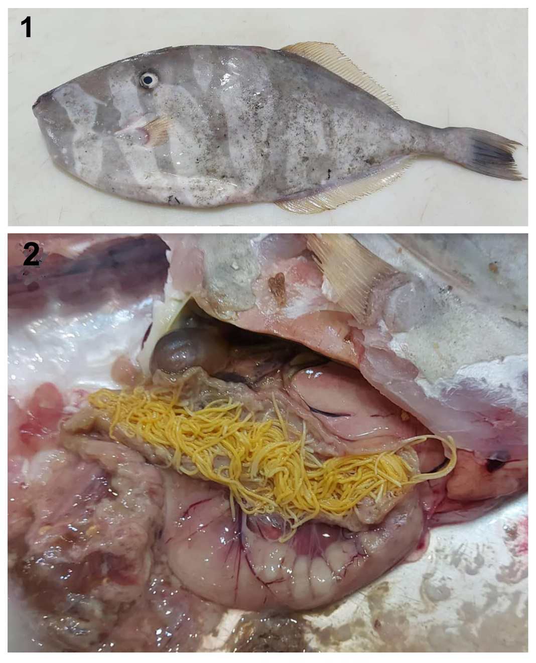
Figures 1 & 2: The host fish and the parasites. 1. An individual leather jacket filefish Aluterus monoceros from the Persian Gulf off the Iranian coast. 2. A collection of specimens of Echinorhynchus abissicola acanthocephalans from the intestine of one infected fish. Five infected fish yielded 3 petri dishes full of acanthocephalans.
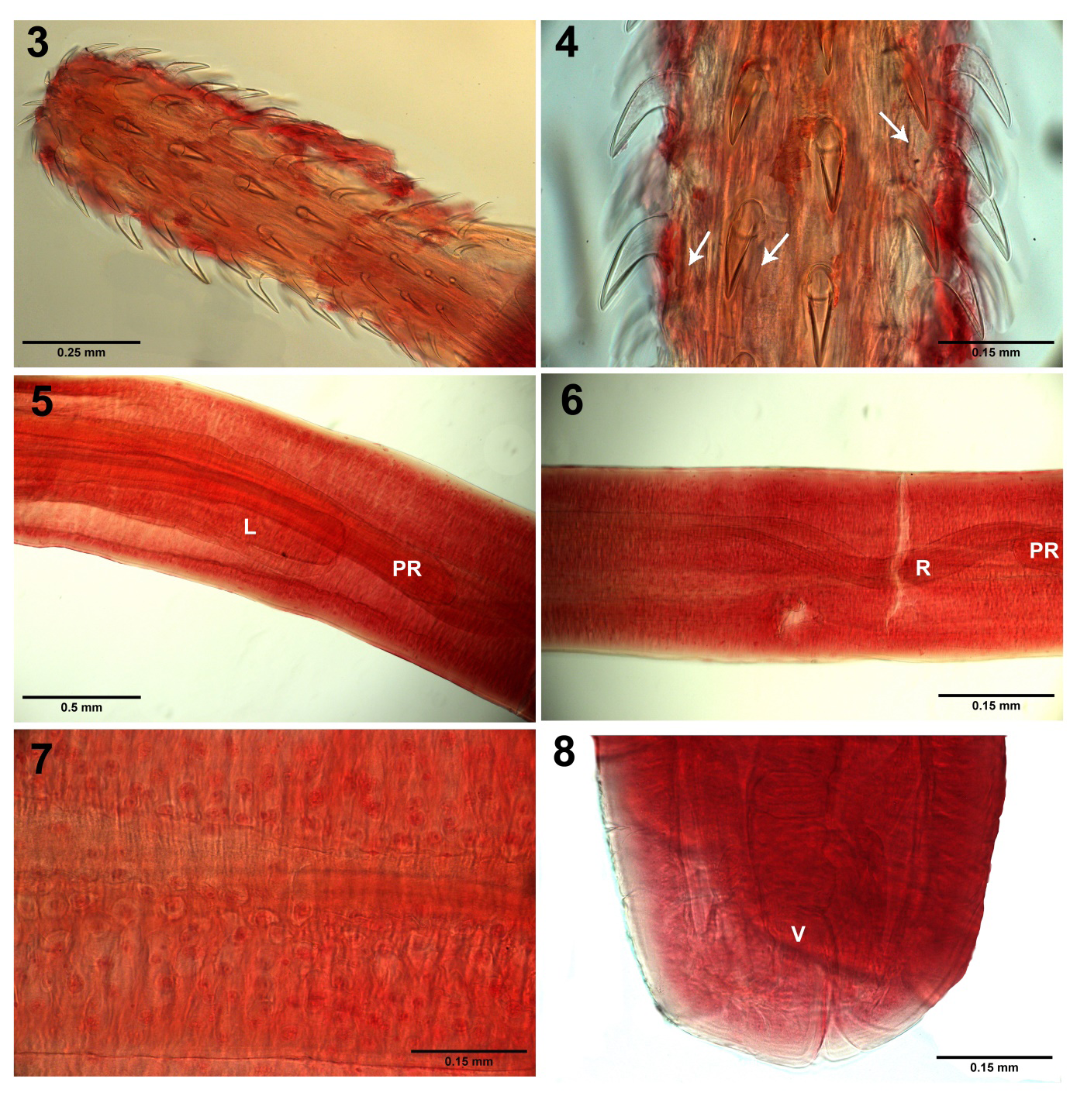
Figures 3-8: Specimens of Echinorhynchus abissicola acanthocephalans from the intestine of leather jacket filefish Aluterus monoceros from the Persian Gulf off the Iranian coast. 3. The organization of hooks on the proboscis of one specimen. 4. A higher magnification of a part of the same proboscis in Fig. 3 shows the larger ventral hooks (left) compared to the smaller dorsal hooks (right) and the truncated roots (arrows), shorter than the blades. 5. An anterior segment of the trunk showing the posterior part of the proboscis receptacle (PR) overlapping the shorter lemniscus (L). 6. A more anterior part of the trunk showing part of the proboscis receptacle (PR) and the prominent retractor muscles (R) extending through it posteriorly. 7. Part of the trunk showing the fragmented hypodermal nuclei. 8. The posterior part of a female trunk shows the terminal gonopore and vagina (V).
A brief Description of the Persian Gulf Females (Figs. 3-8)
With characters of the genus Echinorhynchus Zoega in Müller, 1776. Measurements in Table 1. Main longitudinal lacunar canals lateral. Lemniscal and hypodermal nuclei fragmented (Fig. 7). Trunk long, cylindrical with parallel sides (Fig. 5). Proboscis cylindrical joined basally with receptacle, with 12-13 rows of 10 hooks each, smallest anteriorly and basally (Fig. 3). Hooks dorso-ventrally differentiated with the ventral hooks being relatively longer and more robust than dorsal hooks (Fig. 4) and with simple shorter truncated roots directed posteriorly (Fig. 4, arrows). Proboscis receptacle more than twice the length of the proboscis, double- walled, with undecided position of the cephalic ganglion and retinacula. Lemnisci shorter than receptacle (Fig. 5). Retractor muscles emerge from receptacle and extend into body cavity posteriorly (Fig. 6). Body wall covered with fragmented hypodermal nuclei (Fig. 7). Gonopore terminal and vagina relatively short with reduced sphincter (Fig. 8).
Taxonomic summaryType host: Snubnose eelpout, Pachycara obesa Zugmayer (synonym of Pachycara bulbiceps (Garman) (Zoarcidae). Other hosts: leather jacket filefish, Aluterus monoceros (Linn.) (Monacanthidae).
| Host | Aluterus monoceros (Linn.) | Pachycara bulbiceps (Garman) |
|---|---|---|
| Locality | Persian Gulf | Mediterranean |
| Source | This paper | Dollfus, 1931 |
| Sample size | 2 | 2 |
| Trunk L X W (mm) | 11.00-24.00 X 0.50-0.75 | 45-46 X 0.8 |
| Proboscis L X W (mm) | 0.95-1.04 X 0.31-0.32 | 0.85-0.95 X 0.30 |
| Hook rows | 12-13 | 13 |
| Hooks per row | 10 | 11 (Fig. 1 with some rows of 10) |
| Hook L X W (μm) | 75-115 X 15-45 dorsally | Emerging part of hooks 70 |
| 78-120 X 20-50 ventrally | ||
| Hook roots | Truncated, shorter than hooks | Truncated, shorter than hooks |
| Receptacle L X W (mm) | 2.12-2.70 X 0.27-0.29 | 1.50-1.75 X 0.50 |
| Cephalic ganglion | Indistinct | Unrecognizable |
| Retinacula | Indistinct | Unrecognizable |
| Retractor muscles | Prominent | Not mentioned |
| Lemnisci L X W (mm) | 1.66-2.37 X 0.12-0.20 | Shorter than receptacle |
| Hypodermal nuclei | Prominent | No large (giant) nuclei |
| Gonopore | Terminal | Unrecognizable like genital tract |
Table 1: Comparative measurements of Echinorhynchus abissicola females from the Mediterranean and from the Persian Gulf.
| Hook number | Length of hook blades | Width of hook blades | ||
|---|---|---|---|---|
| Dorsal hooks | Ventral hooks | Dorsal hooks | Ventral hooks | |
| 1 | 75 | 87 | 17 | 22 |
| 2 | 92 | 100 | 20 | 25 |
| 3 | 107 | 107 | 30 | 32 |
| 4 | 112 | 115 | 37 | 45 |
| 5 | 115 | 120 | 45 | 50 |
| 6 | 112 | 117 | 32 | 35 |
| 7 | 107 | 107 | 25 | 25 |
| 8 | 97 | 102 | 17 | 20 |
| 9 | 90 | 90 | 16 | 20 |
| 10 | 78 | 78 | 15 | 20 |
Table 2: Differences in size of dorsal vs. ventral proboscis hook blades of one female Echinorhynchus abissicola from Aluterus mo
Type locality: The Mediterranean at Monaco at depth of 4,785 m. Other locality: The Persian Gulf off the Iranian coast. Site of infection: Intestine. Specimens: In first author’s collection.
Remarks
While the differences between the two sets of females from the Mediterranean and the Persian Gulf appear unremarkable (Table 1), our additions to the original description are significant enough to warrant a special note. For instance, we observed the sexual differentiation in hook sizes being more robust and longer ventrally than dorsally (Table 2, Fig. 4). Dollfus (1931) only measured the emerging parts of the hooks at 70 μm. The cephalic ganglion and the retinacula remain unrecognizable. We noted prominent elements of the retractor muscles (Fig. 6) and the hypodermal nuclei (Fig. 7). We also marked the terminal position of the gonopore and noted the short vagina (Fig. 8). The lessepsian distribution of fish and their parasites across the Suez Canal in the Mediterranean and the Red Sea/Persian Gulf may account for the finding of E. abissicola in A. monocerus in the Persian Gulf. There are at least 63 Lessepsian fish species that are known to have penetrated the Suez Canal either way since its opening in 1869 [7, 8]. If this was the case, then we would not have known if E. abissicola was originally a Mediterranean parasite that spread into the Red Sea and the Persian Gulf or was it the other way around. This Lessepsian migration has been more often documented from the Red Sea to the Mediterranean which may have been the direction of the movement of parasites [9].
Compliance with Ethical Standards
• Conflict of interest: The authors declare conflicts of interest none. • Ethical approval: The authors declare that they have observed all applicable ethical standards. • Author’s contributions: Amin did the research, identified the acanthocephalan parasite, wrote the manuscript, and created the optical microscopy images. Milad helped with fish dissection, administrative tasks, and shipping. Eslahi obtained and dissected fish and managed the parasite collection. • Acknowledgments: This project was supported by an Institutional Grant from the Parasitology Center, Inc. (PCI), Scottsdale, Arizona. Dr. Zahra Gharibi, Qazvin University, collected and dissected fish samples and was a link between Qazvin and Hormozgan universities.
References
-
Dollfus RPh (1931) Acanthocephalide d’un poisson capture par 4.785 m de profondeur. Annal Parasitol Hum Comp 9: 185-187.
-
Petrochenko VI (1956) Acanthocephala of domestic and wild animals. Moscow: Izdatel’stvo Akad Nauk SSSR, 1: 465.
-
Golvan YJ (1969) Systernatique des Acanthocephales (Acanthocephala Rudolphi, 1801). L’ordre des Palaeacanthocephala Meyer, 1931. I. La super-famille des Echinorhynchoidea (Cobbold, 1876) Golvan et Houin, 1963. Mem Mus Nat d’Hist Natur, nouvelle serie, ser. A, Zoologie 57: 1-373.
-
Amin OM (1985) Classification. In: Biology of the Acanthocephala (D.W.T. Crompton & B.B. Nickol edit.), Cambridge Univ Press, Cambridge, USA, pp: 27-72.
-
Amin OM (2013) Classification of the Acanthocephala. Folia Parasitol 60: 273-305.
-
Wayland MT, Sommerville C, Gibson DI (1999) _Echinorhynchus_ _brayi_ n. sp. (Acanthocephala: Echinorhynchidae) from _Pachycara crassiceps_ (Roule) (Zoarcidae), a deep-sea fish. Syst Parasitol pp: 43: 93- 101.
-
Golani D (1998) Impact of Red Sea fish migrants through the Suez Canal on the aquatic environment of the Eastern Mediterranean. Bull Ser Yale Sch Forest Environ Studies (103): 375-387.
-
George CJ, Athanassiou V (1967) A two year study of the fishes appearing in the seine fishery of St. George Bay, Lebanon. Ann Mus Civic Storia Natur Giacomo Doria.
-
Maillard C, Raibaut A (2012) Human activities and modifications of ichtyofauna of the Mediterranean Sea: effects on parasitosis. In: di Castri F, Hansen AJ, Debussche M. (Eds.), Biol Invasions Europe Mediterranean Basin. 65 of Monograph Biol Springer Science & Business Media, pp: 300.
- Mitochondrial Bio-Logistics: Steering Co-Enzyme Q10 and Lycopene Synergies within the Science 4.0 Bio-OS Framework
- Hymenoptera Specimens from the Caño Negro Wetland, of the National Museum Collection, Costa Rica
- Science 4.0: Comprehensive Architecture of the Biological Operating System (Bio-OS) A Framework for Systemic Resilience and Industrialized Bio-Governance
- Rabbit on, or Hare Back? Understanding Climate Change
- Clinical Validation of Science 4.0: Flow Steering and Epigenetic Drift Inversion on a 76-Year-Old Hybrid System
- Seeds Planted by another Mind