Description of Mitochondrial Gene Mutations in Pakistani Patients with Coronary Artery Disease: An Investigation of Genetic Susceptibility
Background: Mutations in mitochondrial DNA (mtDNA) were the important causes of cardiovascular diseases. However, little was known regarding the role of mitochondrial DNA (mt-DNA) mutations in coronary artery disease (CAD). To investigated the association between mitochondrial genes mutations and CAD in Pakistani population. Methods: Approximately, 11 related CAD patients and 29 control subjects were recruited in this study; we performed PCRRFLP to amplify mitochondrial genes and subsequently sequenced the PCR products. In addition, the pathogenicity scoring system was used to evaluate the deleterious roles of these genes mutations. We also used real-time PCR method to determine the mtDNA content in CAD patients carrying these mutations. Results: Three mutations were identified by PCR-Sanger sequencing; these mutations included mt-ND4 12133C>T, mt-ND5 12372G>A and mt-CYB 15884G>C. These mutations were localized at the highly conserved nucleotides, may cause the failure in mt-DNA metabolism. Furthermore, CAD group showed a clear reduction in mitochondrial copy number by comparing with the controls. Conclusions: Mutations in mt-DNA genes were the important causes of CAD, our findings provided novel insight into the pathophysiology of CAD that were manifested by mitochondrial dysfunction.
Rehman A³, Khan S¹, Khan F¹, Lisan F²*, Ullah K², Akhtar A⁴, Ul Ghafoor S¹, and Uddin A¹
¹Department of Biotechnology and Genetic Engineering, Hazara University Mansehra, Pakistan ²Jiangsu Key Laboratory of Sericultural Biology and Biotechnology, School of Biotechnology, Jiangsu University of Science and Technology, China ³Phd. D program for aging, College of medicine China medical university Taichung, Taiwan ⁴Department of Zoology, GC Woman University Sialkot, Pakistan Keywords: Mt-DNA; Mutations; CAD; Copy Number; Mitochondrial Dysfunction
Introduction
Mitochondrial disorders lead to hearing impairment, heart diseases, muscle coordination losses, visual problems, muscle failing, neurological issues, growth reduction, dementia, respiratory disorders and learning disabilities [1]. Mitochondria are one of the unique cellular organelles having ability to perform cellular functions as well as produce energy in form of ATP [2]. Mitochondria is the only organelle that is control by its own and nuclear genome [3]. There are no introns present in human mitochondrial genome. Structurally, mitochondria have double stranded and circular genome that consist of 16569 base pairs [4]. Overall, mitochondria have 37 genes in which 2 genes are for rRNAs, 13 genes are for protein formation and 22 are coded for tRNAs. By nuclear genome, 1500 mitochondrial proteins are encoded approximately which contribute as the part of mitochondrial proteome [5]. The mitochondrial small genome is present in cells as many copies that is maternally inherited [6]. The mitochondrial genome is more susceptible to variation from 10% to 20% due to having limited repair acapabilities, no protective proteins and closely located with memebrane [7]. Mutations in mitochondrial genome lead to missing in metabolism of oxidative energy and other multiple disorders [8, 9]. Some mutations are associated with disorders such as cancer, neurodegenerative diseases and aging [10]. Abnormal oxidative phosphorylation leads 15% to 25% diseases due to pathogenic mutations in mitochondrial DNA [11, 12]. It has been identified that mutated tRNA molecules synthesis protein and make the mitochondrial genome become pathogenic [13, 14]. There exist specific polymorphisms belonging to mitochondrial genes ND3 and CYTB, which are believed to be associated with high-altitude adaptation in the Tajiks population in Tibet native to China [15].
Mitochondrial genes mutations cause a wide range of disorders such as cardiomyopathy and encephalopathy [16, 17]. Defects in blood circulatory system causes cardiac pathogenic conditions represented by cardiovascular diseases. Atherosclerosis, hypertension and coronary artery disease are rapidly growing pathologies [18]. Coronary artery disease is one of the most abundant cardiovascular diseases affecting 502,000 in USA and more than 1000,000 people in China annually [19, 20]. The problematic associations like environmental factors, lifestyle and gene mutations promoted the disease pathology [21, 22]. Whole genome wide study in European countries and central Asia identified numerous genetic loci that are associated with CAD [23, 24]. Coronary Artery Disease is maternally inherited common disorder having association with changes in mtDNA encoded genes. A particular mutation 15928G>A has been accounted in 80% subjects with CAD [25]. We have a systematic mutation screening project for the mitochondrial DNA of subjects from Northern Pakistani families suffering from heart diseases and epilepsy [26]. The current study illustrates the study of a joint family of patients with CAD. The patients were selected having maternally transmitted CAD from the Clinic of Cardialogist in DHQ Hospitals, KP, Pakistan. This study was designed at the screening of mitochondrial protein coding genes for NADH dehydrogenase 4, NADH dehydrogenase 5 and cytochrome b (MT-ND4, MT-ND5 and MT-CYB).
Materials and Methods
Patients and Families Enrollment
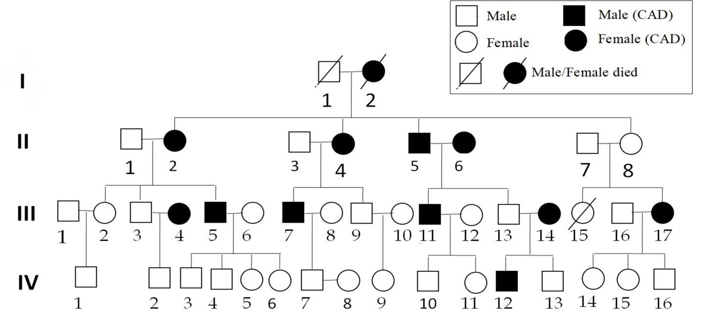
It was analytical and cross sectional study in which patients having CAD were diagnosed through ECG and X-ray and ECG via specialized doctors. The enrolled family members were informed about the aims and objectives of study. To contribute in investigation and data publication, the consent form was filled by family head member. A pedigree was constructed to capture the detaile information about cousin marriages and decease members. The details for each member of family about diseases and treatment were also recorded (Table 1 & Figure 1).
Sampling and PCR Amplification of Target DNA
Blood samples were extracted from patients and normal family members. DNA was extracted using Proteinase K and PCI based method. Nano drop and gel electrophoresis procedures were performed for the determination of quality and quantity of DNA and samples were kept at -20ºC. The following specific primers were used for mt-DNA genes amplification through PCR (Table 2). PCR reaction mixture (15μL) consisted of 1.5µL buffer, 3UTaq polymerase, 1µM dNTPs, 1mM MgCl2, 20 picomole forward and reverse primers, 7.2µL ddH2O and 20ng of DNA template. Thermocycler was adjusted at 95°C for 5 min followed by 35 cycles of denaturation at 95°C for 30s, annealing at 52°C for 1 min, extension at 72°C for 1 min. The final extension time of 7min was adjusted at 72°C.
| Subjects | Gender | Age (Years) | Observation | Diagnosis | Systolic/Diastolic mm/Hg | LVD | RVD |
|---|---|---|---|---|---|---|---|
| II-1 | M | 90 | N | N | 130/85 | 45 | 20 |
| II-2 | F | 80 | CAD | X-ray, ECG | 140/90 | 55 | 30 |
| II-3 | M | 85 | N | N | 135/80 | 40 | 18 |
| II-4 | F | 72 | CAD | X-ray, ECG | 140/90 | 52 | 26 |
| II-5 | M | 75 | CAD | X-ray, ECG | 130/85 | 50 | 29 |
| II-6 | F | 82 | CAD | X-ray, ECG | 140/90 | 53 | 28 |
| II-7 | M | 70 | N | N | 145/95 | 40 | 12 |
| II-8 | F | 78 | N | N | 130/90 | 38 | 15 |
| III-1 | M | 62 | N | N | 125/83 | 39 | 13 |
| III-2 | F | 59 | N | N | 122/81 | 37 | 16 |
| III-3 | M | 63 | N | N | 140/90 | 42 | 15 |
| III-4 | F | 58 | CAD | X-ray, ECG | 110/70 | 50 | 30 |
| III-5 | M | 60 | CAD | X-ray, ECG | 125/82 | 55 | 29 |
| III-6 | F | 56 | N | N | 120/90 | 50 | 20 |
| III-7 | M | 50 | CAD | X-ray, ECG | 150/90 | 49 | 20 |
| III-8 | F | 51 | N | N | 135/95 | 46 | 18 |
| III-9 | M | 48 | N | N | 140/85 | 43 | 20 |
| III-10 | F | 45 | N | N | 130/90 | 38 | 25 |
| III-11 | M | 35 | CAD | X-ray, ECG | 140/90 | 52 | 25 |
| III-12 | F | 30 | N | N | 135/85 | 47 | 18 |
| III-13 | M | 30 | N | N | 130/90 | 41 | 17 |
| III-14 | F | 27 | CAD | X-ray, ECG | 130/70 | 49 | 30 |
| III-15 | M | 40 | N | N | 130/85 | 45 | 12 |
| III-16 | F | 38 | CAD | X-ray, ECG | 150/95 | 50 | 26 |
| IV-1 | M | 19 | N | N | 120/80 | 35 | 18 |
| IV-2 | M | 25 | N | N | 120/85 | 37 | 20 |
| IV-3 | M | 26 | N | N | 120/90 | 45 | 13 |
| IV-4 | M | 24 | N | N | 120/80 | 39 | 12 |
| IV-5 | F | 21 | N | N | 130/85 | 41 | 15 |
| IV-6 | F | 19 | N | N | 120/90 | 47 | 14 |
| IV-7 | M | 25 | N | N | 130/80 | 46 | 10 |
| IV-8 | F | 20 | N | N | 120/85 | 40 | 18 |
| IV-9 | F | 16 | N | N | 120/80 | 39 | 20 |
| IV-10 | M | 22 | N | N | 125/80 | 37 | 25 |
| IV-11 | F | 20 | N | N | 120/80 | 47 | 24 |
| IV-12 | M | 15 | CAD | X-ray, ECG | 130/90 | 48 | 25 |
| IV-13 | M | 13 | N | N | 120/80 | 39 | 16 |
| IV-14 | F | 23 | N | N | 125/80 | 40 | 12 |
| IV-15 | F | 20 | N | N | 120/85 | 43 | 9 |
| IV-16 | M | 18 | N | N | 120/80 | 42 | 13 |
Table 1: Clinical and biochemical data for pedigrees of a Pakistani family.
| Primer sequence (5'-3') | Product | |
|---|---|---|
| mt-DNAND4MT-13F | TTTACCACAACACAATTGGG | 525bp |
| MT-13R | GCTCAGTGTCAGTTCGAGATA | |
| mt-DNACYBMT-21F | ATCGGAGGACAACCAGTAAGC | 320bp |
| MT-21R | TGATGGGTGAGTCAATACTTGG |
Table 2: Primers for mt-DNA genes PCR amplification.
Detection of Mutations by Sequence Analysis
The PCR products were analyzed by 1% agarose gel electrophoresis. Purified PCR products were commercially analyzed for nucleotide sequences from biological technology department of TSINGKE Chengdu (China)http:// foreign.macrogen.co.kr/eng/. Further alignment studies were carried out through online DNA analysis tools like NCBI Blast and Ugene. The nucleotide sequences obtained were compared with rCRS sequence.
Cross-Validation Of Deleterious Effect of Mutations by Computational Tools
PON-mt-tRNA, a multi-factorial probability-based prediction tool, was used for classification of newly observed human mt-tRNA mutations. It integrates machine learning prediction together with evidence of biochemistry, histochemistry, and segregation, to compute the posterior probability of pathogenicity. This method displayed high performance with Accuracy and Matthews Correlation Coefficient (MCC) of 1.00 and 0.99, respectively. It accepts input as the comma separated single query with mitochondrial genome location, reference nucleotide, and new nucleotide; output score ranges from 0 to 1, following increasingly deleterious pattern. Variations are classified into five classes that is, variants of uncertain significance, neutral, likely neutral, likely pathogenic, and pathogenic.
Mitochondrial tRNA Informatics Predictor (MitoTIP, Philadelphia, PA, http:// journals. plos. org/ ploscompbiol/ article? id=10.1371/ journal.pc bi.1005867), is another tool for predicting pathogenicity of novel mitochondrial tRNA variants, which was effectively employed in our analysis to have combinatorial optimization of in silico predictions. MitoTIP is based on multiple sources of information for prediction of the likelihood that novel single nucleotide variants in tRNA encoding sequences would cause disease [27, 28]. Upon query, the predictive algorithm incorporates an estimation of the importance of a position across all known mitochondrial tRNAs using data from publically available databases (like MITOMAP and GenBank); the output ranges from−5.9 to 21.8 (Supplementary data).
Secondary Structure Prediction of Mutated Genes
“RNA structure web server is a tool to predict secondary structures of mutated genes with lowest free energy and base pair probabilities. The server to predict secondary structure combines separate prediction and analyzed algorithms which is, finds structures with most predictable accuracy, expects a lowest free energy structures, calculates the function separately and pseudo knot (if any) prediction. DNA sequence takes by server and generates an extremely possible, annotated cluster of secondary structures, opening with the minimum free energy structure and including furthers with different probabilities of accuracy. Three mutant sequences (m.12133C>T, m.12372G>A and m.15884G>C) were submitted to the server for comparative structure analysis.
Prediction of Secondary and 3D Structures
For the prediction of mitochondrial genes secondary structure, the RNA fold web server was used. The generated 2D structures were used in dot-bracket format for the generation of normal and mutant sequence 3D structures using the automated RNA structure 3D Modeling server (RNA
Composer) was used for the prediction of mitochondrial genes 3D.
Results
Subjects
In present study 40 individuals were selected from the family. All the diagnosed patients had signs of hypertension and CAD in their family history.
Genes and Mutations
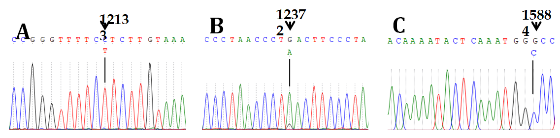
We identified three mutations in CAD patients after mtDNA genes analysis and indicate three point mutations including m.12133C>T, m.12372G>A, and m.15884G>C (Figure 2). The mutations m.12133C>T and m.12372G>A are heteroplasmic while m.15884G>C representing homoplasmic condition. Validation by in Silico Predictive Tools All the three variants were reported to be deleterious by MitoTIP server. The difference in the degree of predicted deleteriousness is due to the fact that both tools work on different algorithms/principles and consider diverse factors. However, the computational predictive tools strongly support that the mutations show different types of variation (Table 3).
| Locus | rCRS Position | Query Position | rCRS NT | Query NT | Mut Type |
|---|---|---|---|---|---|
| ND4 | 12133 | 110 | C | T | Transition |
| ND5 | 12372 | 350 | G | A | Transition |
| Cytb | 15884 | 93 | G | C | Transversion |
Table 3: List of mutations identified in CAD patients and their pathogenicity predictions by MitoTIP and PON-mt-tRNA.
Predicted RNA Secondary Structures of Mitochondrial Genes
The harmful impact of the varients nucleotide could be understood by particular RNA secondary structures. Two to three structures generated by RNA structure Webserver and one of them was selected with free minimum energy. Three mutant models that are m.12133C>T, m.12372G>A and m.15884G>C were observed (Figure 3), with disruptive confirmation that could possibly change the role of mt-DNA genes which lead to pathogenic phenotype. Discussion CAD is the most abundant type of heart diseases disturbing approximately 25% individuals and the primary death cause worldwide [29]. It leads to heart failure, myocardial infarction and rapid loss that is recorded 64% in women and 50% in men [30, 31, 32]. CAD has been linked with a combined or quantitative impact of multifactorial ecological conditions, hereditary factors or nuclear factors and psychological status [33, 34, 35, 36]. Numerous risk factors having linked with CAD include serum lipids, diabetes, blood ceramides, lifestyle and hypertension [37]. Studies on the family history and genetic has an established association with CAD that may enhance the chances of occurrence from 40% to 60%. CAD developed primarily by contribution of genetic factors [38, 39]. Mitochondrial DNA mtDNA4977 deletion, 16189T>C, 15928G>A and T16519C have been associated with CAD [40, 41, 42, 43]. Equally, several tRNA pathogenic mutations have been identified having contribution in CAD patients [44, 45, 46]. We have evaluated a family with history of CAD for mitochondrial gene mutations (Figure 1). Total 40 individuals were recruited in the study after signing the consent forms, 11 of these subjects were diagnosed for CAD (Table 1). Three genes mt-ND4, mt-ND5 and mt-CYB were PCR amplified and fragment size was confirmed for each by agarose gel electrophoresis. Purified PCR product was commercially analyzed for nucleotide sequences and mutations in the above gene sequences were detected. We found mutations m.12133C>T, m.12372G>A and m.15884G>C position in the mitochondrial genes – mt- ND4, mt-ND5 and mt-CYB respectively (Figure 2). None of these mutations was detected in the healthy subjects from this family.
Conclusions
Our findings suggest an association of coronary artery disease with mitochondrial genes mutations that are different from normally published pathogenic mutations. The effect of known mutations on RNA secondary structure that was estimated by online biological softwares. Disruptive structure of mutants confirmed through in silico studies which lead to mitochondrial dysfunction or pathogenic effect. The prediction studies by software based on computer have strongly maintained the pathogenicity level of known mutations signifying an in vivo confirmation.
Ethical Statement
This study was approved by Ethical Committee of Institution and Board of Advanced Studies and Research at Hazara University, Mansehra 21300, Pakistan according to the notification number F.No.73/Ittl/ORlClB C2016/.
Conflicts of Interest
The authors have no competing interests to declare.
References
-
Nunnari J, Suomalainen A (2012) Mitochondria: in sickness and in health. Cell 148(6): 1145-1159.
-
Margineantu DH, Hockenbery DM (2016) Mitochondrial functions in stem cells. CurrOpin Genet Develop 38: 110- 117.
-
Couvillion MT, Soto IC, Shipkovenska G, Churchman LS (2016) Synchronized mitochondrial and cytosolic translation programs. Nature 533: 499-503.
-
Taanman JW (1999) The mitochondrial genome: structure, transcription, translation and replication. BiochimBiophysActa 1410: 103-123.
-
Anderson S, Bankier AT, Barrell BG, de Bruijn MH, Coulson AR, et al. (1981) Sequence and organization of the human mitochondrial genome. Nature 290: 457465.
-
Pagliarini DJ, Calvo SE, Chang BA (2008) Mitochondrial protein compendium elucidates complex I disease biology. Cell 134: 112-123.
-
Sato M, Sato K (2013) Maternal inheritance of mitochondrial DNA by diverse mechanisms to eliminate paternal mitochondrial DNA. BiochimBiophysActa-Cell Mol Res 1833: 1979-1984.
-
Shokolenko I, Venediktova N, Bochkareva A, Wilson GL, Alexeyev MF (2009) Oxidative stress induces degradation of mitochondrial DNA. Nucleic Acids Res 37: 2539-2548.
-
Blakely EL, Yarham JW, Alston CL (2013) Pathogenic mitochondrial tRNA point mutations: nine novel mutations affirm their importance as a cause of mitochondrial disease. Hum Mutat 34: 1260-1268.
-
Young MJ, Copeland WC (2016) Human mitochondrial DNA replication machinery and disease. CurrOpin Genet Dev 38: 52-62.
-
Schon EA, DiMauro S, Hirano M (2012) Human mitochondrial DNA: roles of inherited and somatic mutations. Nat Rev Genet 13: 878-890.
-
Thorburn DR (2004) Mitochondrial disorders: prevalence, myths and advances. J InheriMetabol Dis 27: 349-362.
-
Hellebrekers DM, Wolfe R, Hendrickx AT (2012) PGD and heteroplasmic mitochondrial DNA point mutations: a systematic review estimating the chance o healthy offspring. Hum Reprod Update 18: 341-349.
-
Chinnery PF, Hudson G (2013) Mitochondrial genetics. Br Med Bull 106: 135-159.
-
Wang Y, Zhou XL, Ruan ZR, Liu RJ, Eriani G, et al. (2016) Ahuman disease-causing point mutation in mitochondrial threonyl-tRNA synthetase induces both structural and functional defects. J Biol Chem 291: 6507- 6520.
-
Liu C, Yang Q, Hwang SJ, Sun F, Johnson AD, et al. (2012) Association of genetic variation in the mitochondrial genome with blood pressure and metabolic traits. Hypertension 60: 949-956.
-
Chen Y (2020) Mitochondrial DNA genomes revealed different patterns of high-altitude adaptation in high- altitude Tajiks compared with Tibetans and Sherpas. Sci Rep 10: 10592.
-
Dominic EA, Ramezani A, Anker SD, Verma M, Mehta N, et al. (2014) Mitochondrial cytopathies and cardiovascular disease. Heart 100(8): 611-618.
-
Finsterer J (2012) Stroke and Stroke-like Episodes in Muscle Disease. Open Neurol J 6: 26-36.
-
Marian AJ (2011) Mitochondrial genetics and human systemic hypertension. Circ Res 108: 784-786.
-
Lopez A, Mathers D, Ezzati CD, Jamison M, Murray DT (2001) Global and regional burden of disease and risk factors. Systematic analysis of population health data. Lancet 367: 1747-1757.
-
Zhang X, Lu H, Liu ZL (2008) Coronary heart disease in China. Heart 94: 1126-1131.
-
Wilson PW, D’Agostino RB, Levy D, Belanger AM, Silbershatz H, et al. (1998) Prediction of coronary heart disease using risk factor categories. Circulation 97: 1837-1847.
-
Khot UN, Khot MB, Bajzer CT, Sapp SK, Ohman EM, et al. (2003) Prevalence of conventional risk factors in patients with coronary heart disease. J Am Med Assoc 290: 898-904.
-
PedenJF, Farrall M (2011) Thirty-five common variants for coronary artery disease: the fruits of much collaborative labour. Hum Mol Genet 20: R198-R205.
-
Tregouet DA, Konig IR, Erdmann J, Munteanu A, Braund PS, et al. (2009) Genome-wide haplotype association study identifies the SLC22A3-LPAL2-LPA gene cluster as a risk locus for coronary artery disease. Nat Genet 41: 283-285.
-
Schunkert H, Konig IR, Kathiresan S, Reilly MP, Assimes TL, et al. (2011) Large-scale association analysis identifies 13 new susceptibility loci for coronary artery disease. Nat Genet 43: 333-338.
-
Peden JF, Hopewell JC, Saleheen D, Chambers JC, Hager J, et al. (2011) A genome-wide association study in Europeans and South Asians identifies five new loci for coronary artery disease. Nat Genet 43: 339-344.
-
Nadeem MS, Ahmad H, Mohammed K, Muhammad K, Ullah I, et al. (2018) Identification of variants in the mitochondrial lysine‐tRNA (MT‐TK) gene in myoclonic epilepsy—pathogenicity evaluation and structural characterization by in silico approach. Journal of cellular biochemistry 119(7): 6258-6265.
-
Sonney S, Leipzig J, Lott MT, Zhang S, Procaccio V, et al. (2017) Predicting the pathogenicity of novel variants in mitochondrial tRNA with MitoTIP. PLoS computational biology 13(12): e1005867.
-
Rautou PE, Vion AC, Amabile N, Chironi G, Simon A, et al. (2011) Microparticles, vascular function, and atherothrombosis. Circulation research 109(5): 593- 606.
-
Okrainec K, Banerjee DK, Eisenberg MJ (2004) Coronary artery disease in the developing world. American heart journal 148(1): 7-15.
-
Sundaram V, Bloom C, Zakeri R, Halcox J, Cohen A, et al. (2020) Temporal trends in the incidence, treatment patterns, and outcomes of coronary artery disease and peripheral artery disease in the UK, 2006–2015. European Heart Journal 41(17): 1636-1649.
-
Nabel EG, Braunwald EA (2012) Tale of coronary artery disease and myocardial infarction. New England Journal of Medicine 366: 54-63.
-
Wang Q (2005) Molecular genetics of coronary artery disease. Curr Opin Cardiol 20: 182-188.
-
Khandaker GM, Zuber V, Rees JM, Carvalho L, Mason AM, et al. (2020) Shared mechanisms between coronary heart disease and depression: findings from a large UK general population-based cohort. Molecular psychiatry 25(7): 1477-1486.
-
Nikpay M, Mohammadzadeh S (2020) Phenome-wide screening for traits causally associated with the risk of coronary artery disease. Journal of human genetics 65(4): 371-380.
-
Ghassibe-Sabbagh M, Platt DE, Youhanna S, Abchee AB, Stewart K, et al. (2012) Genetic and environmental influences on total plasma homocysteine and its role in coronary artery disease risk. Atherosclerosis 222(1): 180-186.
-
Poss AM, Maschek JA, Cox JE, Hauner BJ, Hopkins PN, et al. (2020) Machine learning reveals serum sphingolipids as cholesterol-independent biomarkers of coronary artery disease. The Journal of clinical investigation 130(3).
-
Zdravkovic S, Wienke A, Pedersen NL, Marenberg ME, Yashin AI, et al. (2002) Heritability of death from coronary heart disease: a 36‐year follow‐up of 20 966 Swedish twins. Journal of internal medicine 252(3): 247-254.
-
Shadrina AS, Shashkova TI, Torgasheva AA, Sharapov SZ, Klarić L, et al. (2020) Prioritization of causal genes for coronary artery disease based on cumulative evidence from experimental and in silico studies. Scientific reports 10(1): 1-5.
-
Botto N, Berti S, Manfredi S, Al-Jabri A, Federici C, et al. (2005) Detection of mtDNA with 4977 bp deletion in blood cells and atherosclerotic lesions of patients with coronary artery disease. Mutation Research/ Fundamental and Molecular Mechanisms of Mutagenesis 570(1): 81-88.
-
Abu-Amero KK, Al-Boudari OM, Mousa A, Gonzalez AM, Larruga JM, et al. (2010) The mitochondrial DNA variant 16189T> C is associated with coronary artery disease and myocardial infarction in Saudi Arabs. Genetic Testing and Molecular Biomarkers 14(1): 43-47.
-
Rad RG, Saleh SK, Kouchaksaraei AS, Houshmand M, Salehi A, et al. (2016) Association of Mitochondrial T16519C polymorphism with Coronary Artery Disease (CAD) in Iranian patients underwent coronary angiography. International Journal of Medical Research & Health Sciences. 5(9): 132-145.
-
Wang X, Lu J, Zhu Y, Yang A, Yang L, et al. (2008) Mitochondrial tRNAThr 15927G>A mutation may modulate the phenotypic manifestation of ototoxic 12S rRNA A1555G mutation in four Chinese families. Pharmacogenet. Genom 18: 1059-1070.
-
Heidari MM, Mirfakhradini FS, Tayefi F, Ghorbani S, Khatami M, et al. (2020) Novel Point Mutations in Mitochondrial MT-CO2 Gene May Be Risk Factors for Coronary Artery Disease. Applied Biochemistry and Biotechnology 25: 1-4.
- Mitochondrial Bio-Logistics: Steering Co-Enzyme Q10 and Lycopene Synergies within the Science 4.0 Bio-OS Framework
- Hymenoptera Specimens from the Caño Negro Wetland, of the National Museum Collection, Costa Rica
- Science 4.0: Comprehensive Architecture of the Biological Operating System (Bio-OS) A Framework for Systemic Resilience and Industrialized Bio-Governance
- Rabbit on, or Hare Back? Understanding Climate Change
- Clinical Validation of Science 4.0: Flow Steering and Epigenetic Drift Inversion on a 76-Year-Old Hybrid System
- Seeds Planted by another Mind