Acquired Neutropenia and Fever, its Treatment and Complications in an Infant, A Case Report
An acquired neutropenia is a common condition which occurs during some viral infections and usually is a mild, self-limited disorder. This ailment frequently happens in childhood, especially during infancy, and the few reported cases in infants were not associated with any serious complications. In this case report, we present a less than 6 month previously healthy male involved with fever and neutropenia during a viral infection. 3 days after admission, he developed a lower respiratory tract infection associated with fever, tachypnea, and respiratory distress as well as a nodule appeared at a mosquito bite bump in his left forearm and oral candidiasis. On the next day, the case showed a remarkable rising in the absolute neutrophil count following application of intravenous Granulocyte Colony Forming Factor (G-CSF). Fortunately, during the third day of its emergence, the extension of the lesion has stopped and the case got afebrile. In spite of sloughing of the necrotic layer of the skin, and forming a deep sited defect at the underneath tissue, the residual cavity has healed completely without any requirement to skin engraftment, stem cell implant or tissue debridement in about 2 months of its emergence. At the healing area, a scar has formed which was not deformed; otherwise, the morphology, size, and function of his left forearm as well as its growth and development are completely normal as the same as his right forearm.
Introduction
Generally, neutropenia is the result of reducing the number of circulating neutrophils as an absolute neutrophil count (ANC) below 1500/mm3 in the peripheral blood; ANC less than 500/mm3 is defined as severe neutropenia [1]. Acquired neutropenia (AN) is a common condition in childhood frequently emerges following infections, nutritional deficiencies, consuming some drugs or autoimmune diseases [1, 2, 3, 4, 5, 6, 7]. Although viral respiratory tract infections such as Respiratory Syncytial Virus (RSV), Influenza, Para influenza, Corona and Covid 19 viruses, are the most frequent etiology of the infections resulted to AN in childhood, other viruses such as EBV, CMV, Dengue virus, Measles and HIV have been blamed for the disorder with lower incidence [5, 8, 9]. Most related articles have mentioned that post infectious AN in a previously healthy baby is a mild, short term disease with benign, self-limited period which generally recovers without any serious complications [2, 3, 5, 8, 10, 11, 12, 13, 14]. In this case report, we present a less than 6 month previously healthy male with severe leucopenia and neutropenia following a mild viral upper respiratory tract infection.
Case Presentation
A 4 month and 20 day male admitted to our ward with one day history of fever and 3 times vomiting. His symptoms associated with mild coughing and coryza. In physical examination, his vital signs included, T: 39 °C, Respiratory Rate: 33 per minute, Pulse rate: 115 per minute and blood pressure: 90 mmHg / pulse. He was conscious, and in good general condition with no clinical signs or symptoms of dehydration. His weight was 8 kilograms. There were no signs of petechiae, purpura, ecchymosed or bleeding lesions on his skin or mucosal membranes. His other physical examination findings were unremarkable either. In his past personal history, he was delivered full term by cesarean section after 17 years of his mother’s infertility at the result of an in vitro fertilization (IVF). He was under exclusive breast feeding regimen since his birth. His history of routine vaccination was complete and uneventful. Her parents were no relative. In his past medical history, he just had a mild ASD proved with echocardiography in neonatal screening without any positive history of infection or hospital admission. His Laboratory findings included: CBC with WBC: 1000/ml, WBC differential was impossible because of the low count, Hemoglobin: 9.9 g/dl, platelet count: 295,000/ ml, RBC indexes were in normal range, ESR: 92, CRP: >150, alanine amino transferase (ALT): 33 U/L (Normal range: 4- 36 U/L), aspartate aminotransferase (AST): 65 U/L (Normal range: 8 - 33 U/L). His peripheral blood smear (PBS) showed leucopenia and neutropenia, without any evidence of blast cells.
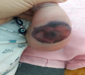
Blood culture sampling for bacteria and fungi in Bactec media were performed 3 times with negative results. His chest x ray (CXR) was normal. With considering fever and a viral infection induced leucopenia and neutrpenia, he was isolated and intra venous (IV) serum and full dose empirical parenteral antibiotics included ceftriaxone, amikacin and ceftazidim were started. Three days after admission, he developed dyspnea and tachypnea with RR: 70/min, T: 38.2°C as well as respiratory distress; coarse pulmonary sounds were heard at his lung auscultation. His O2 saturation without receiving O2 on Pulse oximetry was 97%; however, his ABG result was in normal range. His updated CXR has showed hyper aeration without any special findings. Besides, a small perforated nodule has appeared in a mosquito bite bump in his left forearm with 1cm * 0.5 cm diameter. Smear and culture of the wound were negative. Ultrasonography of the lesion has revealed diffused interstitial edema and inflammation without clear evidence of mass or collection. Moreover, a few candidiasis lesions have appeared on her tongue. Fortunately, no signs of bleeding or any other hemorrhagic lesions has detected over his skin or mucosal membranes. An updated CBC has shown WBC: 2100/ml, Neutrophil count (PMN): 24% (Absolute Neutrophil Count: 504/ml), Lymphocyte count: 70%, Monocyte count: 4%, Eosinophil count: 2%, Hemoglobin: 9.5 g/dl, platelet count: 60,000/ml, ESR: 120, CRP: >150, ALT: 33 U/L, AST: 65 U/L. Vitamin B12 and folate levels were in normal range. With regard to severe neutropenia, emergence of serious lower respiratory tract symptoms, oral candidiasis and intradermal nodule, antifungal agents such as intravenous fluconazole, Voriconazole and Liposomal Amphotericin B have been added for the patient. Moreover, because of critical condition of the patient, 40 microgram (5 microgram/kg) Intra venous (IV) Granulocyte Colony Stimulating Factor (G-CSF) has prescribed for him. His CBC result at the next day showed: WBC: 13400, PMN: 42% (Absolute Neutrophil Count: 5628/ ml), Band cell: 13%, metamyelocyte: 9%, myelocyte: 4%, promyelocyte: 1%, lymphocyte: 25%, monocyte: 5%, Eos: 1%, platelet: 30,000/ml, Hb: 9.3 g/dl. 2 days after G-CSF consuming, he became afebrile and his tachypnea as well as his respiratory distress has been recovered. The oral candidiases were healing; however, an extended ecchymotic lesion (2 cm * 6.5 cm diameter), has appeared in the overlying skin of the place of the nodule, however, there was no tenderness, erythema, ulcer or perforation (Figure 1).
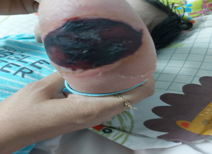
His peripheral pulses such as radial and ulnar pulses at left hand were normal. The Doppler ultrasound test of his left forearm was normal too. The PT, PTT and INR tests were in normal range. The results of Covid 19, adenovirus, parvovirus B19, parainfluenza and Influenza PCR all were negative. Besides, serologic tests for EBV and CMV antibodies included IgM and IgG as well as HIV 1 and 2 antibodies were negative too. Moreover, the results of blood, and urine as well as dermal lesion cultures all were negative. The results of serum Galactomannan antigen test, serum IgM, IgG, IgA and IgE, Nitro Blue Tetrazolium (NBT) and dihydrorhodamine tests, serum C3, C4, CH50, and myeloperoxidase tests all were in normal range. Anti granulocyte antibody was not tested because of not availability of the test in our region. On the third day of G-CSF usage, the expansion of the dermal lesion was stopped and its superficial layer became necrotic. After 7 days of its emergence, the necrotic lesion started to heal in the vicinity (Figure 2).
The peripheral pulses and the movements of the left hand were normal as before. A plastic surgery consultation has performed and the surgeon booked an operation for his skin engraftment for 3 weeks later. After 10 days of admission, he was discharged in good general condition with normal CBC included: WBC: 11700, PMN: 45% (Absolute Neutrophil Count: 5265/ml), Band cell: 3%, lymphocyte:
45%, Monocyte: 5%, Eos: 2%, platelet: 85,000/ml, Hb: 9.0 g/ dl (Table 1).
| WBC/ml | Differential ANC* | Hb(g/dl) | platelet/ml | ESR | CRP | |
|---|---|---|---|---|---|---|
| At admission | 1000 | unreliable | 9.9 | 295000 | 92 | 150 |
| 3rd day after Admission | 2100 | 504 | 9.5 | 60000 | 120 | >150 |
| Next day after G-CSF | 13400 | 5628 | 9.3 | 30000 | 120 | >150 |
| At discharge | 11700 | 5625 | 9 | 85000 | 95 | 120 |
Table 1: The trend of the case’s CBC changes during his stay at the hospital.
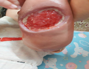
Amoxicillin and cephalexin syrups initiated for him in therapeutic doses because of his necrotic lesion. During his follow up period, a CBC has checked twice a week for 4 successive weeks for ruling out of cyclic neutropenia and all the results were normal. Because of his parents worrisome about the appearance or future probable complications of the necrotic lesion, an out-patient pediatric surgery consultation was performed after a week of his discharge and the case was admitted for removal of the necrotic part plus a wound debridement procedure. Fortunately, the day before achieving the operation (3 weeks after emerging of the ecchymotic lesion) the necrotic lichenified and shrunk superficial layer of the lesion sloughed off the underneath skin (Figure 3).
The residual tissue was a defect with 4 cm * 4.5 cm diameter as well as 4 cm depth. The base and the sides of the defect were clean without any discharge or bleeding. Therefore, the operation was cancelled and twice daily Vaseline petrolatum impregnated sterile dressing after irrigating the lesion with sterile normal saline solution was ordered. A weekly medical visiting and examining the lesion have performed regularly. In successive visits, the lesion was becoming shrunk just as the result of the case’s daily receiving of oral ferrous sulfate, and multivitamin drops as supplemental agents as well as zinc sulfate, vitamin C and folic acid all in therapeutic doses initiating after his primary discharge for better wound healing. At last, after 48 day of his discharge, the defect completely closed spontaneously with a little scar (Figure 4).
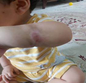
The morphology, size and function of his left forearm were totally normal as the same as his right forearm. Now, he is a 12 kilogram weight 13 month old infant in a good condition with a normal growth and development such as his peers.
Discussion
In this case report, a less than 6 month male infant presented who admitted on Jan 2023 with a probable viral infection induced neutropenia and fever. Actually, there are some interesting viewpoints in the case as follow: 1- The young age of the case. 2- Emergence of lower respiratory infection, oral thrush and a sub dermal nodule in the place of a mosquito’s bite bump a few days after initiating AN. 3- Usage of G-CSF in the case in order to increase the absolute neutrophil count. 4- Applying the broad-spectrum antibiotics and antifungal agents for the patient. 5- Appearance of a necrotic lesion in the place of the sub dermal nodule which its sloughing off resulted in a full thickness dermal defect and healed completely without any requirement to skin engraftment, stem cell implant or debridement procedure. Literature review revealed that AN in previously healthy children mostly emerged following a viral infection mainly involved respiratory tract of the patients [2, 8, 9, 11, 12]; in our case, neutropenia appeared following a probable viral upper respiratory tract infection too, however, we couldn’t find any specific etiology for the symptoms. Albeit, Human Herpes virus 6 or 7, as well as enteroviruses’ infections couldn’t be ruled out, as the Reverse Transcriptase Polymerase Chain Reaction (RT-PCR) tests for these viruses were not available in the country. It seems that viral infections more than bacterial ones result in appearance of AN [1, 2, 4, 6, 8, 9, 10, 11]. According to previous studies and as expected, AN in childhood usually has low risk of inducing severe or serious bacterial or fungal infections [4, 11, 13, 15, 16, 17, 18, 19, 20, 21, 22, 23, 24]; Moreover, all the available references which performed blood cultures for the patients, indicated that the blood cultures’ results were negative [25, 26, 27].
The result of blood, urine, and wound cultures in our patient were negative either. Although, a few reports expressed secondary infections following AN in children such as perirectal, perineal, and oral mucosa infections [4], nosocomial infections, sepsis, shock [22], and Pseudomonas induced shock [26], severe AN in our patient resulted in emerging of lower respiratory tract infection, oral candidiasis and wound infection. Furthermore, a laboratory finding some authors indicated was thrombocytopenia associated with neutropenia which is in favor of probable viral infection as the cause of AN; however, no one mentioned any bleeding manifestations in the patients [14, 21, 22]. Our case also developed thrombocytopenia simultaneously with emerging his symptoms following the AN, although, he didn’t shown any hemorrhagic lesions or bleeding either. Generally, it seems there is no unified guideline in the literature to approach an AN in a previously healthy child; it might be based on published data on the natural history of the admitted patients in different parts of the world which indicated fortunately, most of the patients passed a short-term benign course of the disease and recovered without any serious complications [2, 3, 8, 10, 11, 12, 14, 15, 20, 21, 22, 24, 25, 26, 27, 28, 29]. Actually, a few references frankly mentioned no need to any particular treatment for the patients [13, 16, 29]. A couple of studies advised to use therapeutic agents based on the patient’s clinical judgment, laboratory tests’ and diagnostic imaging procedures’ results without any particular denotation on the treatment [14, 27]. Just in some articles, the authors obviously initiated empirical broad-spectrum antibiotics for all the patients involved with AN since admission [19, 25, 26, 27]. We also prescribed empirical parenteral antibiotics for the patient because of his severe leucopenia since his admission. Despite we initiated a number of antifungal agents for our case after he developed severe lower respiratory tract symptoms, oral candidiasis associated with a small dermal nodule at the place of a mosquito bite bump, no other references mentioned applying these types of drugs in children with AN even in severe ones; albeit, we didn’t have any evident to determine or rule out the fungal nature of the pulmonary or dermal symptoms, which is a natural finding considering case’s severe AN. The above subject confirmed that no other colleague probably has confronted with such suspected fungal lesions in the cases with AN before. It is necessary to point out that we didn’t repeat the case’s CXR, because of rapidly improvement in the patient’s respiratory symptoms after G-CSF prescription. It is worthwhile to mention that the emergence and fast progressive condition of the patient after his admission has shown the critical situation of him emerged at the result of his AN; consequently, the life-threatening complications obliged us to use Intra venous G-CSF immediately for the patient in spite of very confined data of its usage in pediatric AN before [14, 29]. The last challenging problem in the patient was emerging a small nodule at his mosquito bite bump which then developed as an ecchymotic lesion converted to a superficially necrotic lesion after a few days. A similar lesion has not reported before in any articles about children with AN. We didn’t confront with this complication before in previously healthy child with AN admitted to our ward either. Besides, the lesion stopped its expansion 2 days after G-CSF injection; however, we continued oral antibiotics after his discharge until sloughing off the superficial necrotic part. The lesion recovered ultimately without any requirement to skin engraftment, stem cell implant or debridement procedure with a small scar.
Conclusion
Actually, the case was a less than 6 month immunocompetent male who admitted with leucopenia, neutropenia and fever. According to our experience with the patient, we recommend initiating broad-spectrum antibiotics for the very young patient with severe neutropenia. Confronting with abruptly emerging symptoms such as lower respiratory tract infections, oral candidiasis, gastrointestinal symptoms or skin lesions, adding antifungal agents plus applying G-CSF are advised. Furthermore, we propose handling the full thickness dermal defect conservatively without any additional manipulating in case the defect is clean and without any leakage or discharge as well as maintaining the normal morphology and function of the organ. Moreover, it sounds that daily using of supplemental agents such as oral ferrus sulfate, multivitamin, plus vitamin C, zinc sulfate and folic acid for better wound healing, as well as irrigation of the defect with sterile normal saline solution and covering it with Vaseline petrolatum impregnated sterile dressing for preventing of lesion drying or adding any contamination would help the wound healing process. Nevertheless, confirming application of the above therapeutic recommendations for immunocompetent child involved with AN needs to further studies in more cases.
Patient Consent for Publication
Written informed consent for publication of the case report and any accompanying images, without any potential identifying information, was provided by the parents of the patient.
Conflicts of Interest
The author declares no conflict of interest.
References
-
Celkan T, Koç BS (2015) Approach to the patient with neutropenia in childhood. Turk Pediatri Ars 50(3): 136- 144.
-
Ozdemir ZC, Kar DY, Kasaci B, Bor O, Klinikleri IK, et al. (2021) Etiological causes and prognosis in children with neutropenia. North Clin Istanb 8(3): 236-242.
-
Bilić E, Lujić K, Pavlović M (2021) Neutropenia in Children. Arch Dis Child 106: A1-A218.
-
Volokha AP (2021) Neutropenia in Children: Acquired Neutropenia. Modern Pediatrics 5(117): 66-76.
-
Alexandropoulou O, Kossiva L, Haliotis F, Giannaki M, Tsolia M, et al. (2013) Transient neutropenia in children with febrile illness and associated infectious agents: 2 years’ follow-up. Eur J Pediatr 172(6): 811-819.
-
Segel GB, Halterman JS (2008) Neutropenia in pediatric practice. Pediatr Rev 29(1): 12-23.
-
Angelino G, Caruso R, Argenio PD, Carducci FIC, Pascone R, et al. (2013) Etiology, clinical outcome, and laboratory features in children with neutropenia: Analysis of 104 cases. Pediatric allergy and immunology 25(3): 263-270.
-
Kim DH, Lee JH, Yoon HS (2018) Clinical Course of Neutropenia in Previously Healthy Children. Clinical Pediatric Hematology-Oncology 25(2): 87-96.
-
Nguyen SN, Vu LT, Vu QV, Tran TT, Dinh Thi VT (2022) Clinical Epidemiology Characteristics and Etiology of Febrile Neutropenia in Children: Analysis of 421 Cases. Hematol Rep 14(3): 245-252.
-
Özdemir ZC, Kar YD, Kasaci B, Bor O (2021) Etiological causes and prognosis in children with neutropenia. Northern Clinics of Istanbul 8(3): 236-242.
-
Karavanaki K, Polychronopoulou S, Giannaki M, Haliotis F, Sider B, et al. (2006) Transient and chronic neutropenias detected in children with different viral and bacterial infections. Acta Paediatr 95(5): 565-572.
-
Chok R, Price V, Steele M, Corriveau-Bourque C, Bruce A, et al. (2022) Pediatric Benign Neutropenia: Assessing Practice Preferences in Canada. J Pediatr Hematol Oncol 44(6): 318-322.
-
Konieczek J, Bartoszewicz N, Przygońska MR, Cheremska E, Węgrzyn E, et al. (2021) Clinical spectrum of neutropenia in children-analysis of 109 cases. Acta Haematologica Polonica 52(5): 558-565.
-
Vlacha V, Feketea G (2007) The clinical significance of non-malignant neutropenia in hospitalized children. Ann Hematol 86(12): 865-870.
-
Yilmaz D, Ritchey AK (2007) Severe neutropenia in children: a single institutional experience. J Pediatr Hematol Oncol 29(8): 513-518.
-
Lirette MP, Wright N, Evelyne D, Trottier ED, Beck CE, et al. (2023) Management of febrile neutropenia in immunocompetent children and youth. Paediatr Child Health 28(5): 324-330.
-
Wittmann O, Rimon A, Scolnik D, Glatstein M (2018) Outcomes of immunocompetent children presenting with fever and neutropenia. J Emerg Med 54(3): 315- 319.
-
Melendez E, Harper MB (2010) Risk of serious bacterial infection in isolated and unsuspected neutropenia. Acad Emerg Med 17(2): 163-167.
-
Barg AA, Kozer E, Mordish Y, Lazarovitch T, Kventsel I, et al. (2015) The risk of serious bacterial infection in neutropenic immunocompetent febrile children. J Pediatr Hematol Oncol 37(6): e347-51.
-
Husain EH, Mullah-Ali A, Al-Sharidah S, Azab AF, Adekile A, et al. (2012) Infectious Etiologies of Transient Neutropenia in Previously Healthy Children. Pediatric Infectious Disease Journal 31(6): 575-577.
-
Keshav K, Jha G, Singh BK (2021) Profile and Short Term Outcome of Hospitalized Neutropenic Children. International Journal of Scientific Research 10(1):15-17.
-
Mahajan A, Kumar V, Sindhwani SP, Chhapola V (2019) Clinical Profile and Short Term Outcome of Children with Neutropenia. The Indian Journal of Pediatrics 86(11): 1017-1020.
-
Tantawy AAG, Sallam TH, Ibrahim DM, Sallam TM, Ragab IA, et al. (2012) Pathogenesis and Prognosis of Neutropenia in Infants and Children Admitted in a University Children Hospital in Egypt. Pediatric Hematology and Oncology 30(1): 51-59.
-
Larouche V, Pelland-Marcotte MC, Blanchet MÈ, Simonyan D, Bélanger RE, et al. (2020) The Management of Young Children With a Likely Infectious Condition Presenting Moderate to Severe Neutropenia. J Pediatr Hematol Oncol 42(8): e778-e782.
-
Pascual C, Trenchs V, Hernández-Bou S, Català A, Valls AF, et al. (2016) Outcomes and infectious etiologies of febrile neutropenia in non-immunocompromised children who present in an emergency department. EUR J CLIN MICROBIOL 35(10): 1667-1672.
-
Hindie J, Pastore Y, Nguyen U, Cummins-McManus B, Tapiero B, et al. (2014) Severe neutropenia with fever in previously healthy children: Do they all need broad- spectrum antibiotics? Paediatr Child Health 19(6): e40-e41.
-
Jade H, Hervouet-Zeiber C, Uyen-Phuong N, Barbara CMM, Bruce T, et al. (2018) The risk of bacteremia in previously healthy children presenting with severe neutropenia and fever. Ann Pediatr Res 2(1): 1010.
-
Karavanaki K, Polychronopoulou S, Giannaki M, Haliotis F, Sider B, et al. (2006) Transient and chronic neutropenias detected in children with different viral and bacterial infe ctions. Acta Pediatr 95(5): 565-572.
-
Farruggia P, Fioredda F, Puccio G, Onofrillo D, Russo G, et al. (2019) Idiopathic neutropenia of infancy: Data from the Italian Neutropenia Registry. Am J Hematol 94(2): 216-222.
- Assessing the Accuracy of Refractive Prediction of Different IOL Formulas in Medium Long Eyes
- The Effect of Imatinib on the Fetus Growth and Development of a Pregnant Woman Involved with CML, a Case Report and a Literature Review
- Coccidioidomycosis with Laryngeal Nodule and Cavitary Lung Disease: A Case Report
- Hodgkin Lymphoma in a Girl with Common Variable Immune Deficiency: A Case Report and Review of Literature
- Public Administration and the Management of Beliefs in Risks and Dangers in the COVID-19 Era
- Histopathological Study of Placentas of Hypertensive Disorders